
MPL201T — Advanced Pharmacology II
4 hrs/week, 4 credits, 60 hours, 100 marks. Notes follow the M.Pharm syllabus, unit by unit.
MPL201T Advanced Pharmacology II notes for M.Pharm Semester 2 Pharmacology: endocrine pharmacology, chemotherapy and antimicrobial resistance, immunopharmacology, GIT drugs and chronopharmacology, free radicals and recent advances, with free unit wise PDF downloads on PharmacoLearn.
Units in this subject
- Unit 1 — Endocrine Pharmacology
- Unit 2 — Chemotherapy: Antimicrobial Agents and Resistance
- Unit 3 — Chemotherapy: Protozoa, Helminths and Cancer; Immunopharmacology
- Unit 4 — GIT Pharmacology and Chronopharmacology
- Unit 5 — Free Radicals Pharmacology and Recent Advances in Treatment
Download MPL201T PDF Notes
Free PDFs for this subject, one per unit:
Advanced Pharmacology II, subject code MPL201T, is a core Semester 2 subject of M.Pharm Pharmacology. It carries 4 credits and 60 hours of theory, taught at 4 hours per week, for 100 marks.
This subject completes the systemic pharmacology begun in Advanced Pharmacology I, and it works throughout at the cellular and molecular level the PCI scope demands. Unit 1 is endocrine pharmacology: how hormones act on their receptors and signalling pathways (growth hormone, prolactin, thyroid hormones, insulin and the sex hormones), and the drug classes built on that knowledge (antithyroid drugs, oral hypoglycemic agents, oral contraceptives, corticosteroids and the drugs that regulate calcium). Unit 2 opens chemotherapy with the antibacterial agents, studying each class through its molecular target and, just as carefully, through the resistance mechanisms bacteria evolve against it, then adds the antifungal, antiviral and anti-TB drugs. Unit 3 finishes chemotherapy (protozoal infections, helminthiasis, cancer) and turns to immunopharmacology: the mediators of inflammation and immune response, the hypersensitivity reactions, the pharmacotherapy of asthma and COPD, and the immunosuppressants and immunostimulants. Unit 4 covers the drugs of the gastrointestinal tract and then chronopharmacology, the pharmacology of biological time. Unit 5 studies free radical pharmacology and closes with the recent advances in treating Alzheimer's disease, Parkinson's disease, cancer and diabetes mellitus. As in Semester 1, every class is studied the same way: pathophysiology first, then mechanism at the cellular and molecular level, then uses, adverse effects and contraindications.
Unit 1: Endocrine Pharmacology
1.1 Hormones as messengers: classes and receptor mechanisms
The endocrine system coordinates the body with chemical messengers released into the blood. Three chemical classes of hormone use three broad signalling designs, and the design predicts the drug behaviour:
| Hormone class | Examples | Receptor location | Mechanism and time course |
|---|---|---|---|
| Peptides and proteins | Insulin, growth hormone, prolactin | Cell surface | Receptor enzymes or associated kinases (insulin receptor tyrosine kinase, GH and prolactin receptor JAK2 STAT), or G protein coupled receptors (TSH, ACTH, GnRH); fast, minutes, and the hormone cannot be given orally |
| Steroids | Cortisol, aldosterone, oestrogen, progesterone, testosterone, calcitriol | Intracellular (cytoplasm or nucleus) | Nuclear receptors change gene transcription; slow, hours to days, but oral activity is usual |
| Amino acid derivatives | Thyroid hormones (from tyrosine), adrenaline | Nucleus for thyroid hormones; cell surface for catecholamines | Thyroid hormones are the exception among amino acid derivatives: they enter the cell and bind nuclear receptors like steroids |
Two consequences run through this unit. First, surface receptor hormones work through the second messenger and kinase machinery of MPL104T, so their mechanisms can be stated exactly. Second, the hypothalamus and pituitary sit above most axes, releasing and feedback loops decide hormone levels, and many endocrine drugs work by pushing on the loop (suppressing a pituitary hormone, replacing a missing one, or blocking the peripheral receptor) rather than on the gland alone.
1.2 Growth hormone: JAK2 STAT signalling and the somatotropic axis
Growth hormone (GH, somatotropin) is a 191 amino acid peptide secreted in pulses by the anterior pituitary, driven by hypothalamic growth hormone releasing hormone (GHRH, stimulatory) and somatostatin (inhibitory), and by ghrelin from the stomach. Its receptor is a cytokine family receptor with no kinase of its own: GH binding dimerises the receptor, the associated JAK2 tyrosine kinase phosphorylates it, and STAT5 carries the signal to the nucleus. Many growth effects are indirect, through hepatic and local production of insulin like growth factor 1 (IGF-1), and IGF-1 feeds back to restrain GH at pituitary and hypothalamus.
The pharmacology follows the axis point by point. Somatropin, recombinant human GH, treats GH deficiency in children (failure to grow) and adults, given by daily subcutaneous injection because it is a peptide; adverse effects include raised intracranial pressure, glucose intolerance (GH is a physiological insulin antagonist) and, in children, slipped capital femoral epiphysis. Mecasermin is recombinant IGF-1 for the rare patients whose defect is GH insensitivity (Laron syndrome), where GH itself cannot work. In acromegaly, GH excess from a pituitary adenoma, the aims are to cut GH secretion or block its receptor: octreotide and lanreotide are long acting somatostatin analogues acting at somatostatin receptor subtype 2 (sst2) on the tumour, cabergoline helps a minority, and pegvisomant is a modified GH molecule that binds the receptor without activating it, a true receptor antagonist judged by the fall in IGF-1 rather than GH. In deficiency the hormone is replaced; in excess the axis is blocked at pituitary or receptor; the unit's logic in one axis.
1.3 Prolactin and the dopamine brake
Prolactin is also a JAK2 STAT cytokine family hormone, secreted by pituitary lactotrophs, but its control is inverted: the hypothalamus restrains it continuously through dopamine acting at D2 receptors, and prolactin rises whenever that brake is released. Raised prolactin (hyperprolactinaemia) causes galactorrhoea, amenorrhoea and infertility in women and loss of libido and erectile failure in men, and it arises from prolactin secreting adenomas (prolactinomas), from stalk compression cutting off dopamine delivery, and from drugs that block D2 receptors, above all the typical antipsychotics and metoclopramide of earlier units.
Treatment restores the brake. Cabergoline and bromocriptine are dopamine D2 agonists: they shrink prolactinomas and normalise prolactin, cabergoline being better tolerated and dosed once or twice weekly, bromocriptine older, with nausea, postural hypotension and, at the high cumulative doses once used in Parkinson's disease, fibrotic reactions including cardiac valvulopathy through 5-HT2B agonism. The teaching point is the mirror of Unit 3 of Semester 1: a drug that blocks D2 receptors in the pituitary (risperidone is the commonest offender) manufactures the disease that a D2 agonist cures, because the tuberoinfundibular dopamine pathway lies outside the blood brain barrier and sees both drug families at full strength.
1.4 Thyroid hormones: synthesis, nuclear action and the antithyroid drugs
The thyroid concentrates iodide by the sodium iodide symporter, oxidises it with thyroid peroxidase (TPO), and iodinates tyrosine residues on thyroglobulin to form mono and di iodotyrosine, which TPO couples into thyroxine (T4) and triiodothyronine (T3). Proteolysis of thyroglobulin releases the hormones; most T3, the active form, is made outside the gland by deiodinase enzymes removing one iodine from T4 in liver, kidney and target tissues. T3 enters cells and binds nuclear thyroid hormone receptors (TR alpha and TR beta) bound to thyroid response elements, changing transcription of genes for metabolic rate, growth, and cardiac and neural function. T4 is therefore largely a prohormone and a circulating reservoir, with a half life of about a week, which is why levothyroxine dosing is forgiving and why antithyroid drugs act slowly.
THYROID HORMONE SYNTHESIS AND THE DRUG BLOCK POINTS

  Iodide in blood
      |
      |  sodium iodide symporter  [blocked by perchlorate, thiocyanate]
      v
  Follicle cell --iodide--> Colloid
      |
      |  thyroid peroxidase: oxidation and organification
      |  [blocked by thionamides: carbimazole, propylthiouracil]
      v
  MIT + DIT --coupling--> T4 and T3 on thyroglobulin
      |
      |  proteolysis releases stored hormone
      |  [release blocked by high dose iodide: Lugol solution]
      v
  T4 in blood --deiodinases--> T3 in tissues
         [conversion reduced by propylthiouracil, propranolol, steroids]
Hyperthyroidism (Graves disease, toxic nodular goitre) is treated at four points of this scheme. The thionamides, carbimazole (converted to methimazole) and propylthiouracil (PTU), block TPO and so stop new hormone synthesis; they do nothing to the weeks of hormone already stored in the colloid, which is why clinical control takes three to eight weeks. PTU additionally blocks peripheral conversion of T4 to T3 and is preferred in thyroid storm and the first trimester of pregnancy, but carries a risk of severe hepatotoxicity; carbimazole and methimazole carry the rarer but dangerous risk of agranulocytosis, so a sore throat or fever on a thionamide is an emergency blood count, not a minor illness. Inorganic iodide in high dose acutely inhibits organification (the Wolff Chaikoff effect) and hormone release and firms the gland before surgery, but escape occurs in about two weeks, so it is a short term tool. Radioactive iodine (I-131) exploits the symporter to concentrate a beta emitter inside the gland and destroy it, the commonest definitive treatment, with hypothyroidism the expected long term result. Beta blockers (propranolol) control the adrenergic storm of tremor, palpitation and heat intolerance while the slower drugs work.
Hypothyroidism is treated by replacement: levothyroxine, synthetic T4, once daily, titrated against TSH; liothyronine (T3) is faster, shorter and rarely needed outside specialist care. The examination trap is interaction: calcium, iron and bile acid resins block levothyroxine absorption, and enzyme inducing drugs raise its requirement.
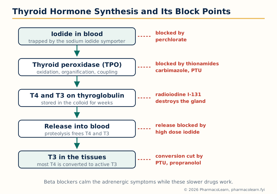
Figure: Thyroid hormone synthesis in the follicle, and the points where thionamides, iodide, radioiodine and beta blockers act.
1.5 Insulin: receptor signalling and the control of secretion
Insulin is secreted by pancreatic beta cells when glucose rises: glucose enters through GLUT2, its oxidation raises ATP, ATP sensitive potassium channels close, the membrane depolarises, calcium enters, and insulin granules are exocytosed. This secretion machinery matters here because the oral hypoglycemic agents of section 1.6 work by forcing it. Insulin's receptor is a receptor tyrosine kinase: insulin binding activates the intracellular kinase, which phosphorylates insulin receptor substrate proteins, and the signal divides between the PI3K Akt pathway (metabolic actions: GLUT4 transporters move to the membrane of muscle and adipose cells and take up glucose, glycogen synthesis is switched on, lipolysis and hepatic glucose output are switched off) and the MAPK pathway (growth effects). Failure of this signalling, insulin resistance in muscle, fat and liver, is the core defect of type 2 diabetes; absolute insulin deficiency from autoimmune beta cell destruction is type 1 diabetes, where insulin replacement is life saving and oral agents have no place.
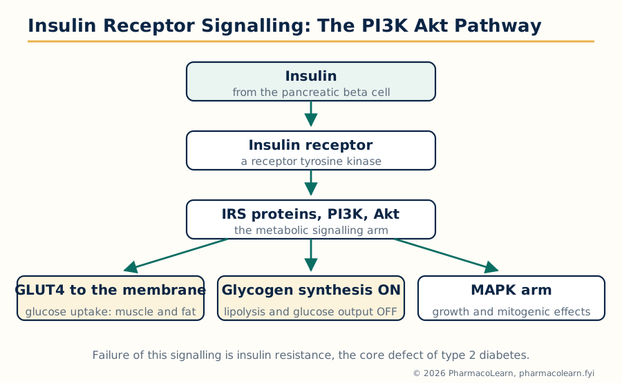
Figure: Insulin receptor signalling. The PI3K Akt arm produces the metabolic actions of insulin, including GLUT4 movement to the cell membrane.
1.6 Oral hypoglycemic agents
Type 2 diabetes is treated orally with classes that each repair a different defect. None replaces insulin in type 1 disease.
| Class and examples | Mechanism | Main advantages | Signature adverse effects |
|---|---|---|---|
| Sulfonylureas: glibenclamide, gliclazide, glimepiride | Close ATP sensitive potassium channels on the beta cell, forcing insulin secretion | Potent, cheap, long experience | Hypoglycaemia (worst with long acting glibenclamide, in the elderly and in renal failure) and weight gain |
| Meglitinides: repaglinide, nateglinide | Same channel, much shorter action, taken with meals | Controls post meal spikes, flexible with irregular meals | Hypoglycaemia, less than sulfonylureas |
| Biguanide: metformin | Reduces hepatic glucose output (AMPK linked) and improves insulin sensitivity; does not stimulate secretion | No weight gain, no hypoglycaemia alone, first line standard | GI upset; lactic acidosis risk in renal failure and acute illness, so it is stopped when patients are acutely unwell |
| Thiazolidinediones: pioglitazone | PPAR gamma nuclear receptor agonists, increase insulin sensitivity in adipose tissue | No hypoglycaemia alone, durable control | Weight gain and fluid retention, worsens heart failure, bone fractures; slow onset over weeks (a gene transcription drug) |
| Alpha glucosidase inhibitors: acarbose, voglibose | Block brush border enzymes, delay carbohydrate digestion | Targets post meal glucose | Flatulence and diarrhoea from undigested carbohydrate reaching the colon |
| DPP-4 inhibitors: sitagliptin, vildagliptin | Block the enzyme destroying incretins (GLP-1 and GIP), so glucose dependent insulin secretion rises and glucagon falls | Weight neutral, low hypoglycaemia risk | Generally well tolerated; pancreatitis signal watched |
| SGLT2 inhibitors: dapagliflozin, empagliflozin | Block sodium glucose cotransporter 2 in the proximal tubule, glucose is lost in the urine | Weight and blood pressure fall; proven heart failure and kidney protection | Genital mycotic infections, volume depletion; rare euglycaemic ketoacidosis |
Two principles organise the table. Secretagogues (sulfonylureas, meglitinides) need working beta cells and can cause hypoglycaemia; sensitisers and the newer classes cannot, used alone. And the modern classes are chosen on more than glucose: the SGLT2 inhibitors and the injectable GLP-1 receptor agonists of section 5.10 have outcome evidence in heart failure, kidney disease and cardiovascular events, which is why guidelines now place them early in patients with those complications. Insulin itself, in type 2 disease, is added when oral therapy fails or during acute illness; its preparations (rapid, short, intermediate and long acting analogues) are dosed in units and titrated against glucose readings, a practical detail students confuse with the oral agents at their peril.
1.7 Sex hormones and oral contraceptives
The sex steroids oestrogen, progesterone and androgens are made from cholesterol under hypothalamic pituitary control (GnRH, LH and FSH) and act on intracellular steroid receptors that regulate transcription, the slow, durable signalling of section 1.1. Oestrogens grow the endometrium, maintain bone and shape lipid profiles; progesterone prepares and holds the endometrium and is secreted in the luteal phase; their cyclical interplay is the menstrual cycle, and its pharmacological override is contraception.
Combined oral contraceptives pair an oestrogen (ethinylestradiol) with a progestin (levonorgestrel, desogestrel and others). Taken daily, the steady steroid signal suppresses pituitary FSH and LH by negative feedback: no follicle matures, the LH surge never fires, and ovulation does not occur; progestin additionally thickens cervical mucus and thins the endometrium. The benefits beyond contraception include lighter, regular, less painful periods and lower risks of ovarian and endometrial cancer. The adverse ledger is dominated by the oestrogen: a several fold rise in venous thromboembolism (highest in the first months, with third generation progestins somewhat above levonorgestrel pills), a small rise in arterial events in smokers over 35, nausea and breast tenderness, and the interaction trap: enzyme inducing drugs (rifampicin, carbamazepine) speed steroid clearance and cause contraceptive failure, while ordinary antibiotics do not. Progestin only pills avoid oestrogen risk but demand strict timing; long acting progestins (implants, injections, intrauterine systems) remove the daily adherence problem entirely. Emergency contraception with levonorgestrel delays ovulation and works only before the LH surge. On the androgen side, testosterone is replaced in hypogonadism, abused as an anabolic steroid with suppression of the axis it mimics, and blocked therapeutically by finasteride (5 alpha reductase inhibitor, benign prostatic hyperplasia and hair loss) and the antiandrogens used in prostate cancer.
1.8 Corticosteroids: the glucocorticoid receptor and its price
Cortisol is secreted by the adrenal cortex under ACTH, itself under hypothalamic CRH, with negative feedback at both levels: the hypothalamic pituitary adrenal (HPA) axis. The glucocorticoid receptor sits in the cytoplasm held by heat shock proteins; cortisol binding frees it, the complex enters the nucleus and changes transcription in two directions: transactivation of metabolic genes (gluconeogenesis enzymes, the source of steroid diabetes and much of the toxicity) and transrepression of the inflammatory transcription factors NF kappa B and AP-1, shutting down cytokines, adhesion molecules, COX-2 and inducible nitric oxide synthase. That second arm is the anti inflammatory and immunosuppressive action for which synthetic corticosteroids (prednisolone, methylprednisolone, dexamethasone, chosen by potency, duration and mineralocorticoid activity) are prescribed across every specialty: asthma, rheumatoid disease, transplant rejection, cerebral oedema, replacement in adrenal failure.
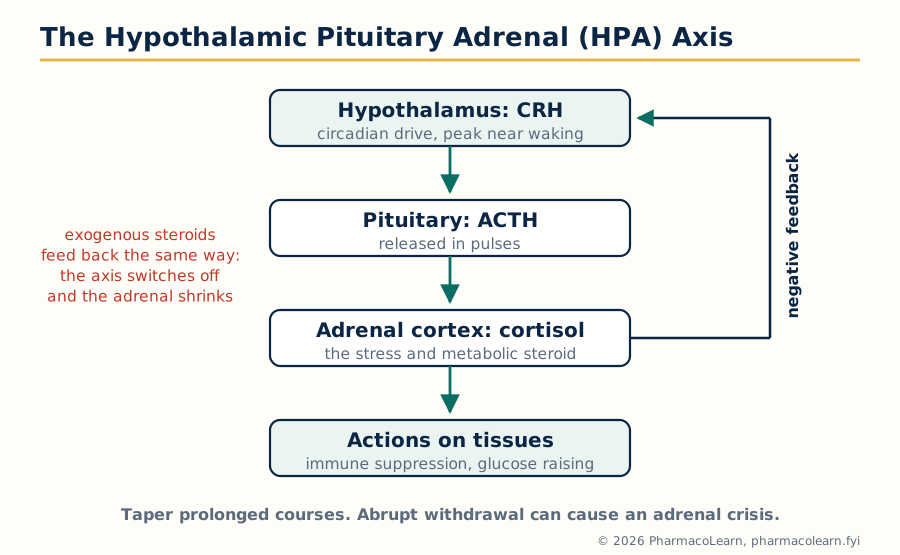
Figure: The hypothalamic pituitary adrenal axis. Exogenous corticosteroids feed back like cortisol and, given for weeks, leave the adrenal unable to respond when they stop.
The price list is the physiology read backwards: hyperglycaemia and diabetes, central obesity and muscle wasting, hypertension and fluid retention (mineralocorticoid spillover), osteoporosis and growth suppression, cataracts and glaucoma, thin skin and poor healing, mood change and psychosis, and two dangers students must never miss. First, immune suppression reactivates latent infections, tuberculosis above all in this country. Second, HPA suppression: exogenous steroid switches off CRH and ACTH, the adrenal cortex atrophies, and abrupt withdrawal after prolonged therapy can produce an adrenal crisis of hypotension and collapse; steroids are therefore tapered, and patients carry steroid cards warning that illness or surgery needs extra cover. Mineralocorticoid replacement (fludrocortisone) is the other half of adrenal replacement, acting at the mineralocorticoid receptor to retain sodium through the epithelial sodium channel, the same channel amiloride blocks from the diuretic side in Semester 1.
1.9 Drugs affecting calcium regulation
Blood calcium is held in a narrow range by three hormones working on gut, bone and kidney. Parathyroid hormone (PTH) raises calcium: it increases bone resorption, renal calcium reabsorption and phosphate excretion, and switches on renal activation of vitamin D. Calcitriol (active vitamin D) raises calcium and phosphate by increasing intestinal absorption. Calcitonin opposes resorption weakly and is a minor physiological player but a usable drug. Drugs exploit each arm:
- Bisphosphonates (alendronate, risedronate, zoledronic acid) bind bone mineral and poison osteoclasts; nitrogen containing members block the mevalonate pathway enzyme farnesyl pyrophosphate synthase inside the osteoclast. They are the mainstay of osteoporosis and of hypercalcaemia of malignancy; poor oral absorption and oesophagitis dictate the fasting, upright dosing ritual.
- Denosumab is a monoclonal antibody against RANK ligand, the osteoclast activating signal, blocking resorption from outside the cell.
- Teriparatide, a PTH fragment, paradoxically builds bone when given as a daily pulse (intermittent exposure favours formation, continuous exposure favours resorption) and is reserved for severe osteoporosis.
- Cinacalcet is a calcimimetic: it makes the parathyroid calcium sensing receptor more sensitive, so the gland behaves as if calcium were higher and PTH falls; used in secondary hyperparathyroidism of renal failure.
- Vitamin D preparations (cholecalciferol for deficiency, calcitriol and alfacalcidol where renal activation fails) and calcium salts complete the set; calcitonin survives mainly for acute hypercalcaemia, where saline, bisphosphonates and glucocorticoids do the heavier work.
Unit 2: Chemotherapy: Antimicrobial Agents and Resistance
2.1 Principles of antimicrobial chemotherapy and the routes to resistance
Antimicrobial chemotherapy rests on selective toxicity: the drug hits a target the pathogen has and the host lacks, or a shared target in a form the host tolerates. Four target families organise the whole field: the bacterial cell wall (absent from human cells), the bacterial ribosome (70S against the host 80S), bacterial enzymes of folate synthesis and DNA handling (different from the host versions), and the fungal membrane sterol ergosterol (cholesterol's counterpart). A drug is bactericidal if it kills and bacteriostatic if it arrests growth and lets immunity finish the work, a distinction that matters in endocarditis, meningitis and the neutropenic patient, where there is no immunity to finish anything.
Resistance is the syllabus's second theme in this unit, and every class below is read through it. Bacteria resist by four general routes:
THE FOUR GENERAL ROUTES TO ANTIMICROBIAL RESISTANCE

  1. Destroy or modify the drug
       beta lactamases hydrolyse beta lactams
       aminoglycoside modifying enzymes acetylate, adenylate, phosphorylate
  2. Change the target
       altered penicillin binding proteins (MRSA), methylated ribosomes
       (erm genes), mutated DNA gyrase, changed dihydrofolate reductase
  3. Keep the drug out, or throw it out
       lost porins (Gram negative outer membrane), efflux pumps
       (mex systems of Pseudomonas, mef pumps of pneumococci)
  4. Bypass the blocked pathway
       an alternative enzyme the drug cannot inhibit (the mecA product
       PBP2a still builds wall when the native PBPs are blocked)

  Spread: mutation under selection, and horizontal gene transfer on
  plasmids, transposons and integrons, which is why resistance travels
  between species and between hospitals faster than mutation alone allows.
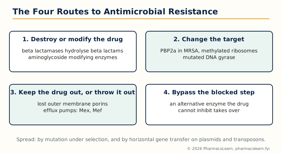
Figure: The four general routes by which bacteria resist antimicrobial drugs. Every class specific resistance in this unit is one of these four, wearing a class uniform.
2.2 Beta lactams: penicillin binding proteins and the beta lactamases
The beta lactams (penicillins, cephalosporins, carbapenems, monobactams) share a four membered beta lactam ring that mimics the D alanine D alanic acid end of the peptidoglycan precursor. They bind the penicillin binding proteins (PBPs), the transpeptidases that cross link the cell wall, and acylate them irreversibly. Wall synthesis stops while autolytic enzymes keep working, and the growing bacterium bursts. Killing therefore needs growth, and beta lactams are the classic time dependent, bactericidal agents. The classes differ in spectrum and stability: benzylpenicillin is narrow and acid labile, amoxicillin adds Gram negative reach, the later cephalosporin generations trade Gram positive for Gram negative power (ceftriaxone, ceftazidime), carbapenems (meropenem, imipenem) are the broadest and most beta lactamase stable, and aztreonam covers Gram negatives only.
Resistance comes by three of the four routes. Beta lactamases hydrolyse the ring: hundreds exist, from simple penicillinases (why flucloxacillin was built penicillinase proof) through the extended spectrum beta lactamases (ESBLs) that destroy third generation cephalosporins, to the carbapenemases (KPC, NDM and others) that threaten the last line. The counter move is the beta lactamase inhibitor: clavulanate, sulbactam, tazobactam, and the newer avibactam, suicide or shield molecules given in fixed combination (co amoxiclav is amoxicillin plus clavulanate). Altered targets give methicillin resistant Staphylococcus aureus its name: the acquired mecA gene encodes PBP2a, a transpeptidase beta lactams bind poorly, so the wall is built anyway; treatment needs vancomycin or the anti MRSA cephalosporin ceftaroline. Porin loss and efflux in Pseudomonas and other Gram negatives complete the picture. The class adverse signature is hypersensitivity, up to anaphylaxis, with partial cross reactivity between penicillins and cephalosporins that modern data put far below the old ten percent teaching.
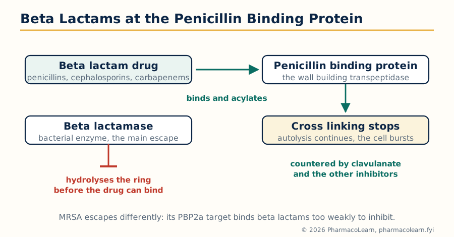
Figure: Beta lactam action at the penicillin binding protein, and destruction of the drug by beta lactamase before it can bind.
2.3 Aminoglycosides: the 30S subunit and oxygen dependent killing
The aminoglycosides (gentamicin, amikacin, tobramycin, streptomycin) are polycationic sugars that must be actively transported into the bacterium, a process needing oxygen and membrane energy, which is why they fail against anaerobes and in acidic, anaerobic abscesses. Inside, they bind the 30S ribosomal subunit at the decoding site: the ribosome misreads messenger RNA, inserting wrong amino acids, and the abnormal proteins damage the membrane, letting still more drug in. The result is rapid, concentration dependent bactericidal killing with a post antibiotic effect, the pharmacokinetic pattern behind once daily dosing. They cover aerobic Gram negatives (and synergise with beta lactams against enterococcal endocarditis, the wall agent opening the door for them), but their toxicity is the price: nephrotoxicity (usually reversible) and ototoxicity (cochlear and vestibular, irreversible, worse with loop diuretics and in patients carrying mitochondrial mutations), plus neuromuscular blockade at high levels.
Resistance is mainly route 1 and route 2: aminoglycoside modifying enzymes (acetyltransferases, phosphotransferases, nucleotidyltransferases, often plasmid borne and different in spectrum, amikacin surviving many of them by its protective side chain), and, most ominously, acquired 16S ribosomal RNA methyltransferases that methylate the binding site itself and defeat the whole class at once.
2.4 Quinolones: DNA gyrase, topoisomerase IV and target mutation
The fluoroquinolones (ciprofloxacin, levofloxacin, moxifloxacin) are synthetic agents that trap the bacterial enzymes DNA gyrase (topoisomerase II, the primary target in Gram negatives, which introduces negative supercoils ahead of the replication fork) and topoisomerase IV (primary in Gram positives, which separates daughter chromosomes) in their DNA cleavage complex; the trapped complexes shatter into double strand breaks and the cell dies. They are concentration dependent, bactericidal, superbly bioavailable, and concentrated in tissues, which made them oral answers to infections once needing intravenous therapy.
Resistance accumulates stepwise: point mutations in the quinolone resistance determining regions of gyrA and parC lower binding, each mutation raising the MIC until clinical resistance emerges; plasmid borne qnr proteins shield the enzymes; efflux pumps and a modifying enzyme variant (aac(6')-Ib-cr) add smaller contributions, important because they let bacteria survive long enough to select the chromosomal mutations. The class cautions are tendinitis and tendon rupture, QT prolongation, dysglycaemia, CNS effects and the aortic aneurysm warning, and ciprofloxacin's inhibition of CYP1A2, which raises theophylline levels.
2.5 Macrolides: the 50S exit tunnel and ribosomal methylation
The macrolides (erythromycin, clarithromycin, azithromycin) are large lactone rings that bind the 50S ribosomal subunit at the nascent peptide exit tunnel, blocking translocation so the growing chain stalls; they are generally bacteriostatic, concentrated inside cells, and therefore strong against the atypical organisms (Mycoplasma, Legionella, Chlamydia) and in respiratory infection. Azithromycin's long tissue half life supports short courses and single dose regimens; clarithromycin adds Helicobacter pylori (Unit 4) and mycobacteria; erythromycin is also a motilin receptor agonist, a prokinetic side effect used deliberately in gastroparesis and suffered as cramping in infection.
Resistance divides neatly by geography and mechanism: erm genes encode methylases that dimethylate a single adenine of 23S ribosomal RNA, producing macrolide lincosamide streptogramin B (MLSB) resistance, inducible or constitutive; mef genes encode efflux pumps giving lower level macrolide only resistance, common in pneumococci in some regions; and 23S mutations occur in Helicobacter and mycobacteria. Drug interactions define clarithromycin and erythromycin in practice: both inhibit CYP3A4 strongly (azithromycin does not), raising levels of statins, warfarin and many others, and all macrolides can prolong the QT interval.
2.6 Antifungal drugs: ergosterol and the fungal wall
Fungi are eukaryotes, so selective toxicity is harder and the targets fewer. Amphotericin B, a polyene, binds ergosterol in the fungal membrane and forms pores; the cell leaks and dies. It remains the broadest, most reliably fungicidal agent for invasive mycoses, paid for with infusion reactions and dose limiting nephrotoxicity, which lipid formulations soften. The azoles (fluconazole, itraconazole, voriconazole, posaconazole) inhibit lanosterol 14 alpha demethylase, a fungal CYP enzyme, starving the membrane of ergosterol; they are mostly fungistatic, orally available, and themselves CYP inhibitors with rich interaction lists. The echinocandins (caspofungin, micafungin) inhibit beta 1,3 glucan synthase, dissolving the fungal cell wall that human cells do not possess at all, the cleanest selective target in the field, invaluable against Candida including many azole resistant strains. Flucytosine is converted inside the fungus to 5 fluorouracil and corrupts its RNA and DNA, used only in combination (with amphotericin for cryptococcal meningitis) because resistance emerges rapidly alone. Terbinafine blocks squalene epoxidase upstream of ergosterol and is the oral standard for nail infection. Resistance, once rare, now matters: azole resistant Aspergillus from agricultural azole use, and Candida species with efflux pump overexpression or target mutation, are reshaping empirical choices.
2.7 Antiviral drugs: hitting a parasite that uses the host
Viruses borrow the host's machinery, so antiviral targets are the few virus specific steps: viral enzymes and entry proteins. The anti herpes agents start the story: aciclovir is a guanosine analogue activated first by viral thymidine kinase (the selectivity trick), then by host kinases to a triphosphate that inhibits viral DNA polymerase and terminates the chain; valaciclovir is its oral prodrug, and ganciclovir extends the pattern to cytomegalovirus at the price of marrow toxicity.
Antiretroviral therapy (ART) for HIV is the field's triumph and its discipline. The classes map onto the viral life cycle: nucleoside reverse transcriptase inhibitors (tenofovir, emtricitabine, zidovudine, abacavir) fake the building blocks of reverse transcription; non nucleoside reverse transcriptase inhibitors (efavirenz, rilpivirine) jam the enzyme's allosteric pocket; protease inhibitors (ritonavir or cobicistat boosted darunavir and others) stop maturation of viral proteins; integrase strand transfer inhibitors (dolutegravir, bictegravir) block insertion of viral DNA into the host genome and now anchor most first line regimens; entry and fusion agents (maraviroc, enfuvirtide) serve salvage situations. Viral replication is error prone and fast, so a single drug selects resistance within weeks: ART is always combination therapy, three active agents, and adherence is pharmacology, not advice, since missed doses are the selection pressure. Elsewhere: oseltamivir inhibits influenza neuraminidase, preventing release of new virions; hepatitis B is suppressed by tenofovir and entecavir; hepatitis C is now cured by direct acting combinations (sofosbuvir based) that inhibit the NS5B polymerase, NS5A and the NS3/4A protease; remdesivir, a nucleotide analogue of an RNA polymerase, represents the same logic in coronaviruses.
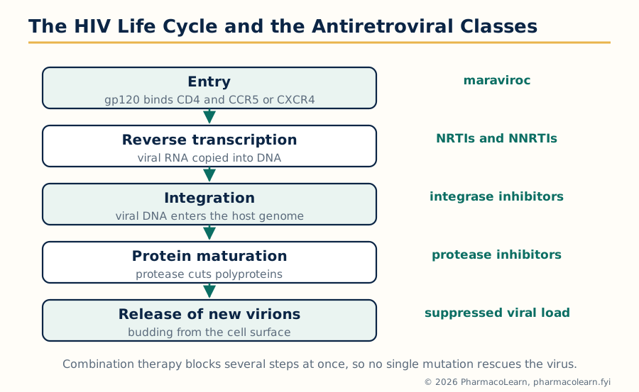
Figure: The HIV life cycle and the antiretroviral classes. Combination therapy blocks several steps at once so that no single mutation rescues the virus.
2.8 Anti-TB drugs: four drugs against persistence and resistance
Tuberculosis combines every chemotherapy difficulty in one disease: a slow growing bacillus with a waxy mycolic acid wall, populations hiding in different states (actively dividing, semi dormant in acidic caseous foci, intracellular), and a huge bacterial load in cavitary disease within which resistant mutants already exist by chance. Standard drug sensitive TB is therefore treated with four drugs for two months, then two drugs for four months (the 2HRZE/4HR pattern), each drug covering the others' resistant mutants and each hitting a different bacterial population:
| Drug | Mechanism | Population it kills best | Signature toxicity |
|---|---|---|---|
| Isoniazid (H) | Prodrug activated by the bacterial catalase peroxidase KatG; the activated form inhibits mycolic acid synthesis (InhA) | Actively dividing bacilli, the most potent early killer | Hepatitis; peripheral neuropathy prevented by pyridoxine (vitamin B6), worst in slow acetylators and diabetics |
| Rifampicin (R) | Inhibits bacterial DNA dependent RNA polymerase | Semi dormant persisters, the great sterilising drug that shortened therapy to six months | Hepatitis, orange urine and tears, and potent enzyme induction collapsing the levels of contraceptives, warfarin and transplant drugs |
| Pyrazinamide (Z) | Prodrug converted by bacterial pyrazinamidase; active against bacilli in acidic environments | Acidic, caseous and intracellular populations, first two months | Hepatitis, raised uric acid and arthralgia |
| Ethambutol (E) | Inhibits arabinosyl transferases building the arabinogalactan wall layer | Protects the regimen while susceptibilities are unknown | Optic neuritis: loss of acuity and red green colour vision, checked during therapy |
Resistance follows predictably when therapy is inadequate: loss of KatG or InhA change defeats isoniazid, rpoB mutation defeats rifampicin, and multidrug resistant TB (MDR TB, resistant to at least isoniazid and rifampicin) and extensively drug resistant disease need second line regimens now built around bedaquiline (which inhibits mycobacterial ATP synthase, a wholly new target), pretomanid, linezolid and the fluoroquinolones, supervised for far longer. The unit's message generalises: in chemotherapy, the regimen is designed around the resistance arithmetic of the pathogen population, not around any single drug.
Unit 3: Chemotherapy of Protozoa, Helminths and Cancer; Immunopharmacology
3.1 Antiprotozoal drugs I: the antimalarials
Malaria teaches chemotherapy against a parasite with a staged life cycle, because each stage has its own drugs. Sporozoites from the mosquito infect liver cells (the silent exoerythrocytic stage); merozoites then cycle through red cells, causing the fever; in Plasmodium vivax and P. ovale some liver forms sleep as hypnozoites and relapse months later; gametocytes return to the mosquito. Chloroquine concentrates in the parasite's food vacuole and blocks detoxification of haem into haemozoin, so free haem poisons the parasite; resistance, spread from two foci to most of the world through the PfCRT efflux transporter, ended its reign against P. falciparum. The artemisinins (artesunate, artemether) carry an endoperoxide bridge that iron from haem splits into carbon radicals, damaging parasite proteins wholesale; they clear parasites faster than any other class and anchor artemisinin based combination therapy (such as artemether lumefantrine), the partner drug guarding against the artemisinin resistance now creeping through Kelch13 mutants in Southeast Asia. Intravenous artesunate is the treatment of severe malaria and has saved more lives than any recent antimalarial decision. Quinine remains a reserve parenteral agent, with cinchonism and hypoglycaemia as its costs. Primaquine alone kills hypnozoites (the radical cure of vivax and ovale) and gametocytes, but it oxidatively stresses red cells and can haemolyse patients with glucose 6 phosphate dehydrogenase deficiency, which must be considered before use. Antifolate combinations (sulfadoxine pyrimethamine) block the parasite's folate pathway at two sequential steps; mefloquine, atovaquone proguanil and doxycycline fill prophylaxis and treatment niches. Every one of these assignments follows the life cycle stage, which is the only way the list makes sense.
3.2 Antiprotozoal drugs II: amoebiasis, leishmaniasis and the rest
Metronidazole is the tissue amoebicide of amoebic dysentery and liver abscess: anaerobic and microaerophilic organisms reduce its nitro group to cytotoxic radicals that shred DNA, a trick that also makes it the workhorse against anaerobic bacteria and Giardia. Because it is absorbed before reaching the colonic lumen, it must be followed by a luminal amoebicide (diloxanide furoate or paromomycin) to clear cysts. Visceral leishmaniasis (kala azar) is treated in India chiefly with liposomal amphotericin B, the polyene of Unit 2 redirected against the leishmania amastigote's membrane sterols; the older pentavalent antimonials are neurotoxic and cardiotoxic and largely abandoned here, and miltefosine, an oral membrane lipid drug, is teratogenic and losing ground to resistance. African trypanosomiasis uses suramin and melarsoprol (an arsenical whose own encephalopathy is feared), Chagas disease benznidazole and nifurtimox, toxoplasmosis in the immunocompromised pyrimethamine with sulfadiazine, and pneumocystis pneumonia, a fungus by taxonomy but treated here by tradition, co trimoxazole. The pattern is less tidy than the antibacterial field because each parasite is its own pharmacology problem.
3.3 Drugs for helminthiasis
Worms are multicellular animals, and the drugs exploit their neuromuscular and energy machinery, paralysing or starving the worm so the gut expels it. The benzimidazoles (albendazole, mebendazole) bind worm beta tubulin far more avidly than the mammalian form, stopping microtubule assembly and glucose uptake; albendazole, with its active metabolite reaching tissues, is the broad agent for roundworm, hookworm, whipworm, threadworm and the tissue diseases hydatid cyst and neurocysticercosis, where killing cysts in the brain demands corticosteroid cover against the inflammatory fallout. Praziquantel raises calcium permeability of the fluke and schistosome tegument, causing spastic paralysis and surface damage that exposes the worm to immunity; it is the drug of schistosomiasis and tapeworm infections and is remarkably safe. Ivermectin opens glutamate gated chloride channels found only in invertebrates, paralysing nematodes and ectoparasites; it transformed onchocerciasis (river blindness) control and treats strongyloidiasis and scabies. Diethylcarbamazine immobilises microfilariae of filariasis and alters their surface so the host destroys them, and pyrantel pamoate depolarises worm muscle as a nicotinic agonist; niclosamide, a luminal tapeworm agent, uncouples worm oxidative phosphorylation. Local reactions after worm killing (the Mazzotti reaction of onchocerciasis) are immunopathology, not drug toxicity, an important bedside distinction.
3.4 Chemotherapy of cancer: principles and the cell cycle
Cancer chemotherapy began with nitrogen mustards and remains governed by three ideas. First, log kill: a given treatment kills a constant fraction, not a constant number, of tumour cells, so repeated cycles are needed to drive a burden of billions of cells toward zero, and small tumours respond better than large ones. Second, most cytotoxic drugs damage DNA or the machinery of division, so they hurt cells in proportion to how fast they divide; the same logic explains the toxicity pattern, since marrow, gut lining, hair follicles and gonads are the body's own fast dividing tissues (myelosuppression, mucositis, alopecia and infertility are the expected class prices). Third, tumours are genetically unstable populations that evolve resistance (by drug efflux through P glycoprotein, target change, DNA repair and blocked apoptosis), so combination regimens of drugs with different mechanisms and non overlapping toxicities are used, exactly the arithmetic of TB therapy in Unit 2.
THE CELL CYCLE AND WHERE THE CYTOTOXIC DRUGS STRIKE

   G1 (growth) ---------------> S (DNA synthesis)
      ^                            |
      |                            |  antimetabolites (methotrexate,
      |                            |  5 fluorouracil, cytarabine)
   G2 (preparation)                v
      ^                         M (mitosis)
      |                            |
      +------- resting G0 ---------+
        vinca alkaloids and taxanes strike in M phase
        alkylating agents and platinum drugs strike in all phases
        (cross linked DNA fails whenever the cell tries to use it)
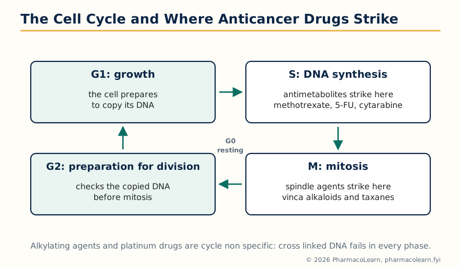
Figure: The cell cycle and the phase specificity of the cytotoxic classes. Phase specific drugs work best against tumours with a high growth fraction and in schedules that expose cells as they enter the sensitive phase.
3.5 The anticancer drug classes
| Class and examples | Mechanism | Signature toxicities and notes |
|---|---|---|
| Alkylating agents: cyclophosphamide, chlorambucil, busulfan | Covalent cross links in DNA; cycle non specific | Cyclophosphamide's metabolite acrolein causes haemorrhagic cystitis, prevented by mesna and hydration |
| Platinum compounds: cisplatin, carboplatin, oxaliplatin | Intrastrand DNA cross links | Cisplatin: severe emesis, nephrotoxicity, ototoxicity; oxaliplatin: cold triggered neuropathy |
| Antimetabolites: methotrexate, 5 fluorouracil, cytarabine, mercaptopurine | Fraudulent substrates starving DNA synthesis; S phase specific | Methotrexate is rescued by folinic acid (leucovorin); 5 fluorouracil toxicity explodes in DPD enzyme deficiency |
| Antitumour antibiotics: doxorubicin, bleomycin, dactinomycin | Intercalation, topoisomerase II trapping, free radical strand breaks | Doxorubicin: cumulative, dose related cardiomyopathy; bleomycin: pulmonary fibrosis, and skin toxicity because skin lacks its inactivating enzyme |
| Microtubule agents: vincristine, paclitaxel | Vinca alkaloids prevent spindle assembly; taxanes freeze disassembly; both arrest mitosis | Vincristine: peripheral neuropathy, dangerous if given intrathecally; paclitaxel: neuropathy and hypersensitivity |
| Topoisomerase inhibitors: etoposide, irinotecan | Trap topoisomerase II or I cleavage complexes | Irinotecan: early and late diarrhoea, the late form from its active metabolite in the gut |
| Hormonal agents: tamoxifen, aromatase inhibitors, androgen deprivation | Block the growth signal of hormone dependent breast and prostate cancers | Tamoxifen is a selective oestrogen receptor modulator, antagonist in breast, agonist in uterus and bone |
| Targeted small molecules: imatinib and successors | Inhibit the driver kinase, here the BCR ABL fusion of chronic myeloid leukaemia | Imatinib converted CML into a chronic oral therapy; resistance by kinase mutation (T315I) drove later generations |
| Monoclonal antibodies: trastuzumab, rituximab | Bind HER2 or CD20 and recruit immune killing as well as signal blockade | Trastuzumab cardiotoxicity, watched by echocardiography |
| Immune checkpoint inhibitors: pembrolizumab, nivolumab | Block PD-1 or PD-L1, releasing T cells from tumour induced braking | Immune related adverse events in any organ: colitis, pneumonitis, endocrinopathies |
3.6 Immunopharmacology: the cells and mediators of inflammation and immune response
The immune system defends with an innate arm (immediate, pattern based) and an adaptive arm (slow, specific, with memory), and inflammation is the visible form of both at work in tissue. The cellular players: neutrophils that phagocytose and degranulate, macrophages that phagocytose, present antigen and secrete cytokines, mast cells and basophils carrying IgE and preformed histamine, eosinophils of allergy and helminth defence, and the lymphocytes, T cells (helper, cytotoxic and regulatory) commanding, B cells becoming antibody factories, and natural killer cells surveying for missing self.
The biochemical mediators are the pharmacology, since most are drug targets or drug mimics:
| Mediator family | Source and signal | Pharmacological handles |
|---|---|---|
| Histamine | Mast cells and basophils; H1 and H2 receptors (Unit 5 of Semester 1) | Antihistamines, H2 blockers |
| Eicosanoids | Arachidonic acid through COX and lipoxygenase (Semester 1) | NSAIDs, corticosteroids upstream, montelukast |
| Cytokines: TNF alpha, interleukins 1, 6, 17, interferons | Macrophages and T cells; protein signals between immune cells | Monoclonal antibodies against TNF (infliximab, adalimumab), IL-6 receptor (tocilizumab), IL-17; JAK inhibitors inside the cell |
| Complement | Plasma cascade, C3a and C5a anaphylatoxins, membrane attack complex | Eculizumab blocks C5 |
| Bradykinin and nitric oxide | Plasma kallikrein system; endothelium and macrophages | Icatibant; corticosteroids suppress iNOS induction |
| Platelet activating factor and chemokines | Membranes; many cell types; direct cell traffic | Fewer direct drugs, but they explain recruitment patterns |
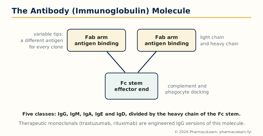
Figure: The antibody molecule. The variable tips bind antigen, the constant Fc stem decides what the immune system does next, and therapeutic monoclonal antibodies are engineered versions of this molecule.
3.7 Allergic and hypersensitivity reactions
The Gell and Coombs classification sorts immune injury into four mechanisms, and drug allergy distributes across all four:
| Type | Mechanism | Time course | Drug examples |
|---|---|---|---|
| I, immediate | IgE on mast cells cross linked by antigen; histamine and leukotriene release | Minutes | Penicillin anaphylaxis, urticaria, angioedema |
| II, cytotoxic | IgG or IgM against cell surface antigens; complement and phagocyte destruction | Hours to days | Drug induced haemolytic anaemia (methyldopa, penicillin hapten type), immune thrombocytopenia (quinine, heparin HIT in its own category) |
| III, immune complex | Antigen antibody complexes deposit in vessels and tissues, complement inflammation | Days to weeks | Serum sickness (antitoxins, some monoclonals), drug induced vasculitis |
| IV, delayed | T cell mediated, no antibody; macrophages and cytotoxic T cells | 48 to 72 hours and beyond | Contact dermatitis (topical agents), Stevens Johnson syndrome and toxic epidermal necrolysis (allopurinol, carbamazepine, lamotrigine), DRESS |
Management repeats one rule: stop the drug, because no antidote outruns continued exposure; anaphylaxis is treated with intramuscular adrenaline first (antihistamines and steroids are supporting acts), and Stevens Johnson syndrome is a burns unit emergency. Cross reactivity questions (penicillin and cephalosporin, sulfonamide antibiotic and non antibiotic sulfonamides) are decided by side chain and structure, not by the word sulphur, a precision examiners test.
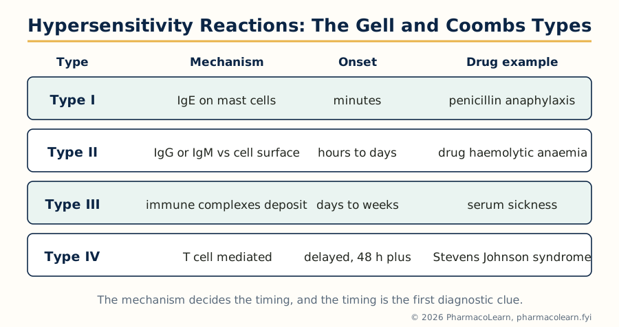
Figure: The four hypersensitivity types. The mechanism decides the timing, and the timing is the first diagnostic clue at the bedside.
3.8 Pharmacotherapy of asthma
Asthma is chronic airway inflammation with reversible bronchoconstriction and hyperresponsiveness; the mediators of section 3.6 (histamine, leukotrienes, eosinophilic cytokines) are its machinery, and treatment divides into controllers (daily anti inflammatory therapy) and relievers (as needed bronchodilators). Inhaled corticosteroids (beclomethasone, budesonide, fluticasone) are the foundation: genomic suppression of airway inflammation, with local adverse effects (oral candidiasis, dysphonia) minimised by spacers and mouth rinsing, and systemic effects small at ordinary inhaled doses. Long acting beta2 agonists (salmeterol, formoterol) relax airway smooth muscle for twelve hours and are never used alone in asthma, only with an inhaled steroid, because bronchodilation without inflammation control masks deterioration; formoterol's fast onset lets a steroid formoterol inhaler serve as reliever too, the modern single inhaler strategy. Short acting beta2 agonists (salbutamol) remain the universal reliever, by inhaler or nebuliser. Leukotriene receptor antagonists (montelukast) suit allergic and aspirin sensitive asthma and exercise induced symptoms. Theophylline, a narrow index phosphodiesterase inhibitor and adenosine antagonist, survives as an add on where resources or responses dictate, with level monitoring. For severe allergic and eosinophilic asthma, biologics aim at single mediators: omalizumab (anti IgE), mepolizumab and benralizumab (anti IL-5 pathway), and dupilumab (anti IL-4 and IL-13 signalling). Acute severe asthma is oxygen, nebulised salbutamol with ipratropium, systemic corticosteroids and intravenous magnesium sulfate, in that order of urgency.
3.9 Pharmacotherapy of COPD
Chronic obstructive pulmonary disease is progressive, largely smoking driven airflow obstruction with less reversibility and a different inflammatory flavour (neutrophils and macrophages more than eosinophils), so the pharmacotherapy leans on bronchodilation. Long acting muscarinic antagonists (tiotropium) and long acting beta2 agonists are the maintenance core, singly or combined; inhaled corticosteroids are added only for patients with frequent exacerbations and higher blood eosinophils, because in COPD they raise pneumonia risk without the broad benefit seen in asthma. Roflumilast, an oral phosphodiesterase 4 inhibitor, helps the chronic bronchitis phenotype with exacerbations, at the cost of weight loss and GI effects. Smoking cessation, vaccination, pulmonary rehabilitation and long term oxygen in chronic hypoxaemia change survival more than any inhaler. Exacerbations are treated with short acting bronchodilators, a short systemic steroid course, antibiotics when infection is likely, and controlled oxygen with ventilatory support when needed.
3.10 Immunosuppressants
Transplantation and autoimmune disease require deliberate weakening of immunity, and each class suppresses a different step of T cell activation. Calcineurin inhibitors (ciclosporin, tacrolimus) block the phosphatase calcineurin, so the transcription factor NFAT never switches on interleukin 2 production and T cell clonal expansion stalls at its start; both are nephrotoxic, level monitored, CYP3A4 substrates with dense interaction lists, tacrolimus being more potent and more diabetogenic. Antiproliferatives: azathioprine, a purine antimetabolite (its breakdown by TPMT decides toxicity, and allopurinol dangerously blocks one of its exits), and mycophenolate, which blocks the de novo purine synthesis lymphocytes depend on and is kinder to kidneys. mTOR inhibitors (sirolimus, everolimus) block the response to interleukin 2 downstream, trading nephrotoxicity for impaired wound healing and mouth ulcers. Glucocorticoids (Unit 1) remain the broad background suppressant. The biologics aim precisely: basiliximab against the IL-2 receptor for induction, antithymocyte globulin to deplete T cells, rituximab against B cells, abatacept blocking costimulation, and the anti TNF agents of section 3.6 in autoimmune disease. The universal costs are infection (including reactivated TB and hepatitis B, screened for before starting) and a raised long term malignancy risk: immunosuppression is a subscription, not a cure.
3.11 Immunostimulants
The opposite prescription is rarer and more specific. Levamisole, an old anthelmintic, restores depressed T cell and phagocyte function and survives in some steroid dependent nephrotic syndrome and adjuvant settings. BCG, the live tuberculosis vaccine strain, instilled into the bladder provokes a local immune attack that remains standard therapy for superficial bladder cancer, immunotherapy's first success. The interferons defend against viruses and modulate immunity (interferon beta in multiple sclerosis). Colony stimulating factors (filgrastim, G-CSF; sargramostim, GM-CSF) rebuild the neutrophil count after chemotherapy, converting section 3.4's dose limiting toxicity into a manageable one. Thymic peptides, inosine derivatives and microbial preparations occupy weaker evidence ground, and intravenous immunoglobulin, though listed with stimulants in some texts, works mainly as replacement and as an immunomodulator in autoimmune cytopenias and Kawasaki disease. Recombinant interleukin 2 (aldesleukin) can cure a small fraction of metastatic melanoma and renal cancer at the price of capillary leak, the sharpest demonstration that stimulating immunity is not gentler than suppressing it.
Unit 4: GIT Pharmacology and Chronopharmacology
4.1 The parietal cell and the regulation of gastric acid
The gastric parietal cell secretes hydrochloric acid through one final machine, the H+/K+ ATPase proton pump in its canalicular membrane, driven by three stimulatory inputs that converge on it: histamine from enterochromaffin like cells acting at H2 receptors (the amplifier, raised by gastrin), gastrin from antral G cells acting at CCK2 receptors directly and through histamine release, and acetylcholine from vagal fibres at M3 receptors. The brakes are prostaglandins E2 and I2 and somatostatin, which restrain histamine release and the parietal cell itself; this prostaglandin brake is why NSAIDs, which remove it, cause ulcers even without extra acid. Acid is both a digestive tool and a threat, held off the mucosa by the mucus bicarbonate barrier, mucosal blood flow and rapid epithelial repair: peptic ulcer disease and reflux are what happen when attack (acid, pepsin, Helicobacter pylori, NSAIDs) outlasts defence.
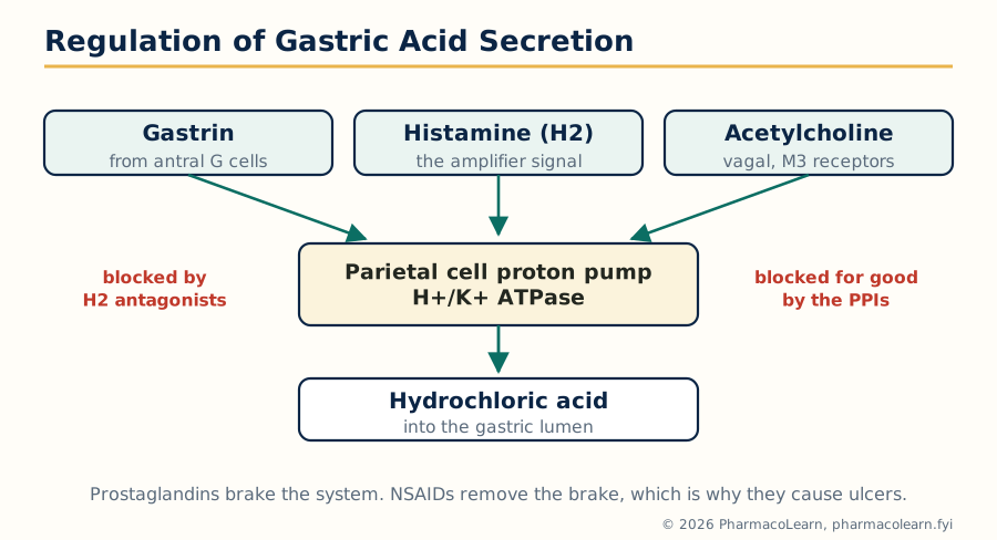
Figure: Regulation of gastric acid secretion. Three stimulants converge on the proton pump, which is why blocking the pump itself beats blocking any single input.
4.2 Antiulcer drugs and the eradication of H. pylori
Treatment either cuts acid or rebuilds defence. Proton pump inhibitors (omeprazole, pantoprazole, esomeprazole) are acid activated prodrugs that concentrate in the canaliculus and bind the proton pump covalently; because the bond is irreversible, acid secretion waits for new pumps, so one daily dose controls acid for a day and a half, and the dose is taken before the first meal, when pumps are inserted and active. They are the strongest and most durable acid suppressants and the backbone of ulcer healing, reflux disease and eradication regimens; long term use carries small but real costs (magnesium and B12 malabsorption, fracture risk, gut infection risk). H2 receptor antagonists (ranitidine's successors famotidine and others) block the histamine amplifier: effective, especially for night acid, but subject to tolerance and weaker than PPIs. Antacids (aluminium and magnesium hydroxides, calcium carbonate) neutralise acid already secreted, fast and brief, useful for symptoms, with diarrhoea (magnesium), constipation (aluminium) and drug chelation as their footprints. Mucosal protectives: sucralfate polymerises in acid into a physical bandage over the ulcer base; misoprostol replaces the missing prostaglandin in NSAID users; colloidal bismuth coats and also kills H. pylori. Helicobacter pylori, the bacterium behind most peptic ulcers and gastric cancer risk, is eradicated, not merely suppressed: a PPI with two antibiotics (clarithromycin and amoxicillin, or metronidazole) for fourteen days, with bismuth quadruple therapy where clarithromycin resistance, now common, defeats the standard triple. Eradication, not lifelong acid suppression, is what changed ulcer disease from a surgical career into a two week prescription.
4.3 Prokinetic drugs
Prokinetics speed the gut by releasing its own excitatory transmitters or blocking its brakes. Metoclopramide blocks dopamine D2 receptors in the gut wall and chemoreceptor trigger zone and, at higher doses, adds 5-HT4 agonism (more acetylcholine release) and 5-HT3 antagonism: it empties the stomach in gastroparesis and doubles as an antiemetic, but crosses into the brain, so dystonias in the young, sedation and, with prolonged use, tardive dyskinesia and hyperprolactinaemia limit its course. Domperidone is the same D2 logic kept outside the blood brain barrier: antiemetic and prokinetic with far fewer central effects, carrying instead a cardiac QT caution. Mosapride and prucalopride are selective 5-HT4 agonists producing motility without dopamine or cardiac baggage (prucalopride is used more for constipation, section 4.6). Erythromycin in low dose is a motilin receptor agonist, the fastest gastric emptier of the set, defeated by tachyphylaxis within weeks. Cisapride, the class's cautionary monument, was withdrawn for fatal QT arrhythmias, and every successor is examined against its ghost.
4.4 Antiemetic drugs
Vomiting is coordinated by a medullary centre fed by four inputs: the chemoreceptor trigger zone in the area postrema, outside the blood brain barrier, tasting blood for toxins through dopamine D2, serotonin 5-HT3, neurokinin NK1 and opioid receptors; the vestibular apparatus, signalling through histamine H1 and muscarine receptors (motion sickness); the gut, through vagal 5-HT3 receptors fired by mucosal serotonin release; and the cortex, anticipation and anxiety. Each drug family guards one gate:
| Family | Gate guarded | Best uses |
|---|---|---|
| 5-HT3 antagonists: ondansetron, granisetron, palonosetron | CTZ and gut vagal afferents | Chemotherapy, radiotherapy, postoperative vomiting; headache and constipation are their costs; QT care with dose |
| NK1 antagonists: aprepitant | CTZ, central | Delayed chemotherapy emesis, added to a setron and dexamethasone |
| D2 antagonists: metoclopramide, domperidone, prochlorperazine | CTZ | Migraine and gastroparesis emesis, general purpose cover, with the central costs of section 4.3 |
| H1 antihistamines: promethazine, meclizine; anticholinergic hyoscine | Vestibular pathway | Motion sickness (hyoscine patch is the classic), vertigo |
| Corticosteroids: dexamethasone | Mechanism debated, central | Chemotherapy combinations |
| Cannabinoids: nabilone | Central | Refractory chemotherapy emesis, where legal |
Highly emetogenic chemotherapy therefore gets a triple shield (NK1 antagonist plus setron plus dexamethasone), the same combination logic as chemotherapy itself.
4.5 Antidiarrhoeal drugs
Diarrhoea kills by water and salt loss, so the first drug is not a drug: oral rehydration solution, glucose driven sodium and water absorption, which has saved more lives than every antimotility agent combined. Drugs then work on secretion and motility. Opioid antimotility agents (loperamide, and codeine or diphenoxylate behind it) act at gut mu receptors to slow transit and increase absorption; loperamide is engineered to stay out of the brain, and all are dangerous in invasive bacterial dysentery and ulcerative colitis, where stopping the gut traps the pathogen and can dilate the colon to perforation. Racecadotril inhibits intestinal enkephalinase, protecting the gut's own antisecretory enkephalins: less secretion with no motility arrest, safe in children. Adsorbents and bismuth (kaolin, bismuth subsalicylate) are modest symptom helpers. Antibiotics are for specific causes (cholera, shigella, traveller's diarrhoea, Clostridioides difficile, which answers to oral vancomycin or fidaxomicin), not for diarrhoea in general, and antimotility drugs never substitute for finding the cause in a sick patient.
4.6 Drugs for constipation
Constipation is treated by adding water, bulk or propulsion, in that order of gentleness. Bulk forming agents (ispaghula, methylcellulose) are indigestible fibre that holds water and stretches the bowel into action, the physiological option, useless without fluid intake. Osmotic laxatives hold water by solute force: lactulose (also trapping ammonia in hepatic encephalopathy), macrogols (polyethylene glycol), and the saline agents magnesium hydroxide and sodium phosphate, the last two risky in renal failure. Stimulant laxatives (senna, bisacodyl) irritate the enteric nerves into propulsive contraction: effective, fast, unfairly feared, since dependence and bowel damage at normal doses are largely myth, though cramping is real. Stool softeners (docusate) are weak detergents of marginal effect. The modern targeted agents work on secretion and receptors: lubiprostone opens chloride channels, linaclotide activates guanylate cyclase C (both pouring fluid into the lumen), and prucalopride (section 4.3) drives motility through 5-HT4, all reserved for chronic constipation that simpler measures fail. Opioid induced constipation answers to the same ladder plus, in palliative care, peripherally acting mu antagonists such as methylnaltrexone.
4.7 Drugs for irritable bowel syndrome
Irritable bowel syndrome is a disorder of gut brain interaction: pain, bloating and disordered bowel habit without structural disease, so therapy is symptom aimed. Antispasmodics (mebeverine, dicyclomine, peppermint oil) relax gut smooth muscle against pain and spasm. For constipation predominant IBS, the secretagogues of section 4.6 (linaclotide, lubiprostone) and simple osmotic laxatives serve; for diarrhoea predominant IBS, loperamide, the bile acid binder colesevelam in bile acid diarrhoea, rifaximin (a non absorbed antibiotic for bloating and diarrhoea), and alosetron, a 5-HT3 antagonist restricted for its ischaemic colitis risk. Low dose tricyclic antidepressants (amitriptyline) quiet visceral hypersensitivity, an analgesic use unrelated to mood, while SSRIs help the pain and the anxiety that travels with the syndrome. Dietary work (a supervised low FODMAP approach) is a genuine co therapy, and the honest pharmacological summary is that IBS drugs manage, they do not cure.
4.8 Chronopharmacology: biological and circadian rhythms
The body is not the same patient at different hours. A master clock in the hypothalamic suprachiasmatic nucleus, set daily by light, drives roughly 24 hour (circadian) rhythms of cortisol (peak at waking), temperature, blood pressure (the early morning surge), airway calibre (narrowest near 4 am), gastric acid (highest at night), and drug handling itself, through clock genes (CLOCK, BMAL1, PER, CRY) running feedback loops in almost every cell. Two pharmacological consequences follow. Chronokinetics: absorption, distribution, metabolism and excretion vary with the hour, so the same dose gives different levels at different times. Chronodynamics: the target's sensitivity varies too: the HMG CoA reductase of cholesterol synthesis works hardest at night, asthmatic airways tighten at night, rheumatoid inflammation flares in the early morning on an interleukin 6 rhythm.
THE MOLECULAR CLOCK, SIMPLIFIED

  Light --> Suprachiasmatic nucleus (master clock)
                  |
                  v
     Peripheral clocks in liver, lung, gut, vessels
                  |
     CLOCK + BMAL1 proteins --switch ON--> Per and Cry genes
                  ^                              |
                  |                              v
                  +--- PER + CRY proteins accumulate,
                       enter the nucleus and switch
                       CLOCK BMAL1 OFF (loop takes ~24 h)
                       |
                       v
     Timed outputs: cortisol rhythm, blood pressure surge,
     airway tone, acid secretion, enzyme activity
4.9 Chronotherapy in practice: timing the dose to the disease
Chronotherapy schedules the dose so peak drug meets peak disease. In cardiovascular disease, events cluster in the morning surge of pressure, platelet activity and sympathetic tone; trials of bedtime dosing of antihypertensives reported large benefits and remain debated, but the principle that timing matters is established, and short acting statins such as simvastatin are dosed in the evening to meet the night peak of cholesterol synthesis (the long acting atorvastatin and rosuvastatin are indifferent to the clock). In asthma, symptoms and airway narrowing peak in the early morning hours, so once daily inhaled steroid and long acting bronchodilator cover is arranged to be strongest overnight, and a single afternoon dose of systemic steroid in exacerbations protects the night. In peptic ulcer, acid secretion peaks at night and the ulcer heals in a quiet stomach, so H2 blockers were classically given as a single bedtime dose and PPIs are timed before the meal that wakes the pumps. In diabetes, insulin regimens mimic the rhythm they replace: long acting basal insulin in the evening or morning matched to the dawn rise in glucose (the dawn phenomenon of early morning growth hormone and cortisol), rapid acting doses with meals. Rheumatoid arthritis answers to modified release prednisone taken at night and releasing near 2 am, ahead of the morning inflammatory peak. The examination point is symmetrical to the rest of pharmacology: dose, route and duration are joined by a fourth variable, time.
Unit 5: Free Radicals Pharmacology and Recent Advances in Treatment
5.1 Free radicals and reactive species: generation
A free radical is an atom or molecule carrying an unpaired electron, which makes it hungry to steal an electron from whatever it meets. The family that matters in medicine is the reactive oxygen species (ROS): the superoxide anion radical, hydrogen peroxide (not a radical but the diffusible parent of worse things), the hydroxyl radical (the most destructive, made when peroxide meets ferrous iron in the Fenton reaction), singlet oxygen, and the reactive nitrogen species nitric oxide and its toxic marriage with superoxide, peroxynitrite. Sources are built into normal life: the mitochondrial electron transport chain leaks electrons onto oxygen (the largest endogenous source, growing with age and hyperglycaemia), the NADPH oxidases of phagocytes make superoxide deliberately as a weapon (the respiratory burst that kills bacteria, absent in chronic granulomatous disease), xanthine oxidase in reperfused tissue, uncoupled nitric oxide synthase, peroxisomes, and cytochrome P450 reactions; outside sources add cigarette smoke, ionising radiation, ultraviolet light, air pollution and redox cycling drugs such as doxorubicin. In small controlled amounts ROS are signals (they tune vascular tone, insulin signalling and immune defence). Disease is oxidative stress: production outrunning defence, so radicals attack membrane lipids (chain reactions of lipid peroxidation), proteins (carbonylation, enzyme inactivation) and DNA (strand breaks and mutagenic bases such as 8 hydroxydeoxyguanosine).
5.2 The antioxidant defence and its pharmacology
Defence is layered, and each layer is a drug target or a drug. Superoxide dismutase converts superoxide to hydrogen peroxide; catalase and glutathione peroxidase (a selenium enzyme fed by reduced glutathione, GSH) convert peroxide to water. Glutathione itself, the cell's main soluble reductant, is regenerated by glutathione reductase using NADPH. Non enzymatic scavengers patrol the phases: vitamin E (alpha tocopherol) breaks lipid peroxidation chains inside membranes, vitamin C scavenges in water and regenerates vitamin E, and urate, albumin and bilirubin add smaller contributions. Metal binding is defence too: transferrin, ferritin and caeruloplasmin keep iron and copper away from the Fenton reaction. The pharmacological entries: N acetylcysteine supplies cysteine, the limiting building block of glutathione, and is the antidote that saves the liver in paracetamol poisoning, where the radical like metabolite NAPQI has exhausted GSH; it is also a mucolytic by breaking disulfide bonds. Melatonin, the pineal night hormone of section 4.8, is itself a scavenger with antioxidant claims. Edaravone, a radical scavenger licensed in some countries for stroke and motor neurone disease, shows the idea translated into a drug. The honest caveat closes the section: the chemistry is certain, the defence system is real, but flooding the system with vitamin antioxidants in trials has repeatedly failed to prevent cardiovascular disease or cancer and sometimes harmed, because ROS signalling is physiological and oral scavengers rarely reach the radical's birthplace at the right concentration. Antioxidant pharmacology works when it is targeted (NAC against a defined glutathione deficit), not when it is a slogan.
5.3 Free radicals in diabetes
Hyperglycaemia floods mitochondria with glucose derived fuel, and the overfed electron transport chain leaks superoxide. That single upstream event activates the four damaging pathways of diabetic complications in the unifying hypothesis: the polyol pathway, advanced glycation end product (AGE) formation, protein kinase C activation, and the hexosamine pathway, each adding its own radicals and inflammatory signalling. The anatomical results are the microvascular complications: retinopathy, nephropathy and neuropathy, and accelerated atherosclerosis. Endothelial nitric oxide is quenched by superoxide into peroxynitrite, so diabetic vessels lose their main dilator and protector, a molecular account of diabetic endothelial dysfunction. The therapeutic reading is not a vitamin: it is glucose control itself (cutting radical production at source), renin angiotensin blockade, statins with their pleiotropic antioxidant effects, and the modern agents of section 5.10 whose organ protection exceeds their glucose lowering.
5.4 Free radicals in neurodegenerative disease
Neurons are radical vulnerable: high oxygen use, membranes rich in peroxidisable polyunsaturated fat, modest antioxidant reserves, and, in the substantia nigra, iron and dopamine chemistry that generate radicals as by products. In Parkinson's disease, dopamine metabolism by monoamine oxidase produces hydrogen peroxide, neuromelanin binds iron, glutathione is lost early, and the complex I defect of mitochondria mirrors the action of the toxin MPP+, which is why oxidative stress sits inside every account of nigral death. In Alzheimer's disease, amyloid beta promotes radical generation and is itself modified by oxidation, metal ions (copper, zinc, iron) concentrate in plaques, and oxidative damage markers precede symptoms. The pharmacological attempts match the theory and share its disappointments: selegiline (section 5.8) was hoped to be protective as an MAO B inhibitor, vitamin E delayed some milestones in one large trial without preventing disease, and edaravone's licence is for motor neurone disease, where excitotoxic and oxidative injury converge. Free radicals in neurodegeneration are best taught as established participants whose blockade has not yet become therapy, which is exactly why they belong in a recent advances unit.
5.5 Free radicals in cancer: a double edged sword
Radicals both cause and cure cancer, and the unit insists on both edges. As causes: chronic oxidative DNA damage mutates tumour suppressor genes and oncogenes, and chronic inflammation (hepatitis, colitis, reflux) is a radical factory, which is one account of why chronic inflammation precedes malignancy; AGEs, smoke and radiation enter by the same door. As cures: radiotherapy kills largely by radical mediated DNA damage, and several cytotoxics (anthracyclines, bleomycin, platinum agents) lean on oxidative injury, so tumours with strong antioxidant up regulation (through the NRF2 pathway) are therapy resistant, and there is real concern that high dose antioxidant supplements during treatment protect the tumour as well as the patient. Prevention claims need the same scepticism as section 5.2: beta carotene supplements increased lung cancer in smokers in two large trials, the antioxidant era's most important negative result. The mature position: oxidative stress is a genuine carcinogenic mechanism and a genuine therapeutic weapon, and indiscriminate scavenging in a patient with cancer is pharmacology applied backwards.
5.6 Recent advances in the treatment of Alzheimer's disease
Alzheimer's pharmacotherapy was for decades symptomatic only: cholinesterase inhibitors (donepezil, rivastigmine, galantamine) raising the failing cholinergic signal, and memantine, an NMDA receptor antagonist damping glutamate excitotoxicity, each buying modest time without altering the disease course. The advance of this decade is the anti amyloid monoclonal antibodies: lecanemab and donanemab bind aggregated amyloid beta and clear plaques measurably on PET imaging, and in early Alzheimer's disease they slow clinical decline by roughly a quarter to a third over eighteen months, the first disease modifying effect ever shown. The costs are equally specific: ARIA, amyloid related imaging abnormalities of brain swelling and microhaemorrhage, riskier in APOE epsilon 4 carriers and patients on anticoagulants, demanding MRI monitoring; intravenous infusions; and a benefit measured in months of slowed decline, not recovery. Aducanumab, the controversial first approval, was withdrawn from the market, a lesson the class carries with it. Tau directed and anti inflammatory agents remain investigational. The correct examination summary: amyloid is now a proven, druggable, modestly rewarding target in early disease, and diagnosis has shifted to biomarkers because treatment must start before dementia is advanced.
5.7 Recent advances in the treatment of Parkinson's disease
No drug yet slows the loss of nigral dopamine neurons, so advances in Parkinson's disease are advances in delivering dopamine replacement more smoothly and in reaching the non motor disease. Levodopa with a dopa decarboxylase inhibitor remains the most effective treatment, and the modern work attacks its fluctuations: COMT inhibitors (opicapone) and MAO B inhibitors (safinamide, rasagiline) extend each dose; istradefylline, an adenosine A2A antagonist, cuts off time by a non dopaminergic route; inhaled levodopa and subcutaneous or intestinal gel infusions (levodopa carbidopa intestinal gel, foslevodopa infusions) rescue patients from oral unpredictability; and amantadine in extended release treats the dyskinesias the treatment itself creates. Deep brain stimulation remains the device counterpart for selected patients. On the horizon, alpha synuclein immunotherapies have so far disappointed in trials, and GLP-1 receptor agonists are under investigation for neuroprotection: the honest verdict is a field superb at symptomatic engineering, still waiting for its first disease modifier.
5.8 Recent advances in the treatment of cancer
Beyond the targeted and immune therapies of Unit 3, three advances define the current era. Immune checkpoint inhibition has widened and moved earlier: combinations and use in adjuvant and neoadjuvant settings, guided by biomarkers (PD-L1 expression, microsatellite instability, tumour mutational burden), and the management of immune toxicity is now a specialty of its own. Antibody drug conjugates deliver chemotherapy on antibody guidance: trastuzumab deruxtecan carries a topoisomerase I poison to HER2 expressing cells (including the newly recognised HER2 low breast cancers) at intracellular doses free chemotherapy could never reach. Cellular therapy manufactures the drug from the patient: CAR T cell therapy re engineers the patient's T cells with a chimeric antigen receptor against CD19 or BCMA and has produced durable remissions in B cell leukaemias, lymphomas and myeloma, at the price of cytokine release syndrome and neurotoxicity that units must be trained to treat. KRAS G12C inhibitors (sotorasib, adagrasib) drugged a target called undruggable for forty years, and PARP inhibitors exploit synthetic lethality in BRCA mutated tumours: the pattern of the era is mechanism first, cancer type second, with basket trials organised by mutation rather than organ.
5.9 Recent advances in the treatment of diabetes mellitus
Diabetes care has been re founded on organ protection. The GLP-1 receptor agonists (semaglutide, dulaglutide, and the dual GIP GLP-1 agonist tirzepatide) amplify glucose dependent insulin secretion, suppress glucagon and appetite, and produce weight loss and cardiovascular outcome benefits that older agents never showed; oral semaglutide exists, weekly injections are standard, and these drugs now treat obesity in their own right. The SGLT2 inhibitors of section 1.6 have grown from glucose drugs into heart failure and chronic kidney disease therapy used even without diabetes. Technology is the third advance: continuous glucose monitors replacing finger prick testing, insulin pumps, and hybrid closed loop systems in which an algorithm doses insulin from sensor readings minute by minute, an applied chronopharmacology of section 4.8. Once weekly basal insulin and glucose responsive smart insulins are in late development. Against all of this, the syllabus's oral hypoglycemics of Unit 1 remain the economic backbone for most of the world's patients, and the examination expects both stories at once.
Frequently Asked Questions
Q: Why do thionamide antithyroid drugs take weeks to control hyperthyroidism if they block hormone synthesis at once? Because they block only new synthesis. The thyroid stores weeks of preformed T4 and T3 in the colloid, and that store is released and used while synthesis is already stopped. Circulating T4 also has a half life of about a week. Clinical control therefore lags biochemical blockade by three to eight weeks, which is why beta blockers carry the patient through the early weeks.
Q: How can metformin lower blood glucose yet almost never cause hypoglycaemia when used alone? Metformin reduces hepatic glucose output and improves insulin sensitivity, but it does not stimulate insulin secretion. When glucose falls toward normal, the body's own counter regulation is intact and there is no forced insulin pushing it lower. Hypoglycaemia is the signature of the secretagogues (sulfonylureas and meglitinides), which close beta cell potassium channels regardless of the glucose level.
Q: Why must corticosteroids never be stopped suddenly after prolonged therapy? Exogenous corticosteroids feed back on the hypothalamus and pituitary exactly like cortisol, suppressing CRH and ACTH, and the unstimulated adrenal cortex atrophies. Stop the tablets abruptly and the patient has no cortisol response to stress: the result can be an adrenal crisis of hypotension, vomiting and collapse. Prolonged courses are tapered, and patients carry steroid cards so that illness or surgery triggers extra cover.
Q: Beta lactamases and MRSA both defeat penicillins. How do the mechanisms differ? Beta lactamases are route 1 resistance: bacterial enzymes that hydrolyse the beta lactam ring before the drug reaches its target, answered by penicillinase proof structures and beta lactamase inhibitor combinations such as co amoxiclav. MRSA is route 2: the mecA gene gives the bacterium a replacement target, the penicillin binding protein PBP2a, which beta lactams bind too weakly to inhibit, so the wall is built regardless of the drug. No inhibitor fixes a changed target; treatment needs an agent that binds PBP2a (ceftaroline) or a different class altogether (vancomycin).
Q: Why is drug sensitive tuberculosis treated with four drugs when one drug can kill the bacillus in a dish? Because a cavitary lesion holds billions of bacilli, among which mutants resistant to any single drug already exist by chance. One drug kills the susceptible majority and selects the resistant minority, which then multiplies into incurable disease. Four drugs with unrelated resistance mechanisms mean a bacillus must carry several rare mutations at once to survive, which essentially never happens. The drugs also divide the work: isoniazid kills dividing bacilli, rifampicin and pyrazinamide kill the persisters that would otherwise relapse.
Q: Why does vivax malaria relapse after chloroquine treatment, and what does primaquine add? Chloroquine and the blood schizonticides clear the red cell stages, but Plasmodium vivax and P. ovale leave dormant liver forms, hypnozoites, untouched, and these wake weeks or months later to restart the blood infection. Primaquine is the drug that kills hypnozoites, the radical cure, given after the blood stages are cleared. Its limit is oxidative haemolysis in glucose 6 phosphate dehydrogenase deficiency, so that risk guides testing and dosing.
Q: How do ciclosporin and tacrolimus suppress immunity at the molecular level, and why are blood levels monitored? Both enter T cells and bind an immunophilin (ciclosporin to cyclophilin, tacrolimus to FKBP), and the complex inhibits the phosphatase calcineurin. Without calcineurin the transcription factor NFAT stays phosphorylated and outside the nucleus, interleukin 2 is never transcribed, and T cell clonal expansion stalls at its first step. Levels are monitored because both drugs have narrow therapeutic indices, CYP3A4 driven interactions, and nephrotoxicity that rises with exposure while rejection rises when levels fall.
Q: Proton pump inhibitors are irreversible blockers, yet they are dosed once a day, before breakfast. Why? The covalent bond is to the pump molecule, not the patient: acid secretion returns as new pumps are made, over about a day and a half. PPIs themselves vanish from plasma within hours and can only bind pumps that are inserted in the membrane and actively secreting, and the largest wave of pumps is activated by the first meal of the day. Dosing shortly before breakfast therefore puts peak drug concentration in front of the maximum number of working pumps.
Q: What does chronopharmacology add to prescribing, beyond dose and route? Timing. The body that receives the drug changes across 24 hours: airways narrow near 4 am, blood pressure surges on waking, cholesterol synthesis peaks at night, and acid secretion peaks at night. Chronotherapy matches the dose to the rhythm: evening dosing of short acting statins such as simvastatin, overnight weighting of asthma controller cover, bedtime dosing of H2 blockers in the classical ulcer regimens, and basal insulin arranged against the dawn rise in glucose. Same drug, same dose, different hour, different result.
Q: Lecanemab is called disease modifying in Alzheimer's disease. What does it actually do, and what limits it? Lecanemab is a monoclonal antibody that binds aggregated amyloid beta and clears plaques from the brain, the first intervention to slow the underlying decline rather than only ease symptoms: in early Alzheimer's disease it slowed clinical decline by roughly a quarter to a third over eighteen months. The limits are that it slows rather than stops or reverses disease, it works only in early stages confirmed by biomarkers, and it carries ARIA, brain swelling and microhaemorrhage, especially in APOE epsilon 4 carriers, so MRI monitoring is part of the treatment.
Complete MPL201T notes, all 5 units in one PDF, free to keep.
Other M.Pharm subjects: MPH101T · MPC101T · MPL101T · MPH102T · MPH103T · MPH104T · MPL102T · MPL103T · MPL104T · MPC102T · MPC103T · MPC104T · MPH201T · MPH202T · MPH203T · MPH204T · MPC201T · MPC202T · MPC203T · MPC204T · MPL201T · MPL202T · MPL203T · MPL204T · MRA101T · MRA102T · MRA103T · MRA104T · MRM301T · MPG101T · MPG102T · MPG103T · MPG201T · MPG202T · MPG203T · MPG104T · MPG204T · MRA201T · MRA202T · MRA203T · MRA204T · M.Pharm