
MPL102T — Advanced Pharmacology I
4 hrs/week, 4 credits, 60 hours, 100 marks. Notes follow the M.Pharm syllabus, unit by unit.
MPL102T Advanced Pharmacology I notes for M.Pharm Semester 1 Pharmacology: pharmacokinetics, pharmacodynamics and receptors, neurotransmission and autonomic drugs, CNS and cardiovascular pharmacology, and autocoids, with free unit wise PDF downloads on PharmacoLearn.
Units in this subject
- Unit 1 — General Pharmacology
- Unit 2 — Neurotransmission and Autonomic Pharmacology
- Unit 3 — Central Nervous System Pharmacology
- Unit 4 — Cardiovascular Pharmacology
- Unit 5 — Autocoid Pharmacology
Download MPL102T PDF Notes
Free PDFs for this subject, one per unit:
Advanced Pharmacology I, subject code MPL102T, is a core Semester 1 subject of M.Pharm Pharmacology. It carries 4 credits and 60 hours of theory, taught at 4 hours per week, for 100 marks.
This subject rebuilds pharmacology from first principles at postgraduate depth. Unit 1 is the engine room: pharmacokinetics, the quantitative account of what the body does to a drug (absorption, distribution, biotransformation and elimination, compartment models and protein binding), and pharmacodynamics, what the drug does to the body (receptors, their structural and functional families, and the quantitation of drug receptor interaction). Unit 2 studies neurotransmission in detail, the autonomic transmitters acetylcholine and adrenaline, the central transmitters, NANC transmission and cotransmission, and then the autonomic drugs themselves. Unit 3 covers central nervous system pharmacology, from anesthetics and sedatives to the drugs of depression, psychosis, epilepsy, neurodegeneration and pain. Unit 4 covers cardiovascular pharmacology: diuretics, antihypertensives, anti-ischemic and antiarrhythmic drugs, heart failure and lipid drugs, and the drugs of blood coagulation. Unit 5 covers the autocoids: histamine, serotonin, kinins, prostaglandins and the endogenous opioids, with the antihistamines and 5-HT antagonists. Throughout, every drug class is studied the same way: the pathophysiology of the disease, the mechanism of action at cellular and molecular level, the pharmacology and toxicology, and the clinical uses, adverse effects and contraindications. A student who completes this subject should be able to work through the kinetics of a dosing problem, read a dose response curve quantitatively, and explain any major autonomic, CNS, cardiovascular or autocoid drug from its receptor to its adverse effects.
Unit 1: General Pharmacology
1.1 Pharmacokinetics: the dynamics of ADME
Pharmacokinetics is the quantitative study of the movement of a drug into, within and out of the body: what the body does to the drug. It rests on four processes, remembered as ADME:
- Absorption: the drug passes from its site of administration into the systemic circulation.
- Distribution: the drug spreads from the blood into tissues, reversibly, according to blood flow, tissue binding and barriers.
- Biotransformation (metabolism): the body chemically alters the drug, usually to more polar products.
- Elimination (excretion): the drug and its metabolites leave the body, mainly in urine and bile.
Four parameters convert these processes into numbers a prescriber can use:
| Parameter | Meaning | Working relation |
|---|---|---|
| Bioavailability (F) | Fraction of the dose reaching the systemic circulation unchanged | Oral AUC compared with intravenous AUC |
| Volume of distribution (Vd) | Apparent volume the drug appears to occupy | Vd = amount in body / plasma concentration |
| Clearance (CL) | Volume of plasma cleared of drug per unit time | CL = rate of elimination / plasma concentration |
| Half life (t1/2) | Time for the plasma concentration to fall by half | t1/2 = 0.693 x Vd / CL |
The half life relation is the most useful equation in this unit: half life rises when Vd rises (more drug hidden in tissues) and falls when clearance rises. A drug reaches a steady state concentration after about four to five half lives of regular dosing, and falls away over the same span when dosing stops.
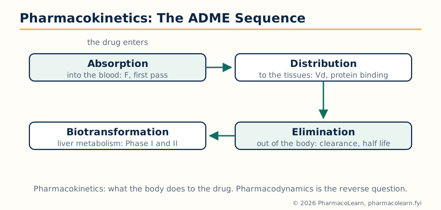
Figure: The ADME sequence. Absorption, distribution, biotransformation and elimination decide the concentration time course of every drug.
1.2 Absorption and bioavailability
Most small drug molecules cross membranes by passive diffusion, moving down a concentration gradient without energy. Only the unionised, lipid soluble fraction crosses easily, so absorption of a weak acid or base depends on the pH of the environment and the pKa of the drug: a weak acid such as aspirin is largely unionised in the acid stomach and absorbed there, while weak bases are ionised and trapped in acid (the basis of urinary acidification strategies and of poor absorption of bases from the stomach). Other routes across membranes include filtration through aqueous pores (small molecules only), facilitated diffusion, active transport by carriers such as P-glycoprotein (an efflux pump that limits absorption of many drugs and is a site of interactions), and endocytosis for very large molecules such as vitamin B12 complexes.
Factors deciding the rate and extent of absorption include the formulation (solutions absorb faster than tablets, which must disintegrate and dissolve), gastric emptying and gut motility, food (which delays most absorption but improves that of some lipophilic drugs), splanchnic blood flow, and disease of the gut wall.
Bioavailability is reduced by incomplete absorption and by the first pass effect: drug absorbed from the stomach and intestine travels in the portal vein to the liver and may be metabolised before it ever reaches the systemic circulation. Glyceryl trinitrate is so completely destroyed on first pass that it is given sublingually, a route that drains directly into the systemic veins; propranolol and morphine also lose a large fraction. Routes that partly or wholly bypass the liver include sublingual, rectal (partly), transdermal and parenteral routes.
1.3 Distribution and plasma protein binding
Once in the blood, a drug distributes according to cardiac output and regional blood flow (highly perfused organs first: liver, kidney, brain, then muscle, then fat), according to its ability to cross barriers (the blood brain barrier of tight endothelium and efflux pumps excludes most polar drugs), and according to binding. Many drugs bind extensively to plasma proteins, chiefly albumin for acidic drugs (warfarin, phenytoin, sulfonamides) and alpha-1 acid glycoprotein for basic drugs (lidocaine, imipramine).
The significance of protein binding, a named syllabus point, is fourfold:
- Only the free (unbound) fraction crosses membranes, reaches receptors and is filtered at the glomerulus. Total plasma concentration therefore misleads when binding changes.
- Binding is a reservoir: bound drug is released as free drug is cleared, prolonging action (one reason highly bound drugs often have long half lives).
- Displacement interactions: a second drug competing for the same sites raises the free fraction of the first. The classic pair is a sulfonamide displacing warfarin, or bilirubin in the newborn (sulfonamides displacing bilirubin from albumin caused kernicterus, a historical warning). Most displacement effects are clinically brief because the freed drug is also cleared faster, but they matter for narrow index drugs like phenytoin and warfarin.
- Disease changes binding: hypoalbuminaemia in liver disease, nephrotic syndrome and old age raises the free fraction of acidic drugs; acute inflammation and trauma raise alpha-1 acid glycoprotein and lower the free fraction of basic drugs. Phenytoin levels in a hypoalbuminaemic patient must be interpreted as free levels, or the patient is overdosed on paper readings alone.
Tissue binding matters too: thiopentone redistributes from brain to muscle and fat, which terminates its anesthetic effect within minutes even though elimination takes hours; tetracyclines deposit in growing teeth and bone; chloroquine accumulates in the retina and liver.
1.4 Biotransformation: Phase I and Phase II
Metabolism usually converts a lipophilic drug, which the kidney would reabsorb, into polar products it can excrete. It can also inactivate an active drug, activate a prodrug (levodopa to dopamine, codeine to morphine by CYP2D6, enalapril to enalaprilat), or create toxic products (paracetamol to the hepatotoxic NAPQI).
| Phase | Reaction types | Enzymes and notes |
|---|---|---|
| Phase I | Oxidation, reduction, hydrolysis | Mostly microsomal cytochrome P450 (CYP) enzymes in the liver: CYP3A4, 2D6, 2C9, 2C19, 1A2 handle most drugs. Introduces or exposes a reactive group |
| Phase II | Conjugation: glucuronidation, sulfation, acetylation, methylation, glutathione and amino acid conjugation | Transferases attach a large polar group; products almost always inactive and readily excreted. Glucuronidation is the commonest route |
Two clinical consequences dominate. Enzyme induction (rifampicin, carbamazepine, phenytoin, chronic alcohol, St John's wort) increases CYP synthesis, speeds the metabolism of other drugs and can cause therapeutic failure, for example of oral contraceptives or transplant immunosuppressants. Enzyme inhibition (cimetidine, ketoconazole, erythromycin, grapefruit juice on intestinal CYP3A4) slows metabolism and raises levels, a route to toxicity with drugs such as theophylline or simvastatin. Superimposed on this is pharmacogenetics: CYP2D6 ranges from absent (poor metabolisers, who get no analgesia from codeine) to duplicated (ultra-rapid metabolisers, who convert codeine dangerously fast); slow acetylators develop more isoniazid neuropathy and hydralazine or procainamide lupus. The neonatal liver is the mirror image: deficient glucuronidation made chloramphenicol accumulate to the fatal grey baby syndrome.
1.5 Elimination and clearance
The kidney eliminates drugs by three processes acting together: glomerular filtration (free drug only, about 120 ml/min in health), tubular secretion (active carriers for acids and bases in the proximal tubule, the route penicillins use and probenecid blocks), and tubular reabsorption (passive, of the unionised fraction, which is why urinary alkalinisation speeds salicylate and phenobarbitone elimination in poisoning). Other routes are biliary excretion with possible enterohepatic recycling (which prolongs the action of drugs such as oral contraceptives whose metabolites are deconjugated by gut bacteria), pulmonary excretion of volatile anesthetics, and minor losses in sweat, saliva and milk (milk matters for the nursing infant).
Clearance is the master elimination parameter. Total clearance is the sum of hepatic, renal and other clearances, and it decides the maintenance dose: at steady state the dosing rate equals CL multiplied by the target steady state concentration. Loading doses, in contrast, depend on Vd, not clearance: loading dose = Vd multiplied by the target concentration. Confusing the two is a classic examination and prescribing error.
1.6 Compartment models: linear kinetics
The body is too complex to model chamber by chamber, so pharmacokinetics uses compartment models: imaginary well mixed spaces between which drug moves at rates proportional to concentration.
ONE AND TWO COMPARTMENT MODELS

  One compartment          Two compartments
  (body = one tank)        (central + peripheral tanks)

   IV dose                    IV dose
      |                          |
      v                          v
  +---------+               +---------+  k12   +------------+
  | Central | --elimination | Central | -----> | Peripheral |
  | (blood) |    (CL)       | (blood) | <----- | (tissues)  |
  +---------+               +---------+  k21   +------------+
                                 |
                              elimination (CL)
In the one compartment model, drug distributes instantly and the plasma concentration after an intravenous bolus falls in a single exponential: a straight line on a log concentration against time plot. Most simple calculations (half life, Vd, clearance) assume this model. In the two compartment model, the log curve bends: an early steep distribution phase, as drug leaves blood for tissues, gives way to a shallower elimination phase once distribution equilibrates. Thiopentone and digoxin behave this way; sampling too early reads the distribution phase and underestimates the half life.
Linear (first order) kinetics means the rate of elimination is proportional to concentration: a constant fraction is removed per unit time, the half life is constant, and doubling the dose doubles the levels predictably. Most drugs behave linearly at therapeutic doses.
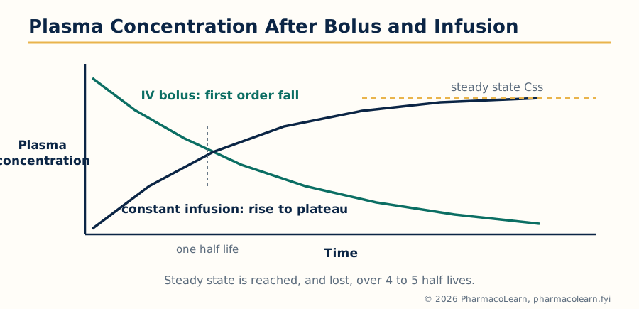
Figure: Plasma concentration after an IV bolus (first order fall) and during constant infusion (rise to the steady state plateau over 4 to 5 half lives).
1.7 Non-linear kinetics: when the rules change
Kinetics become non-linear when an eliminating pathway saturates. The enzymes or carriers reach their maximum rate (Vmax) and elimination becomes zero order: a constant amount per unit time, as with ethanol. The Michaelis-Menten equation governs the transition: at low concentrations the drug behaves first order, and as concentration approaches the Km of the metabolising enzyme it shifts to zero order.
Phenytoin is the clinical example every pharmacologist must command. Within the therapeutic range its hepatic enzymes saturate, so a small dose increase (say from 300 to 400 mg daily) can produce a disproportionate, sometimes toxic jump in plasma level; the apparent half life lengthens as the level rises, steady state takes weeks, and dose changes must be small and guided by plasma concentrations. Salicylates, alcohol and, at high doses, theophylline share the pattern. Non-linearity also arises from saturable protein binding, saturable tubular secretion and autoinduction (carbamazepine speeding its own metabolism over the first weeks of therapy).
1.8 Pharmacodynamics: how drugs produce their effects
Pharmacodynamics studies what the drug does to the body: the mechanism of action and the relationship between drug concentration and effect. Most drugs act on specific macromolecular targets, but the recognised mechanisms form a short list:
- Receptors: the majority of drugs bind receptors, the subject of sections 1.9 to 1.11.
- Enzymes: inhibited (ACE by enalapril, HMG-CoA reductase by statins) or, rarely, activated or used as false substrates (methyldopa).
- Ion channels: blocked (calcium channels by verapamil, sodium channels by local anesthetics) or opened (potassium channels by minoxidil).
- Transporters: blocked (serotonin transporter by SSRIs, Na-K-2Cl cotransporter by furosemide) or exploited for uptake.
- Non receptor mechanisms: antacids neutralise acid chemically, mannitol acts osmotically, chelators bind metal ions, and some drugs act by physical or anti metabolite routes (5-fluorouracil built into nucleic acids).
Even for receptor drugs, the effect is rarely the binding itself: binding triggers a chain, receptor activation, signal transduction, cellular response, organ response, and each link can amplify or saturate.
1.9 Receptors: structural and functional families
A receptor is a macromolecular component of the cell that a drug or endogenous messenger recognises, binds and thereby changes cellular function. The four superfamilies, by structure and signalling route, are:
| Family | Structure | Mechanism | Speed | Examples |
|---|---|---|---|---|
| Ligand gated ion channels (ionotropic) | Multi subunit pore through the membrane | Binding opens the channel, ions flow, membrane potential changes | Milliseconds | Nicotinic acetylcholine, GABA-A, glutamate NMDA and AMPA, 5-HT3 |
| G protein coupled receptors (metabotropic) | Seven transmembrane helices, one protein | Activate G proteins, which regulate enzymes and channels through second messengers | Seconds | Adrenoceptors, muscarinic, dopamine, opioid, angiotensin AT1 |
| Kinase linked receptors | Single transmembrane helix with enzyme activity inside | Binding activates a kinase (often tyrosine kinase), phosphorylating proteins | Minutes to hours | Insulin, growth factors (EGF, VEGF), cytokine receptors (JAK linked) |
| Nuclear receptors | Intracellular, in cytosol or nucleus | The drug receptor complex binds DNA and changes gene transcription | Hours to days | Steroid hormones, thyroid hormone, vitamin D, PPARs |
Functionally, receptors also divide by what the bound drug does: agonists activate, antagonists occupy without activating and block the agonist, partial agonists activate submaximally even at full occupancy, and inverse agonists reduce the receptor's baseline (constitutive) activity below resting level, a property of rimonabant at CB1 and several modern beta blockers at constitutively active receptors. Many receptors exist as subtypes (alpha1 and alpha2, beta1 and beta2, M1 to M5, D1 to D5), and subtype selectivity is the commonest engineering route to a cleaner drug: beta1 selective blockers for the heart, M3 emphasis in bladder drugs, D2 blockade for antipsychosis with 5-HT2A blockade moderating its cost.
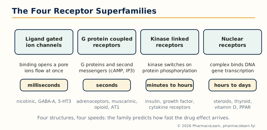
Figure: The four receptor superfamilies and their characteristic speeds of response, from milliseconds for ion channels to days for nuclear receptors.
1.10 Signal transduction: second messengers
Between receptor and response sit the second messenger systems, which amplify a single binding event into a large cellular answer:
- cAMP system: Gs stimulates adenylyl cyclase (beta adrenoceptors), Gi inhibits it (alpha2, opioid, M2). cAMP activates protein kinase A. Phosphodiesterase inhibitors such as milrinone and sildenafil prolong the signal by blocking its breakdown.
- Phosphoinositol system: Gq activates phospholipase C, splitting PIP2 into IP3 (releases calcium from stores) and DAG (activates protein kinase C). Used by alpha1, AT1, H1 and M3 receptors.
- cGMP system: nitric oxide activates soluble guanylyl cyclase; the pathway of nitrates and of sildenafil's target enzyme PDE5.
- Ion channel coupling: Gi/o proteins open potassium channels and close calcium channels directly, the route by which opioids and M2 receptors slow the heart and quieten neurons.
- Enzyme cascades: kinase linked receptors drive phosphorylation cascades (Ras, Raf, MAP kinase), the pathways growth factor drugs and many anticancer agents target.
Amplification explains spare receptors: a maximal tissue response may need only a small fraction of receptors occupied, because each activated receptor generates many second messenger molecules. Insulin and histamine responses show large receptor reserves; irreversible antagonists such as phenoxybenzamine first eat into the reserve (shifting the curve right) before they depress the maximum.
1.11 Quantitation of drug receptor interaction and effects
The graded dose response curve (response against log dose) is sigmoid and yields two independent numbers. Potency, read from the position of the curve (EC50, the concentration giving half the maximal response), says how much drug is needed. Efficacy (Emax, the height of the curve) says how much effect the drug can produce at best. Furosemide is more efficacious than thiazides (a higher ceiling), while bumetanide is more potent than furosemide (works at lower milligram doses) with the same ceiling: potency decides the size of the tablet, efficacy decides whether the drug can do the job at all.
Antagonism is read off curve shifts. A competitive antagonist (surmountable, binding the same site reversibly) shifts the agonist curve to the right in parallel, Emax unchanged; the dose ratio quantifies it, and the Schild analysis converts dose ratios into the antagonist affinity constant pA2. A non competitive or irreversible antagonist depresses Emax once the receptor reserve is consumed. A partial agonist has its own lower Emax and, in the presence of a full agonist, behaves as an antagonist, the dual behaviour of pindolol at beta receptors and aripiprazole at D2 receptors. Repeated exposure changes sensitivity: desensitisation and tachyphylaxis (fading response, as with ephedrine's depleted stores), receptor down regulation with chronic agonist use, and up regulation and supersensitivity with chronic antagonist use, which is why beta blockers are withdrawn gradually, never stopped abruptly in angina.
Safety is quantitated by the quantal dose response curves of a population: the median effective dose ED50, median toxic dose TD50 and median lethal dose LD50. The therapeutic index, TD50/ED50, and the more conservative margin of safety, (TD1 minus ED99)/ED99, express how far treatment sits from harm. Warfarin, digoxin, lithium and phenytoin have narrow indices and demand monitoring; penicillins have a very wide index and forgive dosing error. Finally, variability is itself pharmacology: idiosyncratic reactions (unpredictable, often genetic, such as primaquine haemolysis in G6PD deficiency) differ from dose related toxicity, and both differ from hypersensitivity, which is immune mediated and unrelated to dose.
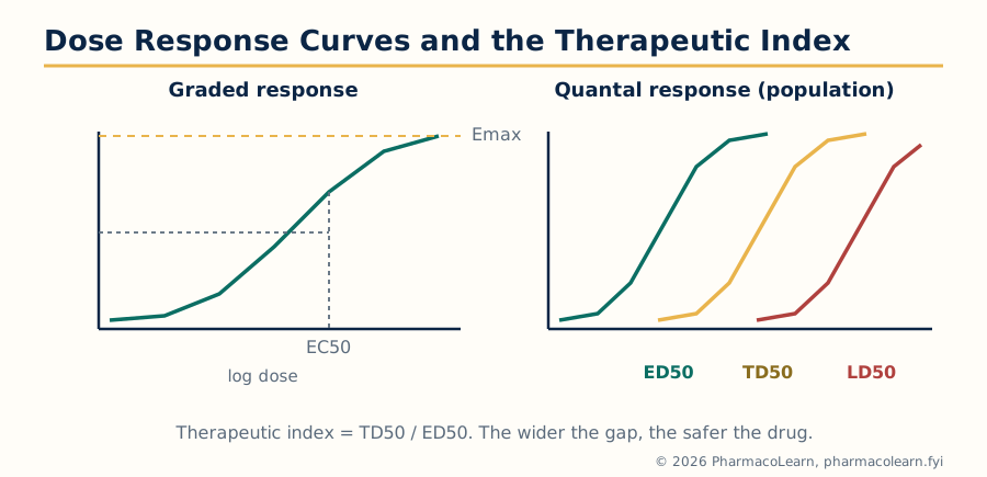
Figure: Graded and quantal dose response curves. The therapeutic index, TD50 divided by ED50, measures the margin between effect and toxicity in a population.
Unit 2: Neurotransmission and Autonomic Pharmacology
2.1 Neurotransmission: general aspects and steps
A neurotransmitter is a substance released from a nerve terminal that carries a signal across a junction to the next cell. The classical criteria are that the substance is synthesised and stored in the neuron, released by stimulation in effective amounts, reproduces the effect of nerve stimulation when applied, and has a mechanism for ending its action. Transmission at a chemical synapse runs through the same steps everywhere, and each step is a drug target:
STEPS OF NEUROTRANSMISSION AND THEIR DRUG TARGETS

  1. Synthesis of transmitter      (hemicholinium blocks choline uptake)
        |
  2. Storage in vesicles           (reserpine empties amine vesicles)
        |
  3. Arrival of the action         (no direct drug target in routine use)
     potential, calcium entry
        |
  4. Release by exocytosis         (botulinum toxin prevents release)
        |
  5. Action on postjunctional      (agonists and antagonists act here)
     and prejunctional receptors
        |
  6. Termination of action         (acetylcholinesterase, amine uptake;
     uptake, metabolism or          anticholinesterases and SSRIs act here)
     diffusion away
Prejunctional (autoreceptor) control makes the system self regulating: alpha2 autoreceptors on noradrenergic terminals brake further noradrenaline release (clonidine exploits this), while similar feedback exists at cholinergic and serotonergic terminals. The two great peripheral transmitters, acetylcholine and noradrenaline, are studied in detail next, exactly as the syllabus orders them.
2.2 Acetylcholine: the cholinergic transmitter
Synthesis: choline is taken up into the terminal by a high affinity sodium dependent transporter (the rate limiting step) and acetylated by choline acetyltransferase using acetyl coenzyme A. Storage: vesicles concentrate acetylcholine (ACh) with ATP by a vesicular transporter. Release: the action potential opens voltage gated calcium channels, calcium triggers fusion of vesicles with the membrane, and quanta of ACh enter the cleft. Action: ACh acts on nicotinic receptors at ganglia and the neuromuscular junction and on muscarinic receptors at effector organs (section 2.6). Termination: acetylcholinesterase in the cleft hydrolyses ACh to acetate and choline within a millisecond, one of the fastest enzymes known; the choline is recycled. Plasma butyrylcholinesterase (pseudocholinesterase) handles circulating esters, including the muscle relaxant suxamethonium, whose action is prolonged in people genetically deficient in that enzyme.
The cholinergic system divides anatomically into the parasympathetic outflow, the sympathetic cholinergic fibres (sweat glands, adrenal medulla), all autonomic ganglia, the neuromuscular junction and much of the brain, where cholinergic loss underlies the memory failure of Alzheimer disease.
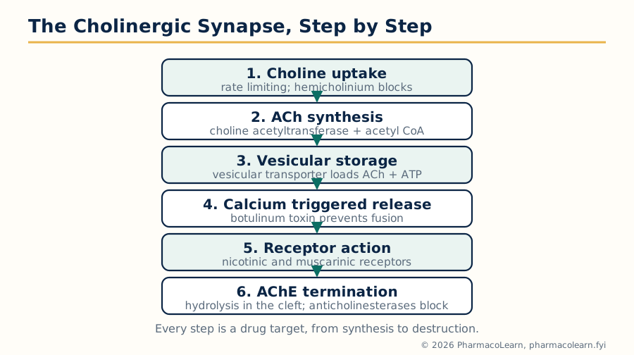
Figure: The cholinergic synapse step by step. Each step is a drug target, from choline uptake to acetylcholinesterase.
2.3 Adrenaline and noradrenaline: the adrenergic transmitters
The catecholamines are built from dietary tyrosine in a fixed sequence: tyrosine to DOPA (by tyrosine hydroxylase, the rate limiting step, inhibited by metyrosine), DOPA to dopamine, dopamine to noradrenaline (inside vesicles), and, in the adrenal medulla and parts of the brain, noradrenaline to adrenaline (by PNMT, an enzyme induced by cortisol draining from the adrenal cortex, a neat anatomical arrangement).
Storage and release mirror the cholinergic pattern, with VMAT pumping amines into granules (reserpine blocks VMAT and depletes them, historically an antihypertensive and a cause of depression). Termination differs fundamentally from ACh: there is no cleft enzyme. Action ends chiefly by reuptake: uptake 1, the neuronal noradrenaline transporter (blocked by cocaine and tricyclic antidepressants), returns most released noradrenaline to the terminal, where it is either restored or destroyed by mitochondrial monoamine oxidase (MAO). Uptake 2 clears catecholamines into non neuronal tissues. Metabolism is by MAO and catechol-O-methyltransferase (COMT), ending in vanillylmandelic acid (VMA), the urinary product once measured to diagnose phaeochromocytoma. Indirect sympathomimetics such as tyramine and amphetamine work inside this machinery, displacing noradrenaline from vesicles and reversing the transporter; the tyramine "cheese reaction" in patients on MAO inhibitors, a hypertensive crisis from dietary tyramine suddenly releasing stored noradrenaline, is the system's famous toxicology.
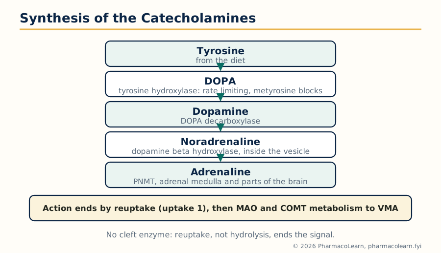
Figure: Catecholamine synthesis from tyrosine. Tyrosine hydroxylase is rate limiting, and reuptake rather than a cleft enzyme ends the signal.
2.4 Neurotransmission in the central nervous system
The brain uses the same chemistry with a wider cast. The syllabus names six for detailed study:
| Transmitter | Type | Principal receptors | Functions and pharmacology |
|---|---|---|---|
| Glutamate | Excitatory, the main CNS excitant | NMDA, AMPA, kainate, metabotropic | Learning and memory; excess causes excitotoxicity in stroke and epilepsy. Ketamine and memantine block NMDA |
| GABA | Inhibitory, the main CNS depressant | GABA-A (ion channel), GABA-B | Benzodiazepines, barbiturates and most anesthetics enhance it (Unit 3) |
| Glycine | Inhibitory, cord and brainstem | Glycine channel | Blocked by strychnine, a convulsant poison |
| Dopamine | Mixed, pathway dependent | D1 family (D1, D5), D2 family (D2, D3, D4) | Movement (nigrostriatal), reward (mesolimbic), prolactin control (tuberoinfundibular); the target of antipsychotics and antiparkinsonian drugs |
| Serotonin (5-HT) | Mixed | 14 subtypes, 5-HT1 to 5-HT7 | Mood, sleep, appetite, gut motility, platelet function; SSRIs, triptans, ondansetron (Unit 5) |
| Histamine | Excitatory in brain | H1, H2, H3, H4 | Wakefulness (sedating antihistamines prove it), appetite; developed fully in Unit 5 |
Alongside these sit the peptide transmitters (substance P, the endogenous opioids), nitric oxide (a gas that diffuses rather than being stored), endocannabinoids (retrograde messengers made on demand) and adenosine, whose receptors caffeine blocks, the pharmacology of the world's commonest drug.
2.5 NANC transmission and cotransmission
Not every autonomic effect is adrenergic or cholinergic. Non adrenergic non cholinergic (NANC) transmission uses other messengers: ATP as a fast cotransmitter in the vas deferens and bladder, nitric oxide in the gut and in penile erection (the target physiology of sildenafil), vasoactive intestinal peptide (VIP), neuropeptide Y and substance P in gut and airways.
Cotransmission is the rule that a neuron releases more than one active substance: sympathetic terminals release noradrenaline with ATP (fast contraction) and neuropeptide Y (slow, prolonged vasoconstriction), so one impulse writes a fast and a slow message. Parasympathetic terminals pair ACh with VIP in salivary glands. Cotransmission explains responses that pure adrenergic or cholinergic blockade leaves behind, and it multiplies drug targets: the purinergic P2X1 blocker, the NK1 antagonist aprepitant (an antiemetic) and CGRP pathway drugs in migraine all grew out of NANC pharmacology.
2.6 The autonomic nervous system as a drug target
The systemic pharmacology of the syllabus begins with the autonomic system. Its two divisions usually oppose each other, with clear exceptions:
| Feature | Sympathetic | Parasympathetic |
|---|---|---|
| Outflow | Thoracolumbar (T1 to L2) | Craniosacral (III, VII, IX, X and S2 to S4) |
| Preganglionic fibre | Short | Long |
| Postganglionic fibre | Long | Short |
| Main transmitter at ganglia | Acetylcholine (nicotinic Nn) | Acetylcholine (nicotinic Nn) |
| Postganglionic transmitter | Noradrenaline (ACh at sweat glands and adrenal medulla) | Acetylcholine |
| Effector receptors | Alpha1, alpha2, beta1, beta2, beta3 | Muscarinic M1 to M5 |
| Dominant role | Stress response: heart rate and force up, bronchodilation, mydriasis, glycogenolysis | Rest and digest: heart slowed, gut and bladder active, miosis, salivation |
The receptor map is the clinical content. Alpha1 (Gq) constricts vessels and the radial muscle of the iris; alpha2 (Gi) is prejunctional and inhibitory; beta1 (Gs) drives the heart and renin release; beta2 (Gs) relaxes bronchi, vessels and uterus; beta3 acts in fat and bladder (mirabegron). Muscarinic M1 sits in glands, CNS and gastric parietal pathways, M2 slows the heart, and M3 contracts smooth muscle and drives secretion. Every autonomic drug in sections 2.7 to 2.12 is a key turned in one of these locks.
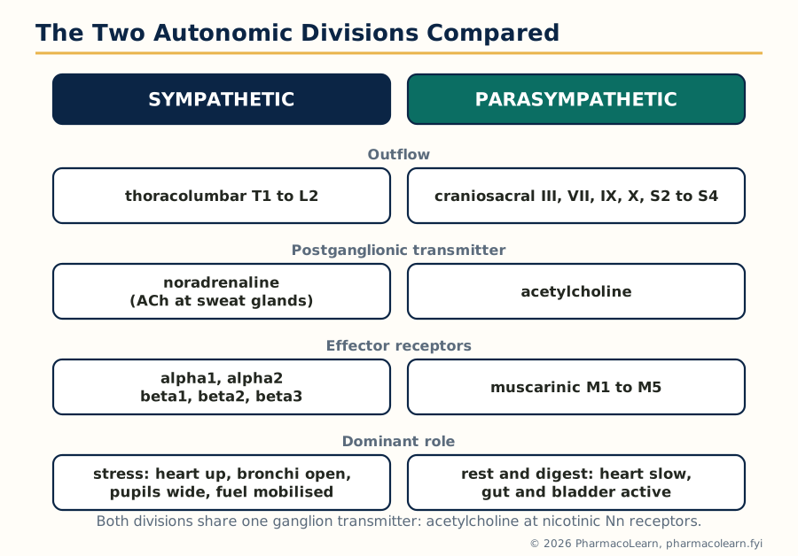
Figure: The two autonomic divisions compared, outflow, transmitter, receptors and dominant role side by side.
2.7 Parasympathomimetics: cholinomimetic drugs
Drugs that mimic ACh divide into direct agonists and indirect anticholinesterases.
Direct agonists. Choline esters are resistant to hydrolysis by design: bethanechol (muscarinic, used for urinary retention and gut atony, given orally or subcutaneously, never intravenously), carbachol (mixed muscarinic and nicotinic, now mainly intraocular for miosis) and methacholine (a bronchial challenge test agent). The alkaloid pilocarpine (M3) constricts the pupil and lowers intraocular pressure in glaucoma and stimulates salivary secretion in dry mouth; cevimeline is a related oral sialogogue. The pattern of muscarinic effects is worth memorising as a system: miosis and accommodation for near vision, salivation and gut activity up, bronchoconstriction, bradycardia, bladder contraction, sweating. Its toxic version is the SLUDGE picture of cholinergic excess (salivation, lacrimation, urination, defecation, gastrointestinal cramps, emesis).
Anticholinesterases are covered as their own class next, since they dominate the clinical use of this area.
2.8 Anticholinesterases and organophosphate poisoning
By inhibiting acetylcholinesterase, these drugs let endogenous ACh accumulate at every cholinergic junction. They divide by chemistry and duration:
- Reversible, short acting: edrophonium (historically the diagnostic test for myasthenia gravis).
- Reversible, medium acting carbamates: physostigmine (tertiary amine, enters the brain; the antidote for atropine and tricyclic poisoning), neostigmine and pyridostigmine (quaternary, kept peripheral; used in myasthenia gravis and to reverse neuromuscular blockade), and the Alzheimer drugs donepezil, rivastigmine and galantamine, which cross into the brain to support its failing cholinergic system.
- Irreversible organophosphates: insecticides (parathion, malathion) and nerve gases (sarin). They phosphorylate the enzyme permanently; the bond then ages, becoming unbreakable.
Clinical uses span myasthenia gravis (the autoimmune destruction of nicotinic receptors, compensated by raising cleft ACh), postoperative ileus and urinary retention, reversal of muscle relaxants, glaucoma (older agents) and Alzheimer disease. Organophosphate poisoning combines muscarinic excess (miosis, salivation, bronchospasm, bradycardia and diarrhoea), nicotinic effects (fasciculations then paralysis) and CNS effects (confusion, convulsions, coma). Treatment is a model of applied pharmacology: atropine in large, escalating doses blocks the muscarinic syndrome, and the oxime pralidoxime reactivates the enzyme if given before ageing; benzodiazepines control the seizures. Atropine does nothing for the paralysis, which is why the oxime and ventilation matter.
2.9 Parasympatholytics: antimuscarinic drugs
Atropine, the prototype from Atropa belladonna, blocks all muscarinic subtypes non selectively and competitively. Its effects are the photographic negative of section 2.7: mydriasis and cycloplegia (loss of accommodation), dry mouth and dry, hot skin, tachycardia, urinary retention, constipation, reduced bronchial secretion and CNS excitation at high doses. Atropine poisoning ("dry as a bone, red as a beet, mad as a hatter, hot as a hare") is reversed by physostigmine, the one anticholinesterase that reaches the brain.
The class has been refined by selectivity and by charge. Quaternary compounds (ipratropium and tiotropium inhaled for airways disease, glycopyrrolate for secretions and bradycardia without CNS effects) stay out of the brain. Hyoscine (scopolamine) crosses into the CNS and is the best drug for motion sickness. Selective members include pirenzepine (M1, peptic ulcer, now historical), oxybutynin, tolterodine and darifenacin (M3 emphasis, overactive bladder), tropicamide (short mydriasis for examination), benztropine and trihexyphenidyl (anticholinergic antiparkinsonian drugs) and the bronchodilator and antisecretory uses scattered through clinical practice. Their shared adverse burden, dry mouth, constipation, urinary retention, confusion in the elderly, is the reason anticholinergic load is now counted as a prescribing risk in older patients.
2.10 Sympathomimetics: adrenergic agonists
Sympathomimetics are read straight off the receptor map of section 2.6:
| Drug | Receptor profile | Main uses |
|---|---|---|
| Adrenaline | Alpha1, alpha2, beta1, beta2 | Anaphylaxis (the life saving drug), cardiac arrest, added to local anesthetics, open angle glaucoma (older use) |
| Noradrenaline | Alpha1, beta1, little beta2 | Septic and cardiogenic shock, to restore vascular tone |
| Dopamine | D1 and D2, then beta1, then alpha1 with rising dose | Shock; the dose dependent receptor recruitment is the teaching point |
| Dobutamine | Beta1 predominant | Cardiogenic shock and acute heart failure, inotropy with less vasoconstriction |
| Phenylephrine | Alpha1 | Nasal decongestant, mydriatic, raises blood pressure, reflex slowing of a fast heart in SVT |
| Salbutamol, terbutaline | Beta2 selective | Asthma and COPD, premature labour (older use) |
| Clonidine, methyldopa | Alpha2 agonists (central) | Hypertension (Unit 4); clonidine also in withdrawal states and ADHD |
| Amphetamine group | Indirect, release noradrenaline | ADHD and narcolepsy (methylphenidate, lisdexamfetamine) |
Direct agonists bind the receptor themselves; indirect agents (tyramine, ephedrine in part, amphetamines) release stored noradrenaline and fade with repetition as stores empty (tachyphylaxis). Mixed agents such as ephedrine do both. The adverse effects are the physiology exaggerated: tachycardia and arrhythmias, hypertension, tremor and hyperglycaemia with beta2 drugs, and, for adrenaline given in error at high dose, hypertensive crisis; beta blockers are the antidote class, with the caveat that blocking beta receptors while alpha stimulation continues (cocaine toxicity) is itself debated territory.
2.11 Sympatholytics: adrenergic antagonists
Alpha blockers. Non selective phenoxybenzamine (irreversible) survives in one niche, preparing a phaeochromocytoma patient for surgery, where alpha blockade must precede any beta blockade. Selective alpha1 blockers (prazosin, doxazosin, terazosin) treat hypertension and, more durably, the urinary symptoms of prostatic enlargement by relaxing bladder neck smooth muscle; their signature adverse effect is first dose postural hypotension. Tamsulosin favours the alpha1A subtype of the prostate and spares blood pressure. Alpha2 antagonists such as yohimbine are of mainly historical and research interest.
Beta blockers are among the most prescribed drugs in the world and are developed fully in Unit 4; here the pharmacology is set. They divide by beta1 selectivity (metoprolol, atenolol, bisoprolol are cardioselective, a relative and dose dependent property), by intrinsic sympathomimetic activity (pindolol, acebutolol partially stimulate while blocking), by membrane stabilising activity (propranolol at high concentrations), by added alpha blockade (labetalol, carvedilol) and by additional actions (nebivolol releasing nitric oxide, sotalol blocking potassium channels). Their dangers follow their receptors: bronchospasm in asthma (beta2), masking of hypoglycaemia warning signs in diabetes, bradycardia and heart block, cold extremities, and rebound events on abrupt withdrawal from the up regulated receptor population of section 1.11.
Ganglion blockers (hexamethonium, mecamylamine) block nicotinic Nn receptors in both divisions at once; the resulting chaos of simultaneous sympathetic and parasympathetic failure retired them from practice, and they remain as a teaching demonstration that ganglia are the crossroads of the whole autonomic system.
2.12 Agents affecting the neuromuscular junction
Skeletal muscle is reached through the somatic motor nerve, whose transmitter is ACh acting on muscle type nicotinic (Nm) receptors. Two families of blockers paralyse by opposite logic:
- Non depolarising (competitive) blockers: tubocurarine historically, and the modern atracurium, vecuronium, rocuronium and pancuronium. They occupy the receptor without opening it; the block is surmountable, and anticholinesterases such as neostigmine reverse it by raising cleft ACh. Rocuronium also has a designed antidote, sugammadex, which encapsulates it.
- Depolarising blockers: suxamethonium (succinylcholine) agonises the receptor and holds the end plate depolarised; fasciculations precede a flaccid paralysis that anticholinesterases cannot reverse (they worsen it). It is very short acting because plasma cholinesterase destroys it, which fails in patients with atypical enzyme, and it can trigger malignant hyperthermia in susceptible patients, treated with dantrolene, and dangerous potassium release in burns and denervation injuries.
Centrally acting relaxants are a different family altogether (baclofen, a GABA-B agonist; tizanidine, an alpha2 agonist; dantrolene, blocking calcium release from the sarcoplasmic reticulum) and are used for spasticity rather than surgical paralysis. Botulinum toxin, which prevents ACh release from terminals, completes the junction's pharmacology: a poison turned therapy for spasticity, dystonia, hyperhidrosis and chronic migraine.
Unit 3: Central Nervous System Pharmacology
3.1 General anesthetics
General anesthesia is a controlled, reversible state of unconsciousness, amnesia, analgesia, immobility and suppression of reflexes. The classical Guedel stages, seen with ether, remain the vocabulary: Stage I analgesia, Stage II excitement (delirium, irregular breathing, the dangerous stage modern induction passes through quickly), Stage III surgical anesthesia in four planes (deepening depression of reflexes), and Stage IV medullary paralysis (respiratory and vasomotor failure: overdose).
Potency is measured by MAC, the minimum alveolar concentration preventing movement in response to a surgical stimulus in 50 percent of patients; MAC falls with age and with adjuvant drugs, which is why anesthetics are combined in "balanced anesthesia". Speed of induction and recovery is governed by blood solubility: agents with a low blood to gas partition coefficient (desflurane, sevoflurane, nitrous oxide) equilibrate fast and wear off fast, while soluble agents such as halothane are slow in both directions. The old Meyer-Overton rule, that potency tracks lipid solubility, pointed at membranes, but modern evidence favours specific protein targets: most intravenous and inhaled agents enhance GABA-A inhibition and many also block NMDA excitation (ketamine, nitrous oxide, xenon).
| Agent | Key properties | Main problems |
|---|---|---|
| Halothane (inhaled, older) | Smooth induction, bronchodilator | Sensitises the heart to catecholamine arrhythmias; immune hepatitis on repeat exposure |
| Isoflurane, sevoflurane, desflurane | Modern maintenance agents, faster and cleaner | Desflurane is pungent and sympathetic stimulating; sevoflurane is the smooth paediatric induction agent |
| Nitrous oxide | Weak (MAC above 100 percent), rapid, analgesic | Expands closed gas spaces; inactivates vitamin B12 pathways with prolonged use; always a carrier, never a sole agent |
| Propofol (intravenous) | Rapid, smooth, antiemetic; the standard induction agent | Pain on injection, dose dependent respiratory and cardiac depression, rare infusion syndrome in prolonged ICU use |
| Etomidate | Hemodynamically the gentlest induction agent | Adrenal suppression after even single doses in the critically ill; myoclonus |
| Ketamine | Dissociative anesthesia, analgesia, bronchodilation, sympathetic stimulation | Emergence delirium and hallucinations; raised intracranial pressure debated; valued in shock and field anesthesia |
| Thiopentone (barbiturate) | Ultra short by redistribution, anticonvulsant | Accumulates with infusion; severe hypotension in hypovolaemia |
Premedication, neuromuscular blockers (Unit 2), opioids and antiemetics complete the anesthetic system; the anesthesiologist's skill is dose titration across all of them at once.
3.2 Local anesthetics
Local anesthetics block voltage gated sodium channels from the inside of the nerve membrane, in their ionised form, preferentially when the channel is open or inactivated (use dependent block). Small, myelinated and rapidly firing fibres (pain, temperature, autonomic) are blocked before large motor fibres, which is why a spinal anesthetic abolishes pain while the patient can still move early on. Chemistry divides the class into esters (procaine, benzocaine, tetracaine, hydrolysed by plasma cholinesterase, with allergy prone PABA metabolites) and amides (lidocaine, bupivacaine, ropivacaine, prilocaine, hepatic metabolism, safer allergy profile). Onset tracks pKa (more unionised drug at tissue pH means faster entry), duration tracks protein binding, and potency tracks lipid solubility. Adrenaline added to the injection constricts local vessels, prolongs the block and lowers peak blood levels.
Systemic toxicity is the class danger: rising plasma levels produce circumoral numbness, tinnitus and convulsions (CNS excitation as inhibitory circuits fail first) and then cardiac arrhythmias and collapse, bupivacaine being the most cardiotoxic; intravenous lipid emulsion is the specific rescue. Prilocaine and benzocaine add methemoglobinaemia to the list. Techniques span surface, infiltration, nerve block, intravenous regional, spinal and epidural anesthesia.
3.3 Sedatives and hypnotics
Sleep drugs act on the GABA-A receptor chloride channel, the brain's main inhibitory switch, but at different sites and with different ceilings:
- Benzodiazepines (diazepam, lorazepam, temazepam, midazolam, chlordiazepoxide) bind the benzodiazepine site and increase the frequency of channel opening in response to GABA. They require GABA to be present, giving them a relative ceiling and a wide safety margin alone; their effects span anxiolysis, sedation, hypnosis, anticonvulsant action, muscle relaxation and amnesia. The antagonist flumazenil reverses them, used cautiously because it can precipitate seizures in dependent patients.
- Barbiturates (phenobarbitone, thiopentone) increase the duration of channel opening and at high concentrations open the channel directly. No ceiling: the dose response runs from sedation through anesthesia to fatal medullary depression, which, with enzyme induction and dependence, has retired them from routine hypnotic use.
- Z drugs (zolpidem, zopiclone, zaleplon) bind the same benzodiazepine site with alpha1 subunit preference, giving hypnosis with less anxiolytic and anticonvulsant baggage, though dependence and complex sleep behaviours still occur.
- Newer mechanisms: melatonin receptor agonists (ramelteon, and melatonin itself) reset the sleep clock rather than forcing inhibition; suvorexant blocks orexin, the wakefulness peptide; sedating antihistamines and low dose sedative antidepressants fill gaps off label.
All GABAergic hypnotics distort sleep architecture (suppressing REM and deep sleep), produce tolerance and dependence, and combine dangerously with alcohol and opioids, the interaction behind many overdose deaths.
3.4 Drugs used to treat anxiety
Anxiety disorders were historically treated with the same sedatives; modern practice separates acute from chronic control. Benzodiazepines act within the hour and remain unmatched for acute anxiety, panic and alcohol withdrawal (where long acting chlordiazepoxide or diazepam cover the withdrawal syndrome), but dependence limits them to short courses. SSRIs and SNRIs are first line for generalised anxiety, panic disorder, social anxiety and PTSD, taking weeks to work. Buspirone, a 5-HT1A partial agonist, treats generalised anxiety without sedation or dependence but with a slow onset and no cross tolerance with benzodiazepines, so it cannot rescue a benzodiazepine dependent patient. Pregabalin (binding the alpha2delta calcium channel subunit, reducing excitatory transmitter release) is licensed for generalised anxiety in many countries; beta blockers blunt the peripheral symptoms of performance anxiety. Hydroxyzine and other sedating antihistamines persist as mild options.
3.5 Depression and antidepressant drugs
The monoamine hypothesis, that depression reflects deficient serotonin, noradrenaline and dopamine signalling, organised fifty years of drug design; the modern reading adds neuroplasticity, since all effective antidepressants take weeks to work while their transporter effects are immediate, and the benefit tracks slower adaptive changes including BDNF signalling. The classes, in the order an examiner expects:
| Class | Mechanism | Examples | Signature problems |
|---|---|---|---|
| SSRIs | Block the serotonin transporter (SERT) | Fluoxetine, sertraline, escitalopram, paroxetine | First line. GI upset, sexual dysfunction, hyponatraemia in the elderly, discontinuation syndrome (worst with paroxetine), serotonin syndrome with MAOIs |
| SNRIs | Block SERT and the noradrenaline transporter | Venlafaxine, duloxetine | Hypertension at higher doses; duloxetine also used in neuropathic pain |
| Tricyclics (TCAs) | Block SERT and NET plus muscarinic, H1 and alpha1 receptors | Amitriptyline, imipramine, nortriptyline | The side effect profile is the receptor profile: dry mouth, sedation, postural hypotension, cardiac conduction block; lethal in overdose |
| MAO inhibitors | Block monoamine oxidase A and/or B | Phenelzine, tranylcypromine (irreversible); moclobemide (reversible A) | Tyramine cheese reaction, serotonin syndrome; dietary and drug interaction discipline required. Selegiline (MAO-B) belongs to Parkinson therapy |
| Atypical agents | Mixed mechanisms | Mirtazapine (alpha2, 5-HT2 and 5-HT3 blockade; sedating, weight gain), bupropion (NET and dopamine transporter; activating, lowers seizure threshold), trazodone, vortioxetine, agomelatine (melatonin agonist) | Each trades the SSRI profile for its own |
| Rapid acting | NMDA antagonism | Esketamine (nasal, for resistant depression under supervision) | Dissociation and abuse potential; important as proof that non monoamine routes work |
The serotonin syndrome, agitation, clonus, hyperreflexia, sweating and hyperthermia from excess serotonergic transmission (classically an SSRI combined with an MAOI, tramadol, linezolid or triptans), is the toxicology of this table and is treated by stopping the agents, sedation and cooling, with cyproheptadine as a 5-HT2 antagonist adjunct.
3.6 Psychosis and antipsychotic drugs
The dopamine hypothesis of schizophrenia holds that positive symptoms (delusions, hallucinations) reflect excess dopamine transmission in mesolimbic pathways, while negative and cognitive symptoms involve cortical dopamine deficiency, which is why simple dopamine blockade treats the first group and not the second. All antipsychotics block D2 receptors; antipsychotic effect appears when roughly two thirds of striatal D2 receptors are occupied, and extrapyramidal effects climb steeply above about four fifths, a narrow mechanistic window.
Typical (first generation) agents such as haloperidol and chlorpromazine are fairly pure D2 blockers, potent against positive symptoms, at the price of extrapyramidal side effects: acute dystonia, akathisia, drug induced parkinsonism and, with time, tardive dyskinesia, plus hyperprolactinaemia (blocking the tuberoinfundibular brake on prolactin) and, for low potency agents, heavy anticholinergic, antihistamine and alpha blocking loads. Atypical (second generation) agents add 5-HT2A antagonism and looser or faster D2 binding: risperidone, olanzapine, quetiapine, amisulpride, aripiprazole (a D2 partial agonist, the mechanistic bridge to Unit 1) and clozapine, the most effective drug for treatment resistant schizophrenia, reserved by its risks of agranulocytosis (mandatory blood monitoring), myocarditis, seizures and severe metabolic effects. The atypical class trades extrapyramidal burden for metabolic syndrome: weight gain, diabetes and dyslipidaemia, worst with olanzapine and clozapine. The class emergency is neuroleptic malignant syndrome: rigidity, hyperthermia, autonomic instability and raised creatine kinase, treated by stopping the drug, cooling, and dantrolene or bromocriptine.
3.7 Mania and mood stabilisers
Bipolar mania is treated acutely with antipsychotics and benzodiazepines, and prevented with mood stabilisers, of which lithium remains the standard. Lithium's mechanism is honestly plural: it modulates inositol signalling (depleting IP3 recycling), inhibits GSK-3 beta, and alters serotonergic and dopaminergic transmission. Its pharmacology is a lesson in narrow indices: a small volume of distribution, no metabolism, renal excretion competing with sodium, and a therapeutic range close to toxicity, so plasma levels, renal function and thyroid function are monitored; thiazides, NSAIDs and dehydration raise lithium levels by increasing its proximal reabsorption. Toxicity progresses from tremor, polyuria (nephrogenic diabetes insipidus) and hypothyroidism to confusion, convulsions and coma. Alternatives and partners include valproate, carbamazepine and lamotrigine (borrowed from epilepsy, lamotrigine favouring the depressive pole), and several atypical antipsychotics with maintenance licences.
3.8 Epilepsy and antiepileptic drugs
A seizure is a paroxysm of abnormal, synchronised neuronal firing; epilepsy is its recurrence. Antiepileptic drugs suppress firing by four main mechanisms:
- Use dependent sodium channel blockade: phenytoin, carbamazepine, lamotrigine, lacosamide. First line for focal seizures; carbamazepine and phenytoin are notably ineffective, indeed aggravating, in absence seizures.
- GABA enhancement: benzodiazepines and phenobarbitone (GABA-A), vigabatrin (irreversible GABA transaminase inhibition, at the cost of visual field defects), tiagabine (GABA uptake blockade).
- Calcium channel effects: ethosuximide blocks T type channels in the thalamus, the absence seizure mechanism; gabapentin and pregabalin bind the alpha2delta subunit of voltage gated calcium channels.
- Mixed and other mechanisms: valproate (several of the above, the broadest spectrum agent), levetiracetam (binds the synaptic vesicle protein SV2A), topiramate, perampanel (AMPA antagonism).
Choice follows seizure type first, then patient factors: valproate is teratogenic (neural tube defects and developmental effects) and is restricted in women of childbearing potential; enzyme inducers (carbamazepine, phenytoin) compromise contraception and other drugs (Unit 1); lamotrigine carries a serious rash risk including Stevens-Johnson syndrome and is titrated slowly. Status epilepticus is a time critical emergency run on a protocol: intravenous lorazepam first, then a loaded second line agent (levetiracetam, phenytoin or valproate), then anesthetic infusion in intensive care.
3.9 Neurodegenerative diseases: Parkinson and Alzheimer
Parkinson disease is the loss of dopaminergic neurons of the substantia nigra, leaving the striatum short of dopamine and relatively overactive in cholinergic terms. Treatment restores the balance: levodopa with a peripheral decarboxylase inhibitor (carbidopa or benserazide) remains the most effective drug, its long term cost being motor fluctuations and dyskinesias as the buffering capacity of the dying terminals is lost; dopamine agonists (ropinirole, pramipexole, rotigotine) serve younger patients but cause impulse control disorders and sleep attacks; MAO-B inhibitors (selegiline, rasagiline, safinamide) and COMT inhibitors (entacapone) prolong dopamine and levodopa respectively; amantadine helps dyskinesia; and anticholinergics (trihexyphenidyl) help tremor in the young at a cognitive price. Blocking dopamine carelessly, with metoclopramide or typical antipsychotics, worsens the disease, and levodopa must never be stopped abruptly.
Alzheimer disease adds cortical cholinergic loss to amyloid and tau pathology. The licensed symptomatic drugs are the cholinesterase inhibitors donepezil, rivastigmine and galantamine (Unit 2) and memantine, a use dependent NMDA blocker that dampens glutamate excitotoxicity while sparing normal transmission. Newer anti amyloid monoclonal antibodies (such as lecanemab) modestly slow decline in early disease at the cost of amyloid related imaging abnormalities (brain swelling and microbleeds), and represent the first disease modifying, rather than symptomatic, approach.
3.10 Narcotic analgesics: opioid drugs
Opioids act on the mu, kappa and delta opioid receptors (Gi/o coupled: less cAMP, more potassium outflow, less calcium entry, so less transmitter release from pain fibres and less postsynaptic excitability). Mu receptor activation gives analgesia, euphoria, respiratory depression, miosis, constipation and physical dependence in one package, because the same receptor serves all those circuits. The class ladder:
- Pure agonists: morphine (the standard), codeine (a CYP2D6 prodrug of morphine), tramadol (weak mu agonism plus serotonin and noradrenaline reuptake blockade, with seizure and serotonin syndrome risks), fentanyl and its analogues (very potent, lipophilic, the anesthetic opioids), pethidine (whose metabolite norpethidine accumulates and causes seizures), methadone (long and variable half life, NMDA antagonism, used in pain and in opioid substitution), oxycodone, hydromorphone, diamorphine.
- Agonist antagonists and partial agonists: buprenorphine (partial mu agonist with a ceiling on respiratory depression, used in substitution therapy and as a patch), pentazocine (kappa agonist, dysphoric), nalbuphine.
- Antagonists: naloxone, short acting, the overdose antidote, dosed to restore breathing without precipitating full withdrawal where avoidable; naltrexone, oral and long acting, for relapse prevention in opioid and alcohol dependence; methylnaltrexone and naloxegol, peripherally restricted antagonists for opioid induced constipation.
Opioid poisoning is the classic triad of coma, pinpoint pupils and respiratory depression. Tolerance develops to most effects but strikingly not to constipation and miosis. Dependence management (methadone and buprenorphine substitution, supervised withdrawal) and the rule never to give an antagonist casually to a dependent patient complete the clinical picture.
3.11 Non-narcotic analgesics: NSAIDs and paracetamol
Non steroidal anti-inflammatory drugs inhibit cyclo-oxygenase, blocking prostaglandin synthesis (the autocoid chemistry is Unit 5). COX-1 is the housekeeping enzyme of gastric mucosa, platelets and kidney; COX-2 is induced at inflammation sites, though it is also constitutive in kidney and endothelium. Non selective agents (aspirin, ibuprofen, diclofenac, naproxen, indomethacin) give analgesia, antipyresis and anti-inflammatory action with a shared adverse triad: gastric ulceration and bleeding, renal impairment (prostaglandins defend renal blood flow when perfusion is threatened) and antiplatelet effects. Aspirin is unique in irreversibly acetylating COX, so its antiplatelet effect lasts the life of the platelet (Unit 4 doses it accordingly). COX-2 selective agents (celecoxib, etoricoxib) spare the stomach but tilt the prostacyclin to thromboxane balance toward clotting, the lesson of rofecoxib's withdrawal for cardiovascular events. Paracetamol is analgesic and antipyretic with negligible peripheral anti-inflammatory action and no gastric or platelet penalty, acting centrally by mechanisms still argued over; its danger is entirely hepatic, the NAPQI metabolite of section 1.4 overwhelming glutathione in overdose, reversed by N-acetylcysteine. NSAIDs also close the ductus arteriosus (indomethacin therapeutically), precipitate asthma in aspirin sensitive patients (leukotriene shunting), and blunt antihypertensive and diuretic action, interactions all readable from the prostaglandin physiology of Unit 5.
Unit 4: Cardiovascular Pharmacology
4.1 Diuretics
Diuretics increase urine output by blocking sodium reabsorption at successive nephron segments; the further downstream the site, the less sodium remains to be claimed, so potency follows anatomy:
| Class | Site and mechanism | Examples | Uses and signature problems |
|---|---|---|---|
| Osmotic | Proximal tubule and loop, water held osmotically | Mannitol | Raised intracranial pressure, acute glaucoma; IV only, volume shifts first |
| Carbonic anhydrase inhibitors | Proximal tubule, block bicarbonate reabsorption | Acetazolamide | Glaucoma, altitude sickness, metabolic alkalosis; weak diuresis, acidosis, paraesthesia |
| Loop diuretics | Thick ascending limb, block Na-K-2Cl cotransporter | Furosemide, bumetanide, torasemide | The most potent class: acute heart failure, oedema, hypercalcaemia. Hypokalaemia, hypomagnesaemia, ototoxicity at high IV doses, gout |
| Thiazides | Distal tubule, block Na-Cl cotransporter | Hydrochlorothiazide, chlorthalidone, indapamide | Hypertension mainstay, mild heart failure; hypokalaemia, hyponatraemia, hypercalcaemia, hyperuricaemia, glucose and lipid effects |
| Potassium sparing: ENaC blockers | Collecting duct, block epithelial sodium channel | Amiloride, triamterene | Added to loop or thiazide to spare potassium; hyperkalaemia is the risk |
| Potassium sparing: aldosterone antagonists | Collecting duct, block mineralocorticoid receptor | Spironolactone, eplerenone | Heart failure with proven survival benefit, resistant hypertension, ascites; hyperkalaemia and, for spironolactone, gynaecomastia |
Site teaching points: loop and thiazide diuretics waste potassium because more sodium reaches the collecting duct and is exchanged for potassium under aldosterone; thiazides reduce calcium excretion (useful in recurrent calcium stones) while loops increase it; and any diuretic fails if the patient eats enough salt to replace the losses, the commonest cause of apparent resistance, alongside NSAIDs.
4.2 The renin angiotensin aldosterone system (RAAS)
Blood pressure and volume are governed by the RAAS cascade. A fall in renal perfusion pressure, sympathetic beta1 drive or low distal sodium delivery makes the juxtaglomerular cells release renin, which cleaves hepatic angiotensinogen to angiotensin I; angiotensin converting enzyme (ACE), largely pulmonary and endothelial, converts it to angiotensin II, the system's effector: a potent vasoconstrictor (AT1 receptors), a stimulus to aldosterone release (sodium and water retention, potassium and hydrogen loss), to ADH release and thirst, to sympathetic facilitation and to cardiac and vascular growth and remodelling. ACE is also the enzyme that destroys bradykinin (Unit 5), so ACE inhibition raises bradykinin, a fact that returns as the class cough. The counter regulatory arm, angiotensin (1-7) acting at Mas receptors and the AT2 receptor, opposes vasoconstriction and growth, and is pharmacology in development rather than in the clinic.
Every binding site in the cascade is drugged: renin directly (aliskiren), ACE (the prils), the AT1 receptor (the sartans), and the mineralocorticoid receptor (spironolactone, section 4.1), giving four pharmacological handles on one physiology.
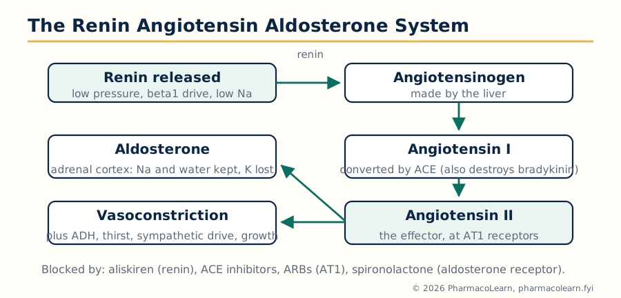
Figure: The renin angiotensin aldosterone system and the four levels at which drugs block it.
4.3 Antihypertensive drugs
Hypertension is treated to targets, usually with combinations, because each class defends against the compensations provoked by the others (a vasodilator's reflex tachycardia and salt retention are answered by a beta blocker and a diuretic).
- ACE inhibitors (ramipril, enalapril, lisinopril, perindopril): block angiotensin II formation. First line, with outcome benefits in heart failure, after myocardial infarction and in diabetic kidney disease, where dilating the efferent glomerular arteriole lowers intraglomerular pressure and proteinuria. Adverse effects: dry cough (bradykinin), angioedema (also bradykinin, commoner in Black patients and potentially fatal), first dose hypotension, hyperkalaemia, a reversible rise in creatinine when renal perfusion depends on angiotensin II (bilateral renal artery stenosis is the danger), and teratogenicity, so they are avoided in pregnancy.
- Angiotensin receptor blockers (losartan, valsartan, candesartan, telmisartan): block AT1 receptors, all the benefits with no cough, the usual substitute when ACE inhibitors are not tolerated. Never combined with an ACE inhibitor for routine hypertension (the combination trials showed harm without gain).
- Calcium channel blockers: dihydropyridines (amlodipine, nifedipine) block L type channels in vascular smooth muscle and dilate arteries, causing ankle oedema, flushing and headache, with short acting nifedipine's reflex tachycardia now avoided; rate limiting agents (verapamil, diltiazem) also slow AV conduction and contractility, are useful in angina and rate control, and must not be combined carelessly with beta blockers. Verapamil constipates.
- Beta blockers (Unit 2 for pharmacology): reduce cardiac output and renin release; no longer first choice for uncomplicated hypertension in many guidelines, but essential when there is angina, post infarction status, heart failure or rate control to treat.
- Thiazide and thiazide like diuretics (section 4.1): cheap, effective, outcome proven.
- Others: alpha1 blockers (doxazosin) as add on therapy; centrally acting alpha2 agonists (methyldopa, the traditional agent in pregnancy; clonidine); direct vasodilators (hydralazine, and minoxidil for resistant cases with hirsutism as its mark); and in hypertensive emergencies, controlled intravenous reduction with agents such as labetalol, nicardipine or sodium nitroprusside, never a precipitous fall.
4.4 Anti-ischemic drugs: treatment of angina
Angina is myocardial oxygen demand outrunning a supply narrowed by atheroma. Drugs work by cutting demand (heart rate, contractility, wall tension through preload and afterload) or, in vasospastic angina, by relieving spasm.
- Organic nitrates (glyceryl trinitrate, isosorbide dinitrate and mononitrate) release nitric oxide, raise cGMP and relax veins more than arteries: preload falls, wall tension and demand fall, and coronary collateral flow improves. GTN sublingual is the acute remedy (and a diagnostic one: pain relieved in minutes supports the diagnosis). Tolerance develops with continuous exposure (a daily nitrate free interval is prescribed), and phosphodiesterase 5 inhibitors such as sildenafil combine with nitrates to cause profound hypotension, an absolute contraindication pairing.
- Beta blockers cut rate and contractility, lengthening diastole when the coronaries fill; a mainstay of effort angina and post infarction care.
- Calcium channel blockers serve both effort angina (rate limiting agents) and vasospastic (Prinzmetal) angina, where they are first line and beta blockers are used cautiously.
- Newer and adjunctive agents: ivabradine blocks the funny current If of the sinoatrial node and slows the heart with no other cardiac action; ranolazine blocks the late sodium current, reducing calcium overload without changing rate or pressure; nicorandil opens potassium channels and donates nitrate like action; trimetazidine shifts cardiac metabolism toward glucose oxidation. All antianginals sit on top of the prognostic treatment of the underlying atheroma: antiplatelet drugs, statins and ACE inhibitors (sections 4.7 and 4.10).
4.5 Antiarrhythmic drugs
The Vaughan Williams classification organises antiarrhythmics by their dominant electrophysiological action, while clinical practice increasingly organises them by the arrhythmia and the state of the heart:
| Class | Mechanism | Examples | Notes |
|---|---|---|---|
| I: sodium channel blockers | Block fast sodium channels, use dependently | Ia quinidine, procainamide (lengthen action potential); Ib lidocaine, mexiletine (shorten); Ic flecainide, propafenone (marked conduction slowing) | The CAST trial taught that suppressing post infarction ectopics with Ic drugs increased mortality; class I is now used cautiously and mainly in structurally normal hearts |
| II: beta blockers | Blunt sympathetic drive to the node and myocardium | Metoprolol, esmolol (IV, ultra short) | Rate control in atrial fibrillation, post infarction protection, inherited arrhythmia syndromes |
| III: potassium channel blockers | Prolong action potential and refractory period | Amiodarone (the broadest agent, also blocks sodium and calcium channels and beta receptors), sotalol (with beta blockade), dofetilide, dronedarone | Amiodarone's toxicology is a syllabus in itself: thyroid both ways, pulmonary fibrosis, corneal deposits, blue grey skin, liver injury, and a half life of weeks to months |
| IV: calcium channel blockers | Block L type channels in nodal tissue | Verapamil, diltiazem | Terminate AV nodal tachycardias and control ventricular rate; never for wide complex tachycardia of unknown origin |
| Others | Various | Adenosine (A1 agonist, opens potassium channels and halts the AV node for seconds; the drug for SVT termination and a famously unpleasant few seconds), digoxin (vagal rate control), magnesium (torsades de pointes), atropine and pacing for bradycardia | Arrhythmia management is as much about anticoagulation, electrolytes and the underlying heart as about these drugs |
The standing lesson is proarrhythmia: every antiarrhythmic can cause the arrhythmias it treats, including torsades de pointes from action potential prolonging drugs combined with hypokalaemia, so the modern trend is toward ablation and devices, with drugs holding specific niches.
4.6 Drugs for heart failure
Heart failure with reduced ejection fraction is a neurohormonal disease: the failing heart's compensations (sympathetic drive, RAAS, vasopressin) become the disease, and the drugs that improve survival all block them.
HEART FAILURE: THE FOUR PILLARS (HFrEF)

  RAAS blockade           Beta blockade
  ACE inhibitor or        bisoprolol, metoprolol
  ARNI (sacubitril /      succinate, carvedilol,
  valsartan)              nebivolol
        \                    /
         \                  /
          >  MORTALITY  <
          /     FALLS     \
         /                  \
  Mineralocorticoid       SGLT2 inhibitor
  antagonist              dapagliflozin,
  spironolactone /        empagliflozin
  eplerenone
- RAAS blockade: ACE inhibitors, or the ARNI sacubitril valsartan (blocking AT1 while inhibiting neprilysin, the enzyme that degrades natriuretic peptides, so the heart's own protective peptides rise; an ACE inhibitor must be stopped 36 hours before starting, because combined ACE and neprilysin blockade invites angioedema).
- Beta blockers titrated slowly upward from low doses (starting during decompensation is an error; continuing through it, where possible, is not).
- Mineralocorticoid antagonists at low, non diuretic doses (spironolactone, eplerenone) with potassium and renal monitoring.
- SGLT2 inhibitors (dapagliflozin, empagliflozin), born as diabetes drugs, now pillars in heart failure with and without diabetes, by mechanisms still debated (natriuresis, metabolic and renal protection).
- Diuretics (section 4.1) relieve congestion and breathlessness but do not change prognosis; dose follows symptoms and weight.
- Digoxin, the old inotrope (Na-K ATPase inhibition raising intracellular calcium, plus vagal effects), now a niche drug for rate control and symptom relief in selected patients, with its narrow index, its toxicity (arrhythmias of nearly every kind, nausea, yellow vision) provoked by hypokalaemia and renal impairment, and its specific antidote, digoxin specific antibody fragments.
- Others: ivabradine when the rate stays high in sinus rhythm; hydralazine with isosorbide dinitrate in selected patients, notably those of African ancestry or intolerant of RAAS blockade; intravenous iron where deficiency limits exercise capacity; and acute decompensation managed with IV loop diuretics, vasodilators and, in low output states, inotropes (dobutamine, milrinone, levosimendan).
4.7 Drugs for hyperlipidemia
Lipid lowering is cholesterol lowering, and the axis is the hepatic LDL receptor: clear more LDL from the blood and events fall.
- Statins inhibit HMG-CoA reductase, the rate limiting enzyme of cholesterol synthesis; the hepatocyte responds by expressing more LDL receptors. They are the foundation (atorvastatin, rosuvastatin the potent pair; simvastatin, pravastatin older), with outcome benefits across primary and secondary prevention. Adverse effects: muscle pain and, rarely, rhabdomyolysis (risk raised by CYP3A4 inhibitors with simvastatin), transaminase rises, and a small increase in diabetes incidence. Their effect is measured in LDL percentage reduction and, in modern practice, LDL targets.
- Ezetimibe blocks the intestinal cholesterol transporter NPC1L1, adding a modest further LDL fall, used when statins are not enough or not tolerated.
- PCSK9 inhibitors (evolocumab, alirocumab, monoclonal antibodies) prevent PCSK9 from marking LDL receptors for destruction, so receptors recycle and LDL falls by more than half again; inclisiran, a small interfering RNA given twice yearly, silences PCSK9 production in the liver.
- Fibrates (fenofibrate, gemfibrozil) activate PPAR alpha and chiefly lower triglycerides; used for severe hypertriglyceridaemia and pancreatitis prevention rather than routine event reduction. Gemfibrozil with statins raises myopathy risk and the pair is avoided.
- Others: bile acid sequestrants (cholestyramine, colesevelam) bind bile acids in the gut and force hepatic cholesterol consumption, safe in pregnancy because they are not absorbed, at the price of gut side effects and binding of other drugs; bempedoic acid inhibits ATP citrate lyase upstream of HMG-CoA reductase and is activated only in the liver, limiting muscle effects; prescription omega 3 preparations and icosapent ethyl serve high triglyceride niches; niacin has effectively retired (flushing, and outcome trials without benefit on top of statins).
4.8 Hematinics
Hematinics supply what red cell production lacks. Iron is given orally as ferrous salts (ferrous sulfate, fumarate, gluconate) for iron deficiency, the commonest anaemia worldwide; absorption is duodenal, helped by vitamin C and an empty stomach, hindered by tea, calcium and proton pump inhibitors, and limited by hepcidin in inflammation. Side effects are gastrointestinal, and black stools are expected. Intravenous iron (ferric carboxymaltose and relatives) serves malabsorption, intolerance, chronic kidney disease and heavy ongoing losses, with modern preparations safe enough for routine use under supervision; acute iron poisoning in children is treated with the chelator desferrioxamine. Vitamin B12 (hydroxocobalamin or cyanocobalamin) treats megaloblastic anaemia of pernicious anaemia and other malabsorption, given parenterally when absorption is the problem; treating B12 deficiency with folate alone corrects the blood while the spinal cord disease (subacute combined degeneration) progresses, a classic examination trap. Folic acid covers increased demand (pregnancy, where it also prevents neural tube defects), malabsorption and antifolate drug effects; folinic acid rescues methotrexate. Erythropoietin (epoetin, darbepoetin) treats the anaemia of chronic kidney disease and chemotherapy, with iron repletion first, and with hypertension and thrombosis as its risks; its abuse in sport is the same pharmacology misapplied.
4.9 Coagulation, anticoagulants and fibrinolytics
Haemostasis balances clotting against bleeding. Vessel injury exposes tissue factor and collagen; platelets adhere, activate and aggregate (section 4.10); and the coagulation cascade converts fibrinogen to a stabilised fibrin mesh: the extrinsic pathway (tissue factor with factor VII) initiates, the intrinsic pathway (XII, XI, IX, VIII) amplifies, and the common pathway (X, V, prothrombin (II), fibrin (I)) executes, with thrombin as the central enzyme and factors II, VII, IX and X dependent on vitamin K for their synthesis. Natural anticoagulants (antithrombin, protein C and S) and fibrinolysis (plasminogen converted to plasmin, which digests fibrin) hold the system in check.
Anticoagulants: - Heparin (unfractionated) binds antithrombin and accelerates its inhibition of thrombin and factor Xa; intravenous, immediate, monitored by aPTT, reversed by protamine. Low molecular weight heparins (enoxaparin) are more Xa weighted, subcutaneous, weight dosed, largely unmonitored, and less prone to heparin induced thrombocytopenia, the immune prothrombotic complication that makes heparin's adverse list unique. - Warfarin blocks vitamin K epoxide reductase, so factors II, VII, IX, X and proteins C and S are made in inactive form. Oral, cheap and effective, with a narrow index managed by the INR, a slow onset (days, and a brief early prothrombotic window as protein C falls first, hence heparin bridging), dozens of CYP2C9 and dietary interactions, teratogenicity, and reversal by vitamin K, prothrombin complex concentrate or plasma. - Direct oral anticoagulants (DOACs): dabigatran (direct thrombin inhibitor, reversed by idarucizumab), and the factor Xa inhibitors rivaroxaban, apixaban and edoxaban (reversal by andexanet alfa or PCC). Fixed dosing, no routine monitoring, fewer interactions and less intracranial bleeding than warfarin; renal function decides eligibility and dose. - Fondaparinux, a synthetic antithrombin binding pentasaccharide, fills niches including heparin induced thrombocytopenia.
Coagulants and antifibrinolytics are the other direction: vitamin K for deficiency and warfarin reversal, tranexamic acid blocking plasminogen activation (trauma, surgery, menorrhagia, dental bleeding in haemophilia), desmopressin releasing von Willebrand factor, and factor concentrates and bypassing agents for the haemophilias.
Fibrinolytics dissolve formed clot: alteplase and its engineered variants tenecteplase and reteplase convert plasminogen to plasmin, given within strict time windows in ST elevation myocardial infarction (where primary angioplasty is unavailable in time) and in selected ischaemic stroke and massive pulmonary embolism. Their price is major bleeding, including intracranial haemorrhage, and their use is a protocol decision, never a casual one. Streptokinase, antigenic and non specific, is now largely historical in high income practice.
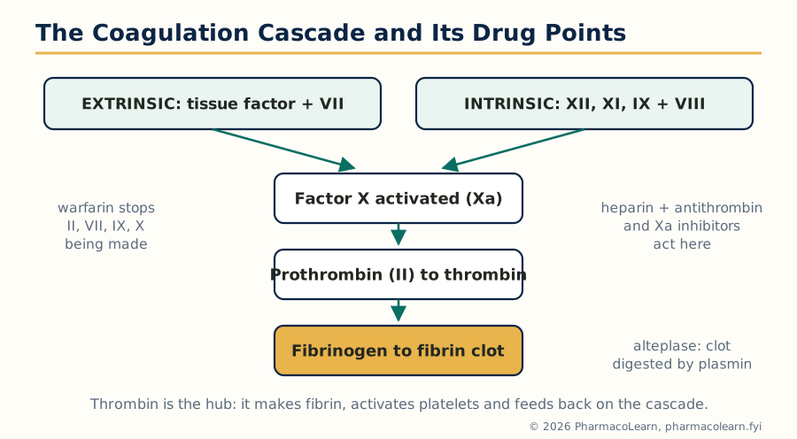
Figure: The coagulation cascade as a single convergent pathway, with the points where warfarin, heparin, the Xa inhibitors and the fibrinolytics act.
4.10 Antiplatelet drugs
Platelets are activated by collagen, thrombin, ADP and thromboxane A2, and aggregate through the glycoprotein IIb/IIIa receptor binding fibrinogen. Each signal is a drug target:
- Aspirin irreversibly acetylates platelet COX-1, abolishing thromboxane A2 production for the platelet's life. Low doses suffice because the effect is cumulative and presystemic. It is the foundation of secondary prevention after myocardial infarction and stroke, given immediately in acute coronary syndromes (chewed, for speed).
- P2Y12 blockers prevent ADP driven activation: clopidogrel and prasugrel (prodrugs, irreversible; clopidogrel needs CYP2C19 activation, which poor metabolisers lack), and ticagrelor (direct, reversible, with dyspnoea and bradycardia among its effects). Combined with aspirin as dual antiplatelet therapy after coronary stenting, for a duration set by the stent and the bleeding risk.
- Glycoprotein IIb/IIIa blockers (abciximab, eptifibatide, tirofiban) block the final common pathway of aggregation, reserved for high risk percutaneous intervention.
- Others: dipyridamole (phosphodiesterase inhibition and adenosine uptake blockade, combined with aspirin for stroke prevention in some practice), cilostazol (peripheral arterial disease), and vorapaxar (thrombin receptor PAR-1 blockade) in narrow niches.
Balancing this section against section 4.9 is the daily work of cardiology: antithrombotic benefit is bought with bleeding, and combinations (dual antiplatelet therapy, or antiplatelet plus anticoagulant) multiply both sides of the bargain.
Unit 5: Autocoid Pharmacology
5.1 Autocoids: local hormones
Autocoids are locally produced, locally acting signalling substances, made on demand rather than stored in endocrine glands, and destroyed near their site of release: histamine, serotonin, the kinins, prostaglandins and related eicosanoids, and, in the syllabus's grouping, the endogenous opioid peptides. They share a pharmacology of inflammation, allergy, pain and vascular control, and their receptors are among the most drugged targets in medicine. This unit takes each in turn: physiology and pathology first, then the drugs.
5.2 Histamine and antihistamines
Histamine is formed from histidine by histidine decarboxylase and stored in mast cells, basophils, gastric enterochromaffin like cells and histaminergic neurons. Release is triggered immunologically (IgE cross linking on mast cells, the mechanism of allergy and anaphylaxis) and by drugs and venoms (morphine, tubocurarine, vancomycin's red man syndrome). Its receptors divide its actions:
| Receptor | Location and action | Drug consequence |
|---|---|---|
| H1 (Gq) | Vessels (vasodilation, increased permeability), bronchi and gut (contraction), skin (itch, wheal and flare), brain (wakefulness) | H1 antihistamines treat allergy; sedation follows brain H1 blockade |
| H2 (Gs) | Gastric parietal cells (acid secretion), heart, vessels | H2 blockers (cimetidine, ranitidine, famotidine) suppress acid; largely replaced by proton pump inhibitors |
| H3 (Gi, presynaptic) | Brain and peripheral nerves, limits histamine and other transmitter release | Pitolisant (H3 antagonist/inverse agonist) treats narcolepsy |
| H4 (Gi) | Immune cells, itch and inflammation | Research target in allergy and pruritus |
Histamine's pathological roles are allergy in all its forms (rhinitis, urticaria, conjunctivitis, anaphylaxis, where adrenaline, not an antihistamine, is the life saving drug), gastric acid secretion, motion sickness and vertigo pathways, and wakefulness.
H1 antihistamines. First generation agents (chlorpheniramine, promethazine, diphenhydramine, hydroxyzine, meclizine) cross the blood brain barrier: sedation and anticholinergic effects are both their burden and, for promethazine and meclizine, part of their antiemetic and antivertigo use. Second generation agents (cetirizine and its enantiomer levocetirizine, loratadine and desloratadine, fexofenadine, bilastine, rupatadine) are kept out of the brain by P-glycoprotein and charge, giving allergy relief (allergic rhinitis, chronic urticaria, conjunctivitis) without meaningful sedation at licensed doses. Fexofenadine is the active metabolite of terfenadine, which was withdrawn for QT prolonging arrhythmias, the standing lesson that antihistamine safety is cardiac as well as sedative. Antihistamines do not treat asthma (the wrong mediators dominate there) and do not reverse anaphylaxis; intranasal and ocular preparations serve local disease, and in chronic spontaneous urticaria, licensed dose escalation of a second generation agent precedes omalizumab.
5.3 Serotonin: physiology and pathology
Serotonin (5-hydroxytryptamine, 5-HT) is made from tryptophan (hydroxylation, then decarboxylation) in enterochromaffin cells of the gut (about 90 percent of the body's store), in serotonergic neurons of the raphe nuclei, and is carried, not made, by platelets, which take it up by SERT. It is destroyed by MAO to 5-HIAA, whose urinary measurement diagnoses carcinoid syndrome, the flushing, diarrhoea, wheezing and right sided valvular fibrosis of a serotonin secreting tumour. Its receptor family is the largest in this unit:
| Receptor | Type | Key actions and drugs |
|---|---|---|
| 5-HT1 (Gi) | Inhibitory; 1B/1D on cranial vessels and nerve terminals | Triptans (sumatriptan and successors) agonise 1B/1D to abort migraine; buspirone is a 1A partial agonist (Unit 3) |
| 5-HT2 (Gq) | Vessels, platelets, CNS; 2A and 2C prominent | Cyproheptadine and ketanserin block; atypical antipsychotics block 2A (Unit 3); 2B activation caused the valvular fibrosis of fenfluramine |
| 5-HT3 | Ligand gated ion channel, gut and chemoreceptor trigger zone | Ondansetron and the setrons block it: the standard antiemetics of chemotherapy, radiotherapy and postoperative vomiting |
| 5-HT4 (Gs) | Gut, prokinetic | Prucalopride treats chronic constipation; older cisapride was withdrawn for arrhythmias |
Pathological roles span migraine (where CGRP now shares the stage: gepants and monoclonal antibodies are the newest antimigraine pharmacology), mood and anxiety (Unit 3's SSRIs raise synaptic serotonin by blocking the same SERT platelets use), gut motility disorders, emesis, carcinoid syndrome (managed with the synthesis inhibitor telotristat and somatostatin analogues) and the serotonin syndrome of excess described in Unit 3. The 5-HT antagonists the syllabus names are therefore a working set: the setrons for emesis, cyproheptadine (also an appetite stimulant and a serotonin syndrome adjunct), methysergide (retired by retroperitoneal fibrosis), and the atypical antipsychotics and mirtazapine, whose 5-HT2 and 5-HT3 blockade is part of their design.
5.4 Kinins: bradykinin and its relatives
The kinins are peptides generated from plasma kininogens by the enzymes kallikreins, themselves activated when factor XII (Hageman factor) contacts damaged surfaces, tying the kinin system to coagulation (Unit 4). Bradykinin (9 amino acids) and kallidin (lysyl bradykinin, 10) act on B2 receptors (constitutive, Gq) to produce intense vasodilation through nitric oxide and prostacyclin, increased vascular permeability, pain (bradykinin is among the most potent algesic substances known, sensitising nociceptors), bronchoconstriction and gut contraction. B1 receptors are absent in health and induced by inflammation and tissue injury, where their ligands (the des-Arg metabolites) sustain inflammatory pain, a division that makes B1 blockade an analgesic research target. Kinins are destroyed within seconds by peptidases, principally ACE, here in its second identity as kininase II, which is why ACE inhibitors raise bradykinin and cause their dry cough and angioedema (section 4.3), and why those risks are worse where bradykinin breakdown is genetically slow.
The clinical pharmacology of the pathway: icatibant, a B2 receptor antagonist, treats acute attacks of hereditary angioedema, a disease of deficient C1 esterase inhibitor in which uncontrolled kallikrein generates bradykinin excess; the kallikrein inhibitors ecallantide and lanadelumab and C1 inhibitor concentrates complete that niche. Aprotinin, a kallikrein inhibitor once used to reduce surgical bleeding, was withdrawn and restricted over safety. The kinins otherwise teach a principle: a mediator can be central to angioedema and inflammatory pain and still be drugged at only a few points, because its physiology is too entangled with blood pressure control to block casually.
5.5 Prostaglandins and the eicosanoid family
Prostaglandins are twenty carbon lipids made on demand from arachidonic acid, liberated from membrane phospholipids by phospholipase A2 (the step corticosteroids suppress through lipocortin/annexin). Cyclo-oxygenase converts arachidonate to the endoperoxides PGG2 and PGH2, from which tissue specific synthases make the family: PGE2 (fever, pain sensitisation, gastric protection, ductus patency), PGI2 prostacyclin (vasodilator and platelet inhibitor from endothelium), TXA2 thromboxane (vasoconstrictor and platelet activator from platelets, prostacyclin's opponent), PGF2 alpha (uterine contraction, luteolysis, aqueous outflow in the eye) and PGD2 (sleep, allergy). The parallel 5-lipoxygenase branch makes the leukotrienes (section 3.11 and Unit 3 of MPC103T chemistry; here: LTB4 the neutrophil attractant, and LTC4, LTD4, LTE4, the slow reacting substance of anaphylaxis driving bronchospasm in asthma, blocked by zileuton at synthesis and by montelukast at the CysLT1 receptor).
Receptors (EP, IP, TP, FP, DP) are all G protein coupled, and the analogues are established drugs: misoprostol (PGE1 analogue: gastric protection against NSAID ulcers, and obstetric uses in induction and postpartum haemorrhage), dinoprostone (PGE2, cervical ripening), carboprost (PGF2 analogue, postpartum haemorrhage), latanoprost and relatives (FP agonists, first line eye drops in glaucoma, with iris darkening and lash growth as their signature), epoprostenol and iloprost (prostacyclin analogues, pulmonary arterial hypertension), and alprostadil (PGE1 keeping the ductus arteriosus open in duct dependent congenital heart disease, and in erectile dysfunction). The physiology explains the pathology list of section 3.11 in reverse: block synthesis with NSAIDs and fever, pain and inflammation improve while stomach, kidney, platelet and ductus pay; the whole of eicosanoid pharmacology is one pathway read from two directions.
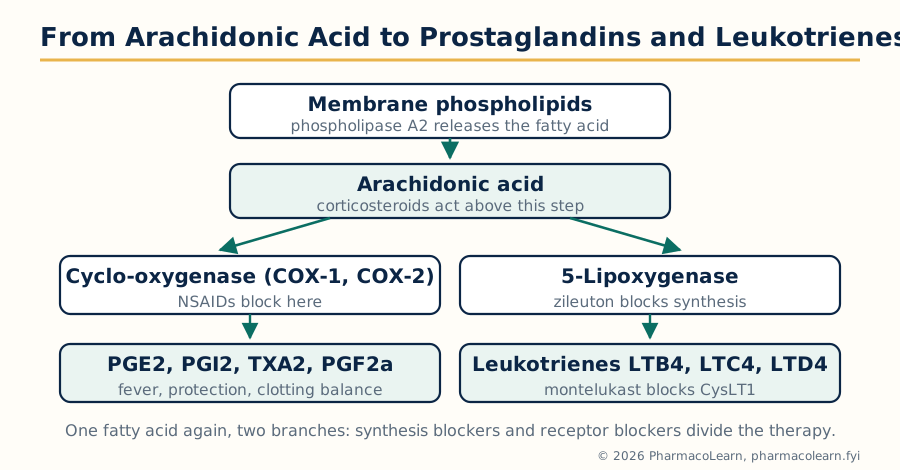
Figure: Arachidonic acid metabolism. The cyclo-oxygenase branch makes the prostaglandins and thromboxane, the lipoxygenase branch makes the leukotrienes.
5.6 Opioid autocoids: the endogenous opioid system
The body runs its own opioid pharmacology. Three precursor proteins are cleaved into peptide families: pro-opiomelanocortin gives beta endorphin (with ACTH and MSH as siblings), proenkephalin gives the met and leu enkephalins, and prodynorphin gives the dynorphins; a fourth pair, the endomorphins, is highly mu selective. The peptides map onto the receptors of section 3.10 with preferences: endorphins at mu, enkephalins at delta, dynorphins at kappa. They are released in stress, exercise, pain and parturition, degrade within minutes by peptidases (enkephalinases), and modulate pain transmission in the dorsal horn and midbrain, reward and mood, gut motility, respiration and the stress hormone axis.
Their roles complete several stories told earlier in this subject. Placebo analgesia is partly endogenous opioid release, reversible by naloxone, one of the neatest demonstrations in all pharmacology. Stress induced analgesia, the analgesia of acupuncture in part, and the runner's high all recruit the system. The peptidase step is drugged: racecadotril inhibits enkephalinase to treat acute diarrhoea by protecting endogenous enkephalins in the gut, opioid effect without an opioid drug. And the antagonist side closes the loop of Unit 3: naloxone and naltrexone block receptors whose natural ligands are these peptides, which is why antagonist therapy in dependence must reckon with a system that also carries normal reward and pain control. With the autocoids, the subject ends where it began in Unit 1: every drug mechanism in these notes is a hand laid on a system the body already owned.
Frequently Asked Questions
Q: What is the difference between clearance and half life, and which one sets the maintenance dose? Clearance is the volume of plasma cleared of drug per unit time; half life is the time for the plasma concentration to halve, and it depends on both clearance and volume of distribution (t1/2 = 0.693 x Vd / CL). The maintenance dose rate is set by clearance alone, because at steady state input must match elimination: dosing rate = CL x target steady state concentration. The loading dose, in contrast, is set by Vd.
Q: Why does phenytoin show non-linear kinetics, and why does it matter at the bedside? Phenytoin is eliminated by hepatic enzymes that saturate within the therapeutic range. Below saturation it behaves first order; once the enzymes approach their maximum rate, elimination becomes zero order, a constant amount per day, so a small dose increase produces a disproportionately large rise in plasma level and the apparent half life lengthens. Dose changes must therefore be small, levels must be measured, and steady state may take weeks rather than days.
Q: A patient on phenytoin develops hypoalbuminaemia. Why can the laboratory total level mislead? Only free phenytoin crosses membranes, acts and is cleared, but the routine assay measures total (bound plus free) drug. Phenytoin is about 90 percent albumin bound, so when albumin falls, the free fraction rises: the patient can be toxic at a total level that looks therapeutic or even low. The correct responses are to measure the free level or apply a correction, and to judge the patient clinically rather than by the total figure alone.
Q: How do competitive and non-competitive antagonists differ on a dose response curve? A competitive antagonist binds the agonist's site reversibly, so enough agonist outcompetes it: the agonist curve shifts right in parallel, potency (EC50) falls, but the maximum response (Emax) is unchanged. A non-competitive antagonist acts at a separate site or irreversibly, so no amount of agonist restores the full response: Emax is depressed once the receptor reserve is used up, while the EC50 of the remaining response may be little changed.
Q: Why must beta blockers never be started before alpha blockade in phaeochromocytoma? The tumour secretes adrenaline and noradrenaline, stimulating both alpha (vasoconstrictor) and beta (including vasodilator beta2) receptors. If beta receptors are blocked first, the beta2 vasodilator escape is removed while alpha vasoconstriction continues unopposed, and blood pressure can rise to a crisis. The correct order is alpha blockade first (phenoxybenzamine or an alpha1 blocker, with volume repletion), and only then a beta blocker for the tachycardia.
Q: How does pralidoxime help in organophosphate poisoning, and why does timing matter? Organophosphates inhibit acetylcholinesterase by phosphorylating it. Pralidoxime is an oxime that pulls the phosphate group off the enzyme and reactivates it, restoring neuromuscular and autonomic function that atropine cannot reach (atropine only blocks muscarinic receptors). With time the phosphorylated enzyme undergoes ageing, a chemical change that makes the bond permanent, after which oximes no longer work; hence pralidoxime is given as early as possible.
Q: Why is aspirin dosed once daily for antiplatelet effect when its plasma half life is only a few hours? Aspirin irreversibly acetylates platelet cyclo-oxygenase 1. Platelets have no nucleus and cannot synthesise replacement enzyme, so thromboxane A2 production is suppressed for the platelet's whole lifespan of about 7 to 10 days. Only the roughly 10 percent of platelets replaced each day restore activity, so a small daily dose keeps the whole population inhibited despite the drug itself disappearing from plasma within hours.
Q: ACE inhibitors cause cough and angioedema, but ARBs largely do not. Why? ACE is also kininase II, the enzyme that destroys bradykinin. ACE inhibitors therefore raise bradykinin levels in the airways and tissues, and bradykinin produces the dry cough and, in susceptible patients, angioedema. ARBs block the angiotensin AT1 receptor and leave ACE free to break down bradykinin, so bradykinin does not accumulate and cough is no more common than with placebo; angioedema is much rarer, though not impossible.
Q: Triptans and ondansetron both act on serotonin receptors. How are their actions opposite in spirit? Sumatriptan is an agonist at 5-HT1B/1D receptors: it constricts dilated cranial vessels and blocks neuropeptide release from trigeminal terminals, aborting a migraine attack. Ondansetron is an antagonist at 5-HT3 receptors, the ion channel receptors of the gut and the chemoreceptor trigger zone, and so prevents chemotherapy and postoperative vomiting. Same transmitter, different receptor subtypes, opposite pharmacological verbs, different diseases entirely.
Q: Why is clozapine reserved for treatment resistant schizophrenia despite being the most effective antipsychotic? Clozapine carries risks the other agents do not: agranulocytosis in about 1 percent of patients, which can be fatal and demands mandatory regular blood monitoring, plus myocarditis, dose dependent seizures, severe constipation that can progress to ileus, and the heaviest metabolic burden of the class. It is therefore reserved for schizophrenia that has failed at least two other antipsychotics, where its superior efficacy, including reduced suicidality, justifies the monitoring system built around it.
Complete MPL102T notes, all 5 units in one PDF, free to keep.
Other M.Pharm subjects: MPH101T · MPC101T · MPL101T · MPH102T · MPH103T · MPH104T · MPL102T · MPL103T · MPL104T · MPC102T · MPC103T · MPC104T · MPH201T · MPH202T · MPH203T · MPH204T · MPC201T · MPC202T · MPC203T · MPC204T · M.Pharm