
MPL104T — Cellular and Molecular Pharmacology
4 hrs/week, 4 credits, 60 hours, 100 marks. Notes follow the M.Pharm syllabus, unit by unit.
MPL104T Cellular and Molecular Pharmacology notes for M.Pharm Semester 1 Pharmacology: cell biology and apoptosis, signalling pathways (cAMP, MAPK, JAK-STAT), PCR and gene therapy, pharmacogenomics, cell culture and biosimilars, with free unit wise PDF downloads on PharmacoLearn.
Units in this subject
- Unit 1 — Cell Biology
- Unit 2 — Cell Signaling
- Unit 3 — Genomic and Proteomic Tools, Recombinant DNA Technology and Gene Therapy
- Unit 4 — Pharmacogenomics and Immunotherapeutics
- Unit 5 — Cell Culture Techniques and Biosimilars
Download MPL104T PDF Notes
Free PDFs for this subject, one per unit:
Cellular and Molecular Pharmacology, subject code MPL104T, is a core Semester 1 subject of M.Pharm Pharmacology. It carries 4 credits and 60 hours of theory, taught at 4 hours per week, for 100 marks.
This subject takes pharmacology down to the level at which drugs actually act: the cell, its genome and its signaling machinery. Unit 1 covers cell biology: the organelles, genome organization, gene expression and its regulation (including siRNA and microRNA), the cell cycle and its checkpoints, and the three routes of cell death, apoptosis, necrosis and autophagy. Unit 2 covers cell signaling: the modes of intercellular signaling, the molecular structure of the four receptor families, the second messengers, and three pathways in detail, the cyclic AMP pathway, the MAP kinase cascade and the JAK/STAT pathway. Unit 3 covers the working tools of molecular pharmacology: DNA electrophoresis, PCR in its reverse transcription and real time forms, gene sequencing, microarrays, SDS-PAGE, ELISA and western blotting, then recombinant DNA technology and gene therapy. Unit 4 covers pharmacogenomics: mapping and cloning disease genes, the polymorphisms that change drug metabolism, transport and GPCR response, the omics family, and immunotherapeutics with the humanisation of antibodies. Unit 5 is the laboratory craft: cell culture techniques from media to cryopreservation, the viability, glucose uptake and calcium influx assays, flow cytometry, and a closing section on biosimilars.
Unit 1: Cell Biology
1.1 Structure and functions of the cell and its organelles
The cell is the smallest unit at which pharmacology happens: every receptor, enzyme, transporter and gene in these notes sits inside one. The plasma membrane, a phospholipid bilayer studded with receptors, channels, transporters and pumps, separates the cytosol from the extracellular fluid and is the first barrier every drug crosses. Inside, each organelle carries work that maps directly onto a drug target or a drug fate.
| Organelle | Main functions | Pharmacological relevance |
|---|---|---|
| Nucleus | Stores DNA as chromatin, site of transcription and ribosome assembly (nucleolus) | Target of anticancer drugs that damage DNA or block replication |
| Mitochondria | ATP production by oxidative phosphorylation, calcium buffering, heat production | Release cytochrome c to start intrinsic apoptosis; injured by some drug toxicities |
| Rough endoplasmic reticulum | Synthesis of membrane and secreted proteins (studded with ribosomes) | Antibody and protein drug production happens on this pathway in producer cells |
| Smooth endoplasmic reticulum | Lipid and steroid synthesis, detoxification | In hepatocytes it carries the cytochrome P450 enzymes of drug metabolism |
| Golgi apparatus | Modifies, sorts and packages proteins, adds carbohydrate chains (glycosylation) | Glycosylation pattern decides the quality of therapeutic proteins and biosimilars |
| Lysosomes | Acid digestion by hydrolytic enzymes, waste and organelle disposal | End point of autophagy; lysosomotropic drugs such as chloroquine accumulate here |
| Peroxisomes | Breakdown of very long chain fatty acids, detoxification of hydrogen peroxide | Their proliferation is switched on by fibrate drugs acting at PPAR alpha |
| Ribosomes | Protein synthesis (translation) on mRNA templates | The target of macrolide, aminoglycoside, tetracycline and oxazolidinone antibiotics in bacteria |
| Cytoskeleton | Shape, transport tracks, cell division spindle (microtubules), contraction (actin) | Microtubules are blocked by vinca alkaloids and stabilised by taxanes in cancer therapy |
Two themes run through the whole subject. First, compartmentation: a drug that cannot reach the right compartment cannot act. Second, turnover: every protein in the table is continuously made and destroyed, so blocking synthesis or accelerating destruction are both workable drug strategies.
1.2 Genome organization
The human genome holds about 3 billion base pairs of DNA and roughly 20,000 protein coding genes, yet only a small fraction of the DNA codes for protein; the rest is regulatory sequence, introns, repetitive elements and structural DNA. The DNA of one cell, stretched out, would run to about two metres, and it fits into a nucleus a few micrometres wide because it is packaged in stages: DNA wraps around an octamer of histone proteins to form the nucleosome, nucleosomes coil into fibres, and the fibres loop and fold into chromosomes. Loosely packed euchromatin is accessible and transcriptionally active; densely packed heterochromatin is silent. Packaging is therefore the first layer of gene control: a gene wound into heterochromatin cannot be read, whatever signals arrive. Each gene itself is organised as a promoter where transcription machinery assembles, exons that survive into the mature messenger RNA, introns that are spliced out, and untranslated regions at both ends that tune how the message is used.
1.3 Gene expression: from DNA to protein
Gene expression is the flow of information from gene to functional product, summarised as DNA to RNA to protein. In transcription, RNA polymerase II (for protein coding genes) binds the promoter with the help of general transcription factors, unwinds the DNA and builds a messenger RNA copy of the coding strand. The primary transcript is processed before export: a 5 prime cap is added, introns are removed by splicing (which can join exons in alternative combinations, so one gene makes several proteins), and a poly A tail is added at the 3 prime end. In translation, the mature mRNA is read at the ribosome in three base codons; transfer RNAs deliver amino acids in the order the codons specify, and the growing chain folds into a protein.
GENE EXPRESSION: DNA TO PROTEIN

  DNA (gene)
    |
    |  transcription (nucleus, RNA polymerase II)
    v
  primary transcript  --  cap, splicing, poly A tail
    |
    v
  mature mRNA  --  exported to the cytosol
    |
    |  translation (ribosome, codons read by tRNA)
    v
  polypeptide  --  folding, modification, targeting
    |
    v
  functional protein (enzyme, receptor, channel, hormone)
Pharmacology intervenes at nearly every arrow: cytotoxic drugs damage the DNA template, antibiotics block bacterial ribosomes, antisense and siRNA drugs (Unit 1.5) destroy the message, and most conventional drugs act on the protein at the end of the line.
1.4 Regulation of gene expression
A liver cell and a neuron carry the same genome; they differ because different genes are switched on. Regulation works at several levels. Transcriptional control is the master level: specific transcription factors bind enhancer and silencer sequences and recruit or block the transcription machinery, and most hormone and drug signals that change cell behaviour end by changing which transcription factors are active. Epigenetic control changes expression without changing the DNA sequence: methylation of cytosine in CpG islands near a promoter usually silences the gene, while acetylation of histones by histone acetyltransferases loosens chromatin and favours expression, and deacetylation by histone deacetylases compacts it again. Both marks are drugged in practice: the hypomethylating agents azacitidine and decitabine are used in myelodysplastic syndromes, and the histone deacetylase inhibitor vorinostat is used in cutaneous T cell lymphoma. Post transcriptional control includes alternative splicing, regulation of mRNA stability and the small RNA silencing of the next section. Translational and post translational control tune how much protein each message yields and how the protein is modified, for example by phosphorylation, after it is made. The point for pharmacology is that the durable drug responses, tolerance, enzyme induction and most hormone actions, are written at the level of gene expression.
1.5 Small regulatory RNAs: siRNA and microRNA
Cells run a silencing system built on short RNA molecules about 21 to 23 nucleotides long, loaded into the RNA induced silencing complex (RISC), whose Argonaute protein uses the short RNA as a guide to find matching messenger RNAs. The two classes differ in origin and in how completely they match their target.
| Feature | siRNA (small interfering RNA) | microRNA (miRNA) |
|---|---|---|
| Origin | Long double stranded RNA, natural (viral defence) or synthetic by design | Endogenous genes, transcribed as hairpin precursors (pri miRNA, then pre miRNA) |
| Processing | Cut by the enzyme Dicer into short duplexes | Drosha in the nucleus, then Dicer in the cytoplasm |
| Match to target | Perfect complementarity to one target mRNA | Partial complementarity, mainly to the 3 prime untranslated region, so one miRNA tunes many mRNAs |
| Main outcome | Argonaute 2 slices and destroys the target mRNA | Translational repression plus deadenylation and decay of the target |
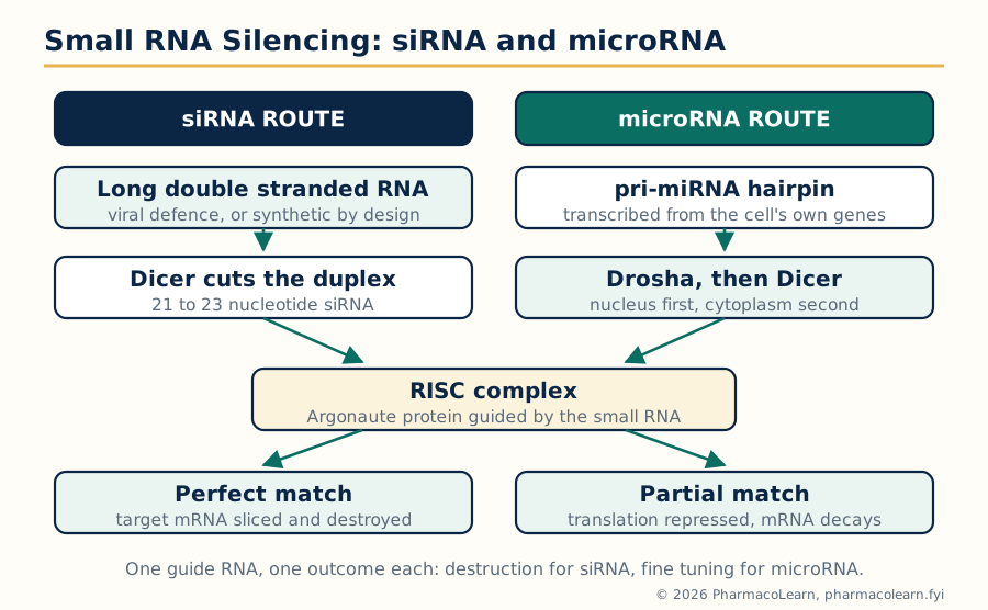
Figure: Two small RNAs, one silencing machine. siRNA arrives as a perfect match and orders destruction; microRNA is home made, matches loosely, and tunes many messages at once.
The importance the syllabus asks for is threefold. In research, synthetic siRNA is the standard tool for knocking down one gene at a time to learn its function and validate drug targets. In physiology, microRNAs fine tune whole programmes of expression, and their disturbance contributes to cancer and cardiovascular disease, making them candidate biomarkers. In therapy, siRNA has become a drug class: patisiran, a lipid nanoparticle siRNA, silences transthyretin production in hereditary transthyretin amyloidosis, and inclisiran silences PCSK9 in the liver to lower LDL cholesterol with twice yearly dosing. The delivery problem, getting a fragile charged RNA into the right cells, is the limiting step, and the liver targeting of the approved drugs shows where the technology works best today.
1.6 Gene mapping and gene sequencing
Gene mapping means fixing the position of a gene on a chromosome. Classical mapping uses linkage: genes close together are inherited together more often than chance predicts, so tracking how a disease travels through families alongside known DNA markers (microsatellites, now single nucleotide polymorphisms) narrows the disease gene to a chromosomal neighbourhood, measured in centimorgans of recombination frequency. Physical mapping then orders the actual DNA, assembling overlapping cloned fragments across the region into a map in base pairs. Gene sequencing reads the base order itself; the principle is introduced here and the laboratory methods are treated in Unit 3.4. Mapping and sequencing together converted genetics into a practical science: once a disease gene is mapped, it can be cloned (Unit 4.1), its protein identified, and the protein becomes a candidate drug target or the basis of a diagnostic test. The Human Genome Project, completed in 2003, delivered the reference sequence against which all mapping is now done.
1.7 The cell cycle and its phases
The cell cycle is the ordered sequence by which a cell duplicates its contents and divides. It runs in four phases: G1 (growth and preparation, the cell decides whether conditions favour division), S (synthesis, the DNA is replicated), G2 (a second gap, the cell checks the replication and prepares the division machinery) and M (mitosis, chromosomes are separated and the cell divides into two). Cells that leave the cycle enter the resting state G0, where most adult cells sit; some, like neurons, stay there permanently, while others, like liver cells, can re-enter on demand. The engine of the cycle is a family of protein kinases, the cyclin dependent kinases (CDKs), which are inactive alone and switched on by partner proteins, the cyclins, whose levels rise and fall in a fixed order: cyclin D with CDK4 and CDK6 drives early G1, cyclin E with CDK2 completes the G1 to S transition, cyclin A with CDK2 works through S phase, and cyclin B with CDK1 drives mitosis. Each cyclin is destroyed once its job is done, which makes the cycle unidirectional.
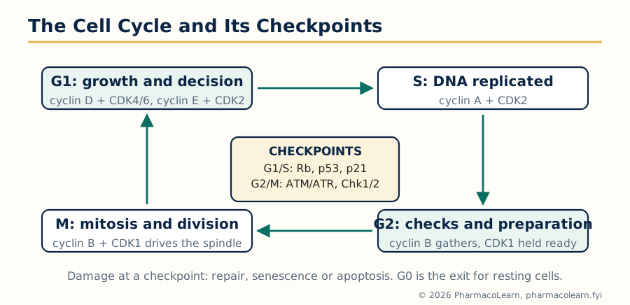
Figure: The cell cycle as a wheel of cyclin and CDK pairs, guarded by three checkpoints. Damage detected at a checkpoint halts the wheel for repair or diverts the cell to apoptosis.
1.8 Regulation of the cell cycle: checkpoints
A cycle that copies three billion base pairs cannot be allowed to run unchecked, so the cell stations checkpoints that halt progress when something is wrong. At the G1/S checkpoint, the retinoblastoma protein Rb is the gatekeeper: unphosphorylated Rb holds the E2F transcription factors silent, and cyclin D and E kinases phosphorylate Rb to release E2F and start S phase. DNA damage activates the protein p53, the guardian of the genome, which switches on the CDK inhibitor p21, freezing the cycle for repair; if the damage is beyond repair, p53 pushes the cell into apoptosis or permanent senescence instead. At the G2/M checkpoint, the ATM and ATR kinases sense unreplicated or damaged DNA and, through Chk1 and Chk2, block the cyclin B and CDK1 complex until replication is complete. The spindle assembly checkpoint in mitosis holds the separation of sister chromatids until every chromosome is correctly attached to the spindle. The pharmacology is direct. p53 is mutated in more than half of human cancers, and Rb pathway failure releases cells from G1 control, which is precisely why CDK4/6 inhibitors (palbociclib, ribociclib, abemaciclib) work in hormone receptor positive breast cancer: they restore the G1 brake that the tumour still depends on losing. Most cytotoxic chemotherapy also exploits the cycle, killing cells preferentially in S phase (antimetabolites) or M phase (vinca alkaloids, taxanes).
1.9 Cell death: the events and regulators of apoptosis
Cells die by controlled demolition or by catastrophe, and the difference decides whether the surrounding tissue is inflamed. Apoptosis, programmed cell death, is an active, energy requiring process with a recognisable sequence of events: the cell shrinks and detaches, the membrane throws out blebs, chromatin condenses against the nuclear envelope, the DNA is cut into regular fragments, and the cell breaks into membrane bound apoptotic bodies that phagocytes engulf at once, so no contents spill and there is no inflammation. Apoptosis sculpts the embryo (separating the fingers), deletes self reactive immune cells, removes cells whose DNA is damaged, and balances cell production in renewing tissues. The executioners are the caspases, cysteine proteases that cut after aspartate residues: initiator caspases (8, 9 and 10) start the cascade and executioner caspases (3, 6 and 7) dismantle the cell. The regulators sit above the caspases: the Bcl-2 family controls the mitochondrial gate (next section), inhibitor of apoptosis proteins restrain caspases directly, and p53 can order apoptosis from the nucleus when damage is unrepairable.
1.10 The intrinsic and extrinsic pathways of apoptosis
Two routes converge on the executioner caspases. The intrinsic (mitochondrial) pathway responds to internal trouble: DNA damage, growth factor withdrawal, oxidative and endoplasmic reticulum stress. These signals activate the BH3 only proteins of the Bcl-2 family (Bid, Bim, Puma, Noxa and others), which tip the balance against the anti apoptotic guardians Bcl-2, Bcl-xL and Mcl-1 and free the effectors Bax and Bak to punch pores in the mitochondrial outer membrane. Cytochrome c escapes to the cytosol, joins Apaf-1 and procaspase 9 to form the apoptosome, and caspase 9 switches on caspase 3. The extrinsic (death receptor) pathway responds to orders from outside: Fas ligand or TNF binds its death receptor (Fas, TNFR1) on the cell surface, the receptor recruits the adaptor FADD, and the resulting death inducing signaling complex activates caspase 8, which activates caspase 3 directly. The pathways talk to each other: caspase 8 also cuts Bid to its active form tBid, which engages the mitochondrial route and amplifies the signal.
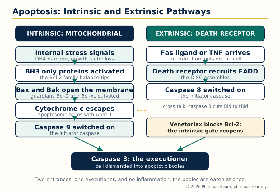
Figure: Two doors into the same execution chamber. The intrinsic pathway reads internal damage through the mitochondria; the extrinsic pathway reads external orders through death receptors; both end at caspase 3.
Cancer is in large part a failure of this machinery: tumours overexpress Bcl-2 or lose p53 and so refuse to die. The refusal can be drugged. Venetoclax is a BH3 mimetic that blocks Bcl-2 itself, freeing Bax and Bak in chronic lymphocytic leukaemia cells that depend on Bcl-2 for survival; it is the clearest example of an apoptosis regulator becoming a medicine.
1.11 Necrosis and autophagy
Necrosis is the catastrophic route: overwhelming injury (ischaemia, toxins, trauma, infection) exhausts ATP, ion pumps fail, the cell and its organelles swell, the membrane ruptures and the contents spill into the tissue, provoking inflammation that damages neighbours. A regulated variant, necroptosis, runs through the kinases RIPK1 and RIPK3 and the pore forming protein MLKL, and can be triggered when apoptosis is blocked; it matters in inflammatory and ischaemic disease.
| Feature | Apoptosis | Necrosis |
|---|---|---|
| Trigger | Programmed signals, damage sensed by the cell | Overwhelming external injury |
| Energy | Required (active process) | Fails as ATP is exhausted |
| Cell volume | Shrinks | Swells |
| Membrane | Intact, blebbing, bodies pinched off | Ruptures early |
| DNA | Cut into regular fragments | Random digestion |
| Inflammation | None, bodies are phagocytosed cleanly | Marked, contents spill |
Autophagy, self eating, is the third route and is as much about survival as death. The cell wraps damaged organelles and protein aggregates in a double membrane autophagosome, marked by the protein LC3, and fuses it with a lysosome so the contents are digested and recycled. The nutrient sensor mTOR normally restrains autophagy; starvation or the drug rapamycin (sirolimus) releases the brake by inhibiting mTOR, and the energy sensor AMPK pushes the same direction. In cancer and infection the process cuts both ways: it keeps stressed cells alive (a resistance mechanism, which is why the lysosome blocking drugs chloroquine and hydroxychloroquine have been studied as chemotherapy partners) and it disposes of intracellular bacteria and damaged mitochondria. For the pharmacologist, Unit 1 ends with a complete set: the cell builds proteins under genomic control, divides under checkpoint control, and dies by three routes, every one of which is now a drug target.
Unit 2: Cell Signaling
2.1 Intercellular signaling: how cells talk to each other
A multicellular organism is a conversation. Intercellular signaling is classified by the distance the message travels:
| Mode | Distance | Examples |
|---|---|---|
| Endocrine | Long, carried in the blood | Insulin, thyroid hormone, cortisol reaching distant organs |
| Paracrine | Short, to neighbouring cells | Histamine, prostaglandins, growth factors in a wound |
| Autocrine | The cell signals itself | Growth factors secreted by tumour cells that feed their own growth |
| Juxtacrine | Direct contact, membrane bound signal | Notch signaling between adjacent cells in development |
| Synaptic | Across a specialised junction, very fast | Neurotransmitters at nerve terminals |
The signal molecule, the first messenger, may be a hormone, neurotransmitter, cytokine, growth factor, gas (nitric oxide) or even a photon or ion flux; what matters is that a receptor exists to read it. Hydrophilic messengers cannot cross the membrane and bind receptors on the cell surface; lipophilic messengers (steroid and thyroid hormones, vitamin D) cross and bind intracellular receptors. That single chemical fact organises the whole of the next four sections.
2.2 Intracellular signaling: the logic of the pathway
Once a surface receptor is occupied, the message must be converted into action inside the cell: intracellular signaling, or signal transduction. Nearly every pathway is built from the same kit of parts. The receptor acts as a transducer, changing shape to activate intracellular proteins. Second messengers are small diffusible molecules that spread the signal through the cytosol and amplify it: each enzyme in a cascade activates many molecules of the next, so a handful of hormone molecules can release millions of product molecules, the classic example being adrenaline mobilising glucose from glycogen. Effector proteins, most often protein kinases that phosphorylate targets and phosphatases that reverse them, change the activity of enzymes, channels and transcription factors, producing the cellular response. Signaling is also actively terminated: messengers are destroyed, receptors are desensitised and internalised, and phosphatases reset the switches. Loss of termination is disease (cholera toxin locks a G protein on, section 2.8), and most pathway drugs work by pressing on an accelerator or restoring a brake.
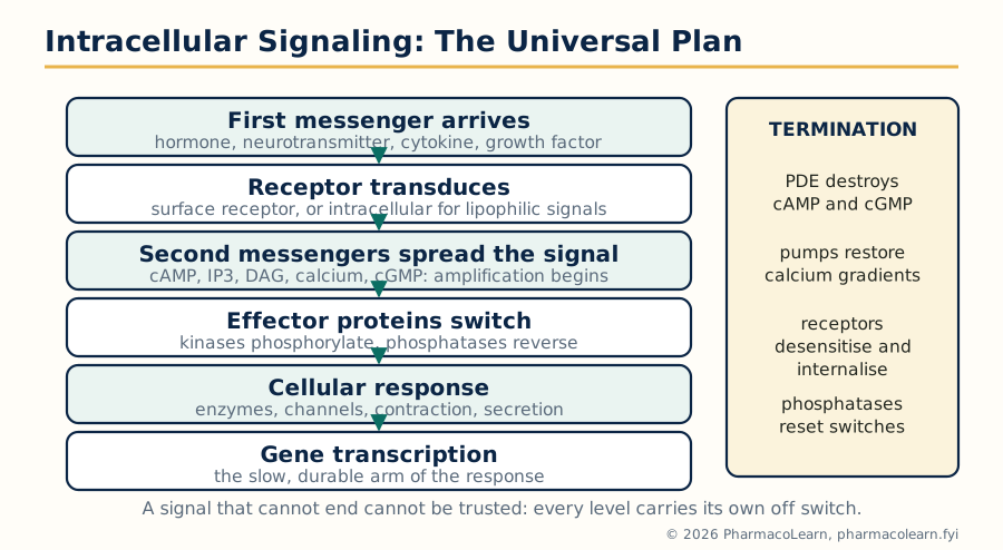
Figure: The universal plan of intracellular signaling. Receptor, transducer, second messenger, effector, response, and the off switches without which no signal can be trusted.
2.3 Ligand gated ion channels: molecular structure
Ligand gated ion channels are receptors that are themselves channels: ligand binding opens a pore and ions flow within microseconds, making these the fastest receptors in the body. The best understood family, the pentameric or Cys loop receptors, assembles five subunits around a central pore. The muscle type nicotinic acetylcholine receptor carries two alpha subunits (which bind acetylcholine) with beta, gamma or epsilon and delta subunits; opening lets sodium and potassium pass and depolarises the muscle fibre. The same architecture serves the inhibitory GABA-A and glycine receptors (chloride channels, targets of benzodiazepines and barbiturates) and the excitatory 5-HT3 receptor (the target of ondansetron). A second family, the ionotropic glutamate receptors (NMDA, AMPA and kainate), is tetrameric; the NMDA receptor is doubly gated, needing both glutamate and removal of a magnesium block by depolarisation, a molecular coincidence detector central to learning and to excitotoxicity. A third small family, the P2X receptors for ATP, is trimeric. Drugs act on these channels as agonists, antagonists, channel blockers and allosteric modulators at sites distinct from the ligand pocket.
2.4 G protein coupled receptors: molecular structure
G protein coupled receptors (GPCRs) are the largest receptor family and the largest single class of drug targets: about a third of marketed medicines act through them. The molecular structure is a single polypeptide folded into seven transmembrane alpha helices, with an extracellular N terminus and an intracellular C terminus; the ligand binds in a pocket among the helices or at the N terminus, and the intracellular loops engage the partner G protein, a heterotrimer of alpha, beta and gamma subunits anchored to the membrane. At rest the alpha subunit holds GDP and the trimer is assembled. Agonist binding makes the receptor act as an exchange factor: GDP leaves, GTP binds, and the alpha GTP and beta gamma parts separate to regulate effectors until the alpha subunit hydrolyses its own GTP back to GDP and the trimer reforms. The alpha subunit defines the family:
| G protein | Main effector | Second messenger change | Example receptors |
|---|---|---|---|
| Gs | Adenylyl cyclase stimulated | cAMP rises | Beta adrenoceptors, H2, dopamine D1 |
| Gi/o | Adenylyl cyclase inhibited, potassium channels opened | cAMP falls | Alpha2, muscarinic M2, opioid, dopamine D2 |
| Gq/11 | Phospholipase C beta stimulated | IP3 and diacylglycerol rise, calcium released | Alpha1, muscarinic M3, AT1, H1 |
| G12/13 | Rho kinase pathway | Cytoskeleton and contraction changes | Thrombin, lysophospholipid receptors |
Signaling is tuned by a control system of its own: GPCR kinases phosphorylate the active receptor, arrestins bind and uncouple it (desensitisation) and route it to internalisation. Arrestins can also start signals of their own, the basis of biased agonism, where a drug favours one output of a receptor over another.
2.5 Tyrosine kinase receptors: molecular structure
Receptor tyrosine kinases are single pass membrane proteins with an extracellular ligand binding domain and an intracellular tyrosine kinase domain. Growth factors such as EGF, VEGF, PDGF and nerve growth factor bind as dimers or drive the receptors together; the paired kinase domains phosphorylate each other on tyrosine residues (autophosphorylation), and the phosphotyrosines become docking sites for proteins carrying SH2 domains. The adaptor Grb2 docks and brings SOS, which starts the MAP kinase cascade of section 2.11. The insulin receptor is the pharmacological exemplar: a preformed dimer whose activation runs through IRS proteins into the PI3 kinase and Akt pathway, ending in translocation of GLUT4 glucose transporters to the membrane of muscle and fat cells, the molecular basis of insulin stimulated glucose uptake measured in the assay of Unit 5.10. Because these receptors drive growth, their overactivity is oncogenic, and the kinase pocket is druggable: gefitinib and erlotinib block the EGF receptor kinase in lung cancer, imatinib blocks the related intracellular kinases BCR-ABL, KIT and PDGF receptor, and the antibody trastuzumab blocks the HER2 receptor from outside.
2.6 Nuclear receptors: molecular structure
Nuclear receptors are transcription factors with a built in ligand pocket, so receptor and effector are one molecule. The structure has four working parts: a variable N terminal region, a highly conserved DNA binding domain built around two zinc fingers that grip specific DNA sequences called hormone response elements, a hinge, and a ligand binding domain that carries the main activation surface. Lipophilic ligands cross the membrane to reach them. In the classic type I pattern (glucocorticoid, oestrogen, progesterone and androgen receptors), the unliganded receptor sits in the cytoplasm held by heat shock proteins; ligand binding releases the chaperones, the receptor dimerises, enters the nucleus and switches gene transcription on or off, recruiting coactivator or corepressor proteins that remodel chromatin. In the type II pattern (thyroid hormone, retinoic acid, vitamin D and PPAR receptors), the receptor already sits on DNA as a heterodimer with the retinoid X receptor, silencing genes until the ligand arrives. Responses take hours to days but last, because they are written into protein synthesis. The drug list is long and central: corticosteroids, sex steroids and their antagonists, the selective oestrogen receptor modulator tamoxifen, thiazolidinediones at PPAR gamma, fibrates at PPAR alpha and calcitriol at the vitamin D receptor.
2.7 Second messengers: the intracellular currency
Second messengers share a design: made on demand in large amounts, small and diffusible, read by specific effector proteins, and destroyed quickly so the signal can end.
| Messenger | Made by | Read by | Ends by |
|---|---|---|---|
| Cyclic AMP (cAMP) | Adenylyl cyclase from ATP (Gs on, Gi off) | Protein kinase A, Epac, cyclic nucleotide gated channels | Phosphodiesterases hydrolyse it to AMP |
| Cyclic GMP (cGMP) | Guanylyl cyclase (soluble form switched on by NO) | Protein kinase G, channels | Phosphodiesterases, including PDE5 |
| Inositol trisphosphate (IP3) | Phospholipase C cutting PIP2 | IP3 receptors on the endoplasmic reticulum, releasing calcium | Dephosphorylation, recycled to inositol |
| Diacylglycerol (DAG) | Phospholipase C cutting PIP2 (same cut as IP3) | Protein kinase C (with calcium and phospholipid) | Phosphorylation to phosphatidic acid |
| Calcium ion (Ca2+) | Enters through channels or is released from the endoplasmic reticulum | Calmodulin and its kinases, troponin, many enzymes | Pumps (SERCA, plasma membrane Ca ATPase) and exchangers restore low cytosolic levels |
| Nitric oxide (NO) | Nitric oxide synthase from arginine | Soluble guanylyl cyclase (it is the ligand) | Scavenged by haemoglobin, oxidised within seconds |
2.8 The cyclic AMP signaling pathway in detail
The cAMP pathway is the prototype against which all others are taught. A Gs coupled receptor (the beta adrenoceptor is the classic) activates adenylyl cyclase, cAMP accumulates, and cAMP binds the regulatory subunits of protein kinase A, freeing its catalytic subunits to phosphorylate target proteins on serine and threonine residues: metabolic enzymes within seconds, and, when PKA enters the nucleus, the transcription factor CREB, whose phosphorylation switches on genes carrying cAMP response elements, giving the pathway a fast and a slow arm. Gi coupled receptors brake the same cyclase. Termination is by phosphodiesterases (PDEs), which hydrolyse cAMP, and by phosphatases. The pathway explains several textbook drug and toxin stories. Cholera toxin permanently ADP ribosylates the Gs alpha subunit, locking it on; intestinal cAMP stays high, chloride and water pour into the gut, and the result is the lethal secretory diarrhoea of cholera. Pertussis toxin disables Gi, removing the brake. Forskolin activates the cyclase directly and is a laboratory standard. PDE inhibitors raise cAMP by blocking its destruction: caffeine and theophylline weakly and non selectively, milrinone (PDE3) in acute heart failure, and roflumilast (PDE4) in severe COPD.
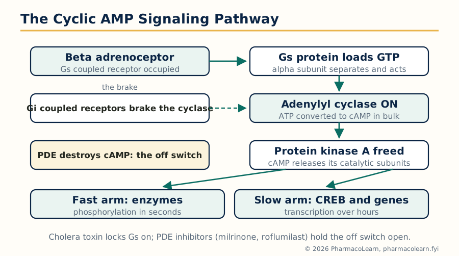
Figure: The cAMP pathway end to end. One receptor, one cyclase, millions of cAMP molecules, two speeds of response: enzymes now, genes later.
2.9 The phosphoinositide pathway: IP3, diacylglycerol and calcium
Gq coupled receptors (alpha1 adrenoceptors, muscarinic M3, angiotensin AT1) activate phospholipase C beta, which cuts the membrane lipid PIP2 into two messengers at once. IP3 diffuses to the endoplasmic reticulum and opens its calcium channels, releasing a pulse of calcium into the cytosol. Diacylglycerol stays in the membrane and, together with the calcium pulse, activates protein kinase C. Calcium itself is a complete signaling system: at rest the cytosol holds it near 100 nanomolar against a millimolar exterior, so opening any calcium channel, in the plasma membrane or the ER, produces a steep local signal. Calcium is read by calmodulin, which wraps around four calcium ions and then activates kinases (including myosin light chain kinase, the trigger of smooth muscle contraction), phosphatases and nitric oxide synthase. Pumps and exchangers then restore the gradient and the store refills. The pathway is the working mechanism of vasoconstriction, glandular secretion and platelet activation, and it gives one famous psychiatric drug clue: lithium inhibits inositol monophosphatase, the enzyme that recycles inositol, which may damp overactive phosphoinositide signaling in bipolar disorder.
2.10 Cyclic GMP and nitric oxide signaling
Nitric oxide is a messenger that ignores the usual rules: a gas, made on demand by nitric oxide synthase from the amino acid arginine, diffusing freely through membranes and living for seconds. Endothelial NOS and neuronal NOS are switched on by calcium calmodulin; inducible NOS, written into macrophages and smooth muscle during inflammation, produces large sustained amounts. NO enters the neighbouring smooth muscle cell and binds the haem group of soluble guanylyl cyclase, which makes cyclic GMP; cGMP activates protein kinase G, which lowers cytosolic calcium and relaxes the muscle. Every step is drugged. Glyceryl trinitrate and the organic nitrates are NO donors. Sildenafil blocks PDE5, the enzyme that destroys cGMP in the corpus cavernosum and the pulmonary circulation, and so prolongs the signal, the basis of its use in erectile dysfunction and pulmonary hypertension. Riociguat stimulates the cyclase directly.
2.11 The MAP kinase signaling pathway
The mitogen activated protein kinase (MAPK) pathway carries growth and differentiation orders from the cell surface to the nucleus, and it is built as a three storey cascade of kinases, each storey amplifying and offering a control point. A growth factor receptor (or a GPCR reaching across) recruits Grb2 and SOS, which load GTP onto the small G protein Ras. Ras switches on Raf (the first kinase, a MAPK kinase kinase), Raf phosphorylates MEK (the MAPK kinase), and MEK phosphorylates ERK1 and ERK2 (the MAPKs themselves). ERK enters the nucleus and phosphorylates transcription factors such as Elk-1, switching on the genes of cell division. Parallel arms of the family, JNK and p38, answer stress and inflammatory signals rather than growth. Because the cascade shouts "divide", its components are frequent oncogenes: activating BRAF mutations (V600E) appear in about half of melanomas and Ras mutations in a large share of pancreatic, colorectal and lung cancers. The cascade is correspondingly drugged at several levels: vemurafenib and dabrafenib block mutant BRAF, trametinib and cobimetinib block MEK, and the multikinase inhibitor sorafenib includes Raf among its targets. Resistance, usually by reactivating the cascade downstream or around the block, is the standing problem of this drug class.
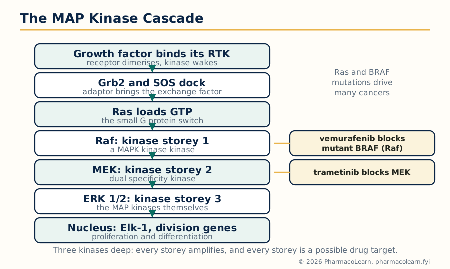
Figure: The MAPK cascade: three kinases deep, each level an amplifier and a possible drug target. BRAF and MEK inhibitors are marked where they strike.
2.12 The JAK/STAT signaling pathway
Many cytokine receptors have no enzyme activity of their own; they borrow one. The Janus kinases (JAK1, JAK2, JAK3 and TYK2) sit permanently attached to the cytoplasmic tails of receptors for erythropoietin, growth hormone, prolactin, the interferons and many interleukins. Ligand binding brings two receptor chains together, the paired JAKs phosphorylate each other and then the receptor tails, and the phosphotyrosines dock STAT proteins (signal transducers and activators of transcription). The JAKs phosphorylate the docked STATs, which pair up as dimers, travel to the nucleus and switch on the genes of the response: red cell production for erythropoietin, immune cell programmes for the interleukins. It is the shortest route in this unit from membrane to gene, with no second messenger at all. Control is by phosphatases and by the SOCS proteins, which the pathway itself induces as a feedback brake. One mutation shows the pathway's power: JAK2 V617F, a constitutively active kinase, drives the myeloproliferative neoplasms. The JAK inhibitors are now a drug family: ruxolitinib (JAK1 and JAK2) in myelofibrosis and polycythaemia vera, and tofacitinib, baricitinib and upadacitinib in rheumatoid arthritis, ulcerative colitis and other immune diseases, traded against a class burden of infection (including herpes zoster reactivation), lipid changes and regulatory warnings on major cardiovascular events and malignancy in older patients with cardiovascular risk.
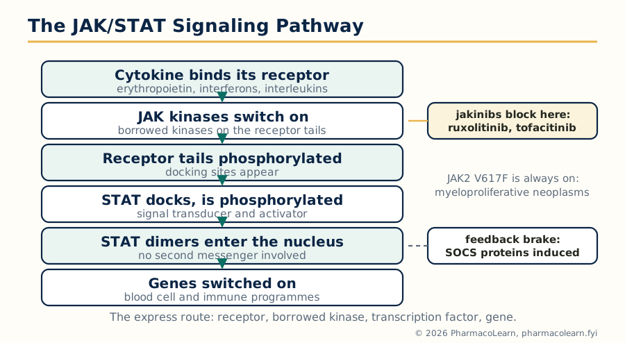
Figure: JAK/STAT: the express route from cytokine receptor to gene, with no second messenger. The jakinib drugs block the borrowed kinase at the top of the pathway.
Unit 3: Genomic and Proteomic Tools, Recombinant DNA Technology and Gene Therapy
3.1 DNA electrophoresis
Gel electrophoresis is the basic separation tool of the molecular laboratory. DNA carries a uniform negative charge on its phosphate backbone, so in an electric field it migrates toward the positive electrode through a gel that acts as a sieve: short fragments thread through the pores faster than long ones, and fragments separate strictly by size. For DNA the gel is agarose, its concentration setting the size range resolved; the run is read by staining with a fluorescent dye that binds DNA (ethidium bromide classically, safer modern alternatives now) and comparing band positions against a ladder of fragments of known size. The pharmacological uses are everyday ones: checking that a PCR worked and made a product of the expected size, verifying that a restriction enzyme cut a plasmid where it should, separating the alleles of a length polymorphism in genotyping, and the older forensic and paternity technique of DNA fingerprinting.
3.2 The polymerase chain reaction
PCR copies a chosen stretch of DNA in a tube, a billion fold, from starting amounts too small to detect. The reaction holds template DNA, two synthetic primers that bracket the target sequence, the four deoxynucleotide building blocks, buffer with magnesium, and a heat stable DNA polymerase (Taq, from the hot spring bacterium Thermus aquaticus) that survives the temperatures used to melt DNA apart. A thermal cycler repeats three steps:
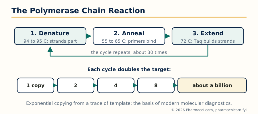
Figure: The PCR cycle. Heat separates the strands, primers mark the target, polymerase extends them; each cycle doubles the product, so about 30 cycles make about a billion copies.
Denaturation at about 94 to 95 degrees C separates the strands; cooling to roughly 55 to 65 degrees C lets the primers anneal to their complementary sites; at 72 degrees C the polymerase extends each primer along the template. Because the products of one cycle become templates of the next, growth is exponential. The applications define modern diagnostics: detecting pathogen DNA in blood and tissue, identifying mutations, quantifying templates in the real time form below, and preparing DNA for sequencing and cloning.
3.3 Reverse transcription PCR and real time PCR
Two extensions of PCR carry separate names that examinations love to confuse. Reverse transcription PCR (RT-PCR) adds a first step in which the enzyme reverse transcriptase copies an RNA template into complementary DNA, after which ordinary PCR amplifies it. It is the only way to PCR an RNA target, so it detects RNA viruses and measures which genes a cell is expressing. Real time PCR (quantitative PCR, qPCR) watches the reaction as it happens: a fluorescent signal grows with the product, either from a dye such as SYBR Green that lights up on binding any double stranded DNA, or from a sequence specific probe (the TaqMan design, in which polymerase activity separates a reporter dye from its quencher only when the true target is amplified). The cycle at which fluorescence crosses a threshold, the Ct value, is inversely related to the starting amount: a low Ct means a lot of template. Combined, reverse transcription real time PCR is the standard for viral load measurement in HIV and hepatitis C, for the respiratory virus testing that became familiar during the COVID-19 pandemic (targeting SARS-CoV-2 RNA), and for quantitative gene expression studies.
3.4 Gene sequencing
Sequencing reads the base order of DNA. The classical Sanger method runs a copying reaction spiked with small amounts of dideoxynucleotides, modified bases that terminate the chain wherever they are incorporated; the result is a ladder of fragments each ending at a known base, separated by size and read off in order, in modern instruments by four fluorescent labels and capillary electrophoresis. It remains the accuracy standard for a single gene or a variant confirmation. Next generation sequencing changed the scale: millions of fragments are sequenced in parallel on a flow cell, the short reads are assembled by computer against the reference genome, and a whole exome or genome becomes a routine clinical order. The pharmacological yield is direct: sequencing finds the mutation behind a genetic disease, panels the mutations of a tumour to select targeted therapy (EGFR in lung cancer is the prototype), and supplies the pharmacogenomic genotypes of Unit 4.
3.5 DNA microarray technique
A DNA microarray measures the activity of thousands of genes in one experiment. A chip carries a grid of microscopic spots, each holding many copies of a probe DNA for one gene. Messenger RNA from the sample is converted to labelled complementary DNA and washed over the chip; each labelled molecule hybridises to the spot whose probe matches it, and a scanner reads the fluorescence at every spot as a measure of how strongly that gene was expressed. Comparing two samples, tumour and normal tissue, or cells with and without a drug, gives an expression signature across the genome at once. Microarrays classified leukaemias and breast tumours into molecular subtypes with different prognoses, identified the genes a drug switches on or off, and genotyped single nucleotide polymorphisms across the genome for the association studies of Unit 4. RNA sequencing now does the expression job with greater range, but the microarray remains the clearer teaching example of hybridisation based measurement.
3.6 SDS-PAGE: separating proteins by size
Proteins vary in charge as well as size, so the protein gel first forces them into a common shape. In SDS-PAGE, the detergent sodium dodecyl sulfate coats each protein with a uniform negative charge per unit length, and a reducing agent breaks disulfide bonds, so every protein becomes an unfolded rod whose migration through the polyacrylamide gel depends on size alone: small proteins run far, large proteins lag. Staining (Coomassie blue, or silver for sensitivity) reveals the bands, sized against a marker ladder. The technique answers everyday questions of molecular pharmacology: is the recombinant protein the right size and how pure is it, which subunits make up a receptor preparation, has a protease cut its target, and it is the obligatory first half of the western blot. Two dimensional electrophoresis, separating first by charge and then by size, was the original engine of proteomics, resolving thousands of protein spots from one sample.
3.7 ELISA
The enzyme linked immunosorbent assay (ELISA) measures an antigen or an antibody by turning its binding into a colour. One partner is immobilised on the wells of a plastic plate; the sample is added and the target, if present, is captured; an enzyme labelled detection antibody (horseradish peroxidase or alkaline phosphatase) completes the complex; washing removes everything unbound; and a substrate is added that the enzyme converts into a coloured product, read as absorbance in a plate reader and quantified against standards. The format varies with the job:
| Format | Arrangement | Typical use |
|---|---|---|
| Direct | Labelled antibody detects antigen fixed on the plate | Quick screens, less specific |
| Indirect | Unlabelled primary antibody, labelled secondary antibody | Detecting antibodies in a patient sample, signal amplified |
| Sandwich | Capture antibody on the plate, sample antigen, labelled detection antibody | Most specific and sensitive; cytokines, hormones, viral antigens |
| Competitive | Sample analyte competes with a labelled reference | Small molecules and haptens, thyroid hormones, some drug assays |
ELISA is the workhorse serology of medicine: screening blood donations for HIV antibodies and hepatitis B surface antigen, the urine pregnancy test's laboratory ancestor for hCG, antibody titres after vaccination, cytokine measurement in research, and the screening tier of therapeutic drug and antidrug antibody monitoring for the biologics of Unit 4.
3.8 Western blotting
Western blotting identifies a specific protein within a mixture and reports its size at the same time. The sample is first separated by SDS-PAGE (section 3.6); the protein bands are then transferred electrically onto a membrane of nitrocellulose or PVDF that preserves their pattern; the membrane is blocked with protein solution so antibodies cannot stick non specifically; a primary antibody against the target protein is applied, then an enzyme or fluorescent labelled secondary antibody against the first; and a substrate reaction reveals a band exactly where the target sits. The blot answers questions no other single technique combines: is the protein present, is it the expected size (so is it cut, modified or the wrong isoform), and roughly how much is there compared with a control. It was the classic confirmatory test for HIV antibodies, is routine for verifying receptor and signaling protein expression (phosphorylated ERK and STAT blots are the standard readout of the pathways of Unit 2), and remains the credibility check behind antibody based claims in research. It is slow and semi quantitative, which is why ELISA and blotting are taught as a complementary pair: ELISA quantifies in bulk, the blot verifies identity.
3.9 Recombinant DNA technology: restriction enzymes and vectors
Recombinant DNA technology joins DNA from different sources in a carrier and lets a host cell copy it. The cutting tools are restriction endonucleases, bacterial enzymes that recognise short, usually palindromic, DNA sequences and cut them, many leaving short single stranded sticky ends that anneal to matching ends cut by the same enzyme; DNA ligase seals the joins permanently. The carrier is a vector, a DNA molecule able to enter a host cell and replicate there, carrying selectable markers (usually antibiotic resistance genes) so the rare cells that took it up can be found:
| Vector | Host | Approximate insert size | Notes |
|---|---|---|---|
| Plasmid | Bacteria (E. coli) | Up to about 10 kb | The everyday cloning and protein production vector |
| Bacteriophage lambda | Bacteria | About 15 to 20 kb | Efficient delivery by infection |
| Cosmid | Bacteria | About 35 to 45 kb | Plasmid with phage packaging signals |
| Bacterial artificial chromosome (BAC) | Bacteria | About 100 to 300 kb | Stable carriage of whole genes with their control regions |
| Yeast artificial chromosome (YAC) | Yeast | Up to about 1 Mb | Largest capacity, more fragile and harder to handle |
A cloning experiment is then simple in plan: cut vector and foreign DNA with matching enzymes, ligate, introduce into host cells, select on antibiotic medium, and screen the colonies for the insert. The pharmacology begins when the insert is a human gene and the host becomes a factory.
3.10 Applications of recombinant DNA technology
Recombinant production replaced extraction from animal tissues and cadavers with fermentation, and several foundation medicines exist only because of it:
MAKING A RECOMBINANT MEDICINE

  human gene identified (Unit 4.1 cloning)
        |
  gene inserted into a plasmid vector
        |
  vector introduced into host cells (E. coli, yeast, mammalian cells)
        |
  fermentation: the host copies the gene and expresses the protein
        |
  protein purified, checked (SDS-PAGE, western blot), formulated
        |
  medicine: insulin, growth hormone, vaccine antigen, antibody
- Recombinant human insulin (1982) was the first medicine of the technology, made in E. coli, ending dependence on pig and cattle pancreases and their allergy and supply problems.
- Recombinant growth hormone replaced hormone extracted from cadaver pituitary glands, which had transmitted the prion disease Creutzfeldt-Jakob disease to some recipients.
- Hepatitis B vaccine is the viral surface antigen made in yeast, a subunit vaccine with no virus at any stage.
- Erythropoietin, granulocyte colony stimulating factor, interferons, tissue plasminogen activator and the monoclonal antibodies of Unit 4 all come from recombinant cell lines, usually mammalian cells such as Chinese hamster ovary cells where human style glycosylation matters.
3.11 Gene therapy: gene transfer techniques
Gene therapy treats disease by delivering a working gene, or a gene modifying tool, to the patient's cells. Strategy decides the technique. In ex vivo therapy, cells are removed, corrected in the laboratory and returned, which suits blood and immune cells that are easy to harvest and reinfuse. In in vivo therapy, the vector is given directly to the patient, by injection into a tissue such as the retina or muscle, or intravenously to reach the liver. The delivery vehicles divide into viral and non viral:
| Vehicle | Properties | Strengths and limits |
|---|---|---|
| Retrovirus | Integrates into the host genome, needs dividing cells | Permanent expression; integration carries a cancer risk |
| Lentivirus | Integrates, infects dividing and non dividing cells | The ex vivo standard for blood stem cells and CAR-T manufacture |
| Adenovirus | Stays outside the genome, strongly immunogenic | High expression but transient; immune reactions limit repeat use |
| Adeno associated virus (AAV) | Stays mainly outside the genome, weakly immunogenic | Small cargo (about 4.7 kb); the in vivo standard for eye, muscle, liver and nerve targets |
| Non viral (plasmid, lipid nanoparticles, electroporation) | No viral proteins | Safer and repeatable, but delivery is far less efficient |
3.12 Clinical applications and recent advances in gene therapy
Clinical gene therapy now has licensed products, and each one teaches a principle. Voretigene neparvovec (an AAV carrying the RPE65 gene, injected under the retina) restores a missing enzyme in an inherited blindness, showing in vivo therapy in an immune sheltered site. Onasemnogene abeparvovec (AAV9 carrying the SMN1 gene, one intravenous dose) treats spinal muscular atrophy in infants, showing systemic delivery across the blood brain barrier at a young age. Ex vivo therapy began with ADA deficient severe combined immunodeficiency, corrected in the patient's own blood stem cells, and matured into the CAR-T cell therapies, in which a patient's T cells are lentivirally equipped with a synthetic receptor against CD19 and returned to attack B cell cancers. The field's standing cautions come from its history: early retroviral trials in SCID-X1 cured the immune defect but caused leukaemia in some children when the vector integrated beside a growth gene, and a fatal immune reaction to an adenoviral vector in 1999 reset safety practice across the field. The recent advance the syllabus asks for is genome editing: in 2023 the first CRISPR based therapy, exagamglogene autotemcel, was approved for sickle cell disease and transfusion dependent beta thalassaemia, editing the patient's stem cells ex vivo to restart fetal haemoglobin production, the same delivery logic with a far more precise tool.
Unit 4: Pharmacogenomics and Immunotherapeutics
4.1 Gene mapping and cloning of a disease gene
Unit 1 mapped genes in principle; this section is the hunt itself, because most pharmacogenomic targets began as disease genes. The classical method is positional cloning: first, linkage analysis in affected families (Unit 1.6) narrows the gene to a chromosomal region, without any knowledge of what the gene does; then the region is searched candidate by candidate until mutations are found that segregate with the disease and are absent from healthy controls; the cloned gene is sequenced, its protein deduced, and the disease mechanism can be worked out from the protein. The landmark hunts still organise how the method is taught: the CFTR chloride channel gene for cystic fibrosis (1989), found by positional cloning and immediately explaining the disease as a channel defect (and, decades later, giving the target for the CFTR modulator drugs), the huntingtin gene (1993), whose expanded CAG repeat causes Huntington disease, and BRCA1 (1994), which turned breast cancer risk into a testable, manageable trait. Modern sequencing (Unit 3.4) has shortened the hunt from years to weeks, but the logic is unchanged: position first, identity next, mechanism and drug target last.
4.2 Genetic variation and its role in health and pharmacology
No two genomes are identical. Human genetic variation comes in graded sizes: single nucleotide polymorphisms (SNPs), one base changed, are the most common, numbering in the millions across each genome; small insertions and deletions; variable repeats; and copy number variations, whole stretches present in differing numbers of copies, which for drug metabolism genes can mean zero, one, two or many gene copies. A variant matters when it changes a protein's sequence or amount, or alters a regulatory region; most variants do neither, so variation studies must prove function, not just association. In health, variation underlies susceptibility to common disease, which genome wide association studies map by comparing variant frequencies between patients and controls. In pharmacology, the same variation, landing in the genes for drug metabolising enzymes, transporters and targets, is the main inherited reason patients differ in drug response: the same dose can be ineffective in one patient, standard in a second and toxic in a third. The discipline that studies this is pharmacogenomics (gene by gene, pharmacogenetics in the older term), and the rest of this unit works through its three classical territories: metabolism, transport and receptors. A special case is immune mediated reaction, where HLA variants decide who develops severe hypersensitivity: abacavir is prescribed only after excluding HLA-B57:01, and carbamazepine induced Stevens-Johnson syndrome is strongly linked to HLA-B15:02 in patients of Asian ancestry.
4.3 Polymorphisms affecting drug metabolism
This is the most clinically mature part of pharmacogenomics, because the enzymes of Unit 1's smooth endoplasmic reticulum are highly polymorphic. Patients fall into metaboliser classes for each enzyme: poor (little or no activity), intermediate, normal (extensive) and ultrarapid (extra gene copies or high activity).
| Gene and enzyme | Drug affected | Consequence of the variant |
|---|---|---|
| CYP2D6 | Codeine (and tramadol) | Ultrarapid metabolisers convert codeine to morphine dangerously fast (fatal respiratory depression has occurred, including in breastfed infants); poor metabolisers get no analgesia. Poor metabolisers also fail to activate tamoxifen to endoxifen |
| CYP2C19 | Clopidogrel | Loss of function alleles (such as *2) leave the prodrug unactivated, platelet inhibition fails and stent thrombosis risk rises; testing now guides antiplatelet choice |
| CYP2C9 and VKORC1 | Warfarin | Slow CYP2C9 variants plus sensitive VKORC1 variants cut the required dose several fold; genotype guided dosing algorithms exist |
| TPMT | Azathioprine, mercaptopurine | Deficient patients cannot detoxify the drugs and develop life threatening bone marrow suppression; testing or dose reduction precedes therapy |
| UGT1A1 | Irinotecan | The *28 variant reduces glucuronidation of the active metabolite SN-38, increasing severe diarrhoea and neutropenia |
| NAT2 | Isoniazid, hydralazine, procainamide | Slow acetylators accumulate the parent drugs: more isoniazid neuropathy, more drug induced lupus |
| G6PD (enzyme deficiency) | Primaquine, rasburicase, some sulfonamides | Deficient red cells cannot withstand oxidative stress and haemolyse; an enzyme defect, screened before these drugs where risk is high |
Guideline bodies (the CPIC consortium) now publish genotype to prescription tables for dozens of drug and gene pairs, which is the practical form in which this section reaches the bedside.
4.4 Genetic variation in drug transporters
ABCB1 encodes P glycoprotein, the efflux pump of the gut wall, blood brain barrier, liver and tumour cells; its common variants have been studied against substrates from digoxin to anticancer drugs, with real but often modest and context dependent effects, an honest contrast with the metabolism genes. SLCO1B1 encodes OATP1B1, the transporter that pulls statins from blood into hepatocytes; a reduced function variant raises simvastatin blood levels and with them the risk of muscle injury, demonstrated in the SEARCH genome wide study, and regulatory guidance now caps simvastatin dose or advises testing in carriers. ABCG2 (the breast cancer resistance protein, BCRP) limits rosuvastatin absorption and raises its levels in variant carriers. In the kidney, variants of the organic cation transporter OCT2 alter the handling of metformin and the nephrotoxicity of cisplatin. The teaching point is comparative: transporter pharmacogenomics is genuine and occasionally decisive, but its predictive value today is generally weaker than that of the metabolic enzymes, and claims should be scaled accordingly.
4.5 Genetic variation in G protein coupled receptors
Unit 2.4 established GPCRs as the largest drug target family, so variation in their genes is the natural third territory. The best studied example is the beta2 adrenoceptor gene ADRB2: common variants at positions 16 (arginine or glycine) and 27 change how the receptor downregulates, and early studies reported different bronchodilator responses to salbutamol between genotypes; larger trials have softened the claim, and routine genotyping before inhaler therapy is not standard practice. The beta1 receptor gene ADRB1 variant Arg389Gly changes coupling efficiency, and the heart failure drug bucindolol showed genotype dependent benefit in a dedicated trial, with metoprolol response also reported to vary. The mu opioid receptor gene OPRM1 variant A118G modestly alters opioid dose requirements in several studies. The balanced summary: GPCR pharmacogenomics is mechanistically sound and occasionally important in trial design and drug labelling, but unlike CYP2C19 and clopidogrel or TPMT and azathioprine it has not yet produced many bedside tests, because receptor variants usually shift response by small amounts against a background of disease and adherence variation. That calibration, knowing which genetic claims are practice ready, is itself a pharmacogenomic skill.
4.6 The omics family: genomics, proteomics, metabolomics, functionomics and nutrigenomics
Pharmacogenomics sits inside a family of whole system sciences that the syllabus lists together, each defined by the level of the cell it surveys:
| Science | What it surveys | Core methods | Pharmacological use |
|---|---|---|---|
| Genomics | The whole DNA sequence and its variation | Sequencing, microarrays, association studies | Finding disease genes and response variants |
| Proteomics | The whole protein complement of a cell or fluid | Two dimensional gels, mass spectrometry | Section 4.7 |
| Metabolomics | The whole set of small molecule metabolites | Mass spectrometry, NMR spectroscopy | Reading the biochemical state a drug leaves behind; toxicity signatures |
| Functionomics (functional genomics) | What genes do, across the genome | Knockout and knockdown screens (including siRNA, Unit 1.5), expression profiling | Assigning function to new genes and targets |
| Nutrigenomics | Interactions between diet and genes | Genotyping with dietary intervention studies | Explaining individual responses to nutrients, for example folate handling with MTHFR variants and the inherited trait of adult lactose intolerance |
The levels are increasingly close to the clinical phenotype as the table descends from DNA to metabolites: the genome says what could happen, the metabolome reports what is happening now, which is why the omics are usually combined rather than played against each other.
4.7 Applications of proteomics in pharmacology and drug discovery
Proteomics, the large scale study of proteins, earns its place in this unit through applications rather than technique. Its first application is biomarker discovery: comparing the protein pattern of blood or tissue between disease and health, or before and after treatment, nominates proteins that can diagnose or stage disease, or report early whether a drug is working; a biomarker that predicts response (HER2 for trastuzumab, section 4.10) converts directly into a patient selection test. Its second is target identification: proteins overexpressed or modified specifically in diseased tissue become candidate targets, and chemical proteomics, using drug like probes to fish out every protein a compound touches, maps a drug's true target spectrum, including the off targets behind its adverse effects. Its third is toxicity prediction: protein signatures of liver or kidney injury in preclinical samples flag dangerous compounds earlier than waiting for organ damage. Proteomics is technically the hardest omics, because proteins span a far wider concentration range than transcripts and cannot be amplified like DNA; its findings pass through the validation tools of Unit 3 (ELISA and western blotting) before they are believed.
4.8 Immunotherapeutics: types
Immunotherapeutics treats disease by directing the immune system rather than by acting on the diseased tissue directly, and it divides by strategy. Passive immunotherapy supplies ready made immune effectors: monoclonal antibodies (the dominant class, sections 4.9 and 4.10) and adoptive cell therapies such as CAR-T cells. Active immunotherapy provokes the patient's own immune system: therapeutic cancer vaccines and the checkpoint inhibitors, antibodies that release the brakes (the PD-1, PD-L1 and CTLA-4 checkpoints) which tumours exploit to switch T cells off. Cytokine therapy doses the immune system's own messengers, interferon alpha and high dose interleukin 2 being the historical examples, potent but toxic. The same toolbox inverted gives immunosuppressive immunotherapy for transplantation and autoimmune disease: antibodies against CD25 on activated T cells (basiliximab), against TNF (infliximab, adalimumab), against the B cell marker CD20 (rituximab) and against IgE (omalizumab). Immunotherapeutics is therefore not a drug class but a direction of aim, and its adverse effects mirror its mechanism: immune activation brings inflammatory, autoimmune like toxicity, and immune suppression brings infection.
4.9 Humanisation of antibody therapy
The first therapeutic antibodies were mouse proteins, and the human immune system read them as foreign: muromonab, the mouse antibody against CD3, provoked human anti mouse antibody responses that neutralised it and caused reactions on repeat dosing. Humanisation is the engineering ladder built to escape that problem, and antibody names record the rung: the international naming stems mark the source, -o- for mouse, -xi- for chimeric, -zu- for humanised and -u- for human, before the -mab ending.
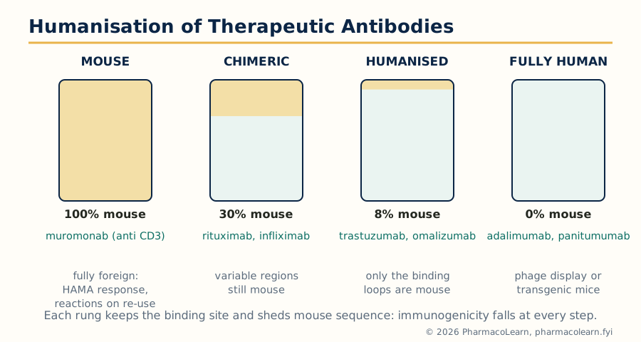
Figure: The humanisation ladder. Each rung replaces more mouse sequence with human, cutting immunogenicity while keeping the binding site that makes the drug.
A chimeric antibody (rituximab, infliximab) grafts the whole mouse variable regions onto human constant regions, leaving about a third of the molecule mouse. A humanised antibody (trastuzumab, omalizumab) grafts only the complementarity determining regions, the binding loops themselves, into a human antibody framework, leaving only about 5 to 10 percent mouse sequence. Fully human antibodies come from phage display libraries or from transgenic mice carrying human antibody genes (adalimumab, panitumumab). Humanisation reduces but does not abolish immunogenicity: patients can still raise anti drug antibodies, especially against chimeric molecules, losing response or reacting at infusion, which is why the antidrug antibody assays of Unit 3.7 are part of biologic therapy monitoring.
4.10 Immunotherapeutics in clinical practice
In the clinic, immunotherapeutics is organised around targets, and target testing comes first. Trastuzumab treats only the minority of breast and gastric cancers that overexpress HER2, so the HER2 test is the prescription's first half; its characteristic toxicity, cardiac dysfunction, follows from HER2 signaling in heart muscle. Rituximab deletes CD20 positive B cells and serves both B cell lymphomas and autoimmune diseases such as rheumatoid arthritis. The TNF blockers (adalimumab, infliximab, etanercept) transformed rheumatoid arthritis and inflammatory bowel disease, at the price of reactivating latent tuberculosis, so screening precedes treatment. The checkpoint inhibitors (pembrolizumab and nivolumab against PD-1, ipilimumab against CTLA-4) produce durable remissions in melanoma and lung cancer in a minority of patients, and their immune related adverse events, colitis, pneumonitis, thyroiditis and others, are managed by stopping the drug and suppressing the immune system the treatment released. Bevacizumab (against VEGF) starves tumours of blood vessels, and omalizumab (against IgE) treats severe allergic asthma. The practice principles are uniform: confirm the target, screen for the infections and organ risks the mechanism predicts, expect infusion and immune reactions rather than classical dose related toxicity, and plan for costs that reflect cell based manufacture, the problem the biosimilars of Unit 5.12 exist to soften.
Unit 5: Cell Culture Techniques and Biosimilars
5.1 The cell culture laboratory: basic equipment
Cell culture keeps living cells growing outside the body, which demands sterility, stable warmth and gas, and gentle handling. The basic equipment follows from those demands:
| Equipment | Purpose |
|---|---|
| Class II biosafety cabinet (laminar airflow) | Sterile working space; filtered air protects both the culture and the worker |
| CO2 incubator | Holds cultures at 37 degrees C with 5 percent CO2 and high humidity; the CO2 sets the pH of bicarbonate buffered media |
| Inverted microscope | Examines cells growing on the base of flasks and plates from below, without opening them |
| Centrifuge | Pellets cells for media changes, isolation and subculture |
| Autoclave | Sterilises media components, glassware and waste with pressurised steam |
| Water bath | Warms media and thaws frozen cells gently and quickly |
| Refrigerators, minus 80 freezer and liquid nitrogen storage | Staged cold storage, ending at minus 196 degrees C for the cell bank |
| Haemocytometer and cell counter | Counts cells and checks viability for seeding and experiments |
| pH meter, balance, sterile filtration units | Media preparation and quality control |
5.2 Cell culture media
A culture medium must replace blood: salts for osmotic balance, glucose and amino acids as fuel and building blocks, vitamins and trace elements, a buffering system, and usually a growth supplement. The standard basal media are named recipes, MEM, DMEM and RPMI-1640 among them, differing in richness and intended cell types (RPMI favours blood and suspension cells, DMEM adherent lines). Buffering is by bicarbonate in equilibrium with the incubator's CO2, and the pH indicator phenol red turns the medium into a visible gauge: pink at the working pH, yellow as growing cells acidify it, purple if it turns alkaline. The decisive supplement is serum, usually fetal bovine serum at 5 to 10 percent, supplying growth factors, hormones, attachment factors and carrier proteins; its undefined composition and batch variation are also the medium's weakest point, driving the move to serum free and chemically defined media for production work. Glutamine is added fresh or in stabilised form because it degrades in storage. Antibiotics such as penicillin and streptomycin are available but good laboratories use them sparingly: they mask low grade contamination instead of preventing it.
5.3 Types of cell culture
Cultures divide first by origin. A primary culture is taken directly from tissue (section 5.5): the cells keep much of their specialised character but divide a limited number of times before senescing. An established or continuous cell line has escaped that limit, spontaneously or by transformation, and divides indefinitely: HeLa, from a human cervical carcinoma in 1951, was the first and remains the most used; the pharmacologist's working set includes CHO cells (production and toxicology), HEK293 (transfection and channel expression, host of the hERG safety assay), HepG2 (liver metabolism models) and Caco-2 (intestinal absorption models). Cultures divide again by habit: adherent cells must attach to a treated surface and grow as a monolayer, while suspension cells, mostly of blood origin, grow floating. Organ culture keeps a tissue fragment's architecture for short studies, and the modern extensions, three dimensional spheroids and organoids, restore some structure that flat monolayers lose.
5.4 The general procedure for cell culture
The general procedure is a discipline more than a recipe, built on aseptic technique: work in the cabinet with surfaces and gloved hands disinfected with 70 percent alcohol, flame or filter sterilise what enters, minimise the time vessels stand open, and never let a pipette pass over an open sterile vessel. The working rhythm runs:
A WORKING DAY IN THE CELL CULTURE LAB

  observe cultures under the inverted microscope
  (confluence, morphology, contamination check)
        |
  warm fresh medium and reagents to 37 degrees C
        |
  feed: replace spent medium, or subculture if confluent (5.6)
        |
  return vessels to the incubator, record passage and observations
        |
  experiments seeded from counted, healthy, log phase cells
Only healthy cells in the logarithmic growth phase give reproducible assays; an overconfluent, starved or quietly mycoplasma contaminated culture will fail an experiment weeks before the failure is traced back to it.
5.5 Isolation of cells
Isolation converts a tissue into a suspension of its cells. The tissue is washed free of blood in sterile balanced salt solution, minced finely to increase surface area, and then disaggregated. Enzymatic digestion is the standard route: trypsin breaks the protein links between cells, collagenase digests the collagen scaffold of solid tissues, and dispase gives a gentler release; enzymes are stopped with serum containing medium once the tissue softens. A mechanical route (sieving, gentle pipetting) suits soft tissues such as spleen and liver. The suspension is filtered to remove debris, centrifuged, washed and resuspended in complete medium, and counted (section 5.6) before seeding. Cells then sort themselves in part: in a mixed primary culture the adherent cells attach within hours while blood cells float, and differential attachment is itself a first purification.
5.6 Subculture: passaging the culture
Subculture, or passaging, is the routine transfer of cells into fresh vessels that keeps a culture alive and expanding. Adherent cells are passaged when they reach about 70 to 80 percent confluence, before crowding stops their growth: the medium is removed, the layer is washed with phosphate buffered saline, and trypsin EDTA is added briefly; the protease cuts the attachment proteins and the EDTA chelates the calcium adhesion depends on, so the cells round up and float free. Serum containing medium stops the enzyme, the suspension is centrifuged and counted, typically on a haemocytometer with trypan blue to exclude dead cells, and a defined fraction is seeded into new vessels (the split ratio, or an exact seeding density where the assay demands it). Suspension cultures are simpler: dilute or centrifuge and reseed. Every passage is recorded with its number, because cells drift in growth rate, karyotype and drug sensitivity with passage count, and an experiment done at passage 40 is not the same experiment done at passage 8.
5.7 Cryopreservation
Cryopreservation banks cells against loss, contamination and drift. Healthy log phase cells are suspended in freezing medium, standardly complete medium with about 10 percent dimethyl sulfoxide (DMSO), a cryoprotectant that limits the ice crystal formation which would otherwise shred membranes. The vials are cooled slowly, about one degree per minute in an insulated freezing container, to minus 80 degrees, then transferred to liquid nitrogen at minus 196 degrees, where biological time effectively stops. Banks are organised as a small master stock and a larger working stock, so the master vials are rarely disturbed. Revival inverts the logic: thaw fast in a 37 degree water bath, because slow warming lets crystals regrow, dilute the DMSO away at once (it is toxic at room temperature), wash, seed into fresh medium, and confirm viability and identity before the cells are trusted.
5.8 Characterisation of cells and their application
A culture is only useful when its identity and health are proven, so characterisation runs on a checklist: morphology under the microscope (shape, growth pattern, the look of a healthy culture of that line); growth characterisation, the growth curve and doubling time; viability (section 5.9); karyotyping to confirm the chromosome complement has not drifted; identity testing, in human lines by short tandem repeat profiling, which exists because cross contamination between lines is a historically common and embarrassing error; marker expression, confirming the cell still displays the proteins of its tissue of origin, by immunostaining or flow cytometry (section 5.11); and sterility testing, including the PCR based hunt for mycoplasma, the invisible contaminant that alters cell behaviour without clouding the medium. The applications in pharmacology justify the whole apparatus: screening compounds for cytotoxicity and activity, expressing a single receptor or channel for clean drug testing (the hERG assay on which cardiac safety screening depends), modelling absorption and metabolism with Caco-2 and liver cells before animal studies, producing recombinant medicines (Unit 3.10), and replacing animal experiments at the earliest stages of drug evaluation.
5.9 Cell viability assays: principles and applications
A viability assay reports how many cells in a culture are alive, or behaving as if alive, after treatment, and it is the readout behind most cell based drug screening. The assays measure different definitions of alive, and the syllabus expects the principles distinguished:
| Assay | Principle | What it really measures |
|---|---|---|
| Trypan blue exclusion | The dye cannot cross an intact membrane; dead cells stain blue | Membrane integrity, counted manually on a haemocytometer |
| MTT assay | Living cells' dehydrogenases reduce the yellow tetrazolium salt MTT to purple formazan crystals, dissolved and read as absorbance | Metabolic activity as a proxy for viable cell number |
| Resazurin (Alamar Blue) | Living cells reduce blue resazurin to pink, fluorescent resorufin | Metabolic activity, read without killing the cells, so it can be repeated |
| ATP luminescence | Luciferase light output is proportional to ATP present | The energy charge of the culture; the most sensitive format |
| LDH release | Lactate dehydrogenase leaking from damaged cells is measured in the medium | Membrane damage (a death readout, complementary to the others) |
The applications are the daily currency of screening: cytotoxicity ranking of anticancer candidates and IC50 determination, safety testing of new compounds on normal cells, and quality control of cultures themselves. The critical skill is caveat recognition: the MTT assay reports metabolism, so a drug that sedates mitochondria without killing cells falsifies it, coloured test compounds can absorb at the reading wavelength, and the honest experiment confirms a hit with a second assay built on a different principle.
5.10 Glucose uptake and calcium influx assays
Two functional assays extend the laboratory from counting cells to watching pathways work. The glucose uptake assay measures transport of glucose into cells, classically in insulin responsive models, the adipocyte line 3T3-L1 and the muscle line L6. The tracer is either radiolabelled 2-deoxyglucose, which enters through the glucose transporters and is trapped inside after phosphorylation, or the fluorescent analogue 2-NBDG, read on a plate reader. Insulin sharply raises uptake by moving GLUT4 transporters to the surface (Unit 2.5's pathway, measured end to end), and the assay, with cytochalasin B as the transporter blocking control, screens for insulin sensitising and antidiabetic compounds and probes the mechanism of insulin resistance in cells made resistant by chronic insulin or fatty acid exposure. The calcium influx assay watches the messenger of Unit 2.9 in real time: cells are loaded with a calcium sensitive fluorescent dye (Fura-2, whose signal is ratiometric and self calibrating, or the brighter single wavelength Fluo-4, both entering as membrane permeant ester forms) and fluorescence reports cytosolic calcium second by second. Run on a fluorescence plate reader of the FLIPR type, it turns Gq coupled receptor activation and ion channel opening into a high throughput signal, and it is one of the standard primary screens of industrial drug discovery for GPCR and channel targets.
5.11 Flow cytometry: principles and applications
Flow cytometry measures cells one at a time, thousands per second, as they stream past lasers. The fluidics system uses hydrodynamic focusing to line the cells up in single file. The optics then record, for every cell, forward scatter (roughly, size), side scatter (roughly, internal granularity) and the fluorescence of any labels the cell carries: antibodies tagged with fluorochromes against surface markers, DNA dyes, or functional probes. Software plots the population, and the analyst gates, selecting subpopulations by their scatter and fluorescence to count and characterise them. In the sorting form (FACS), droplets containing single chosen cells are charged and deflected into separate tubes, physically purifying the selected population.
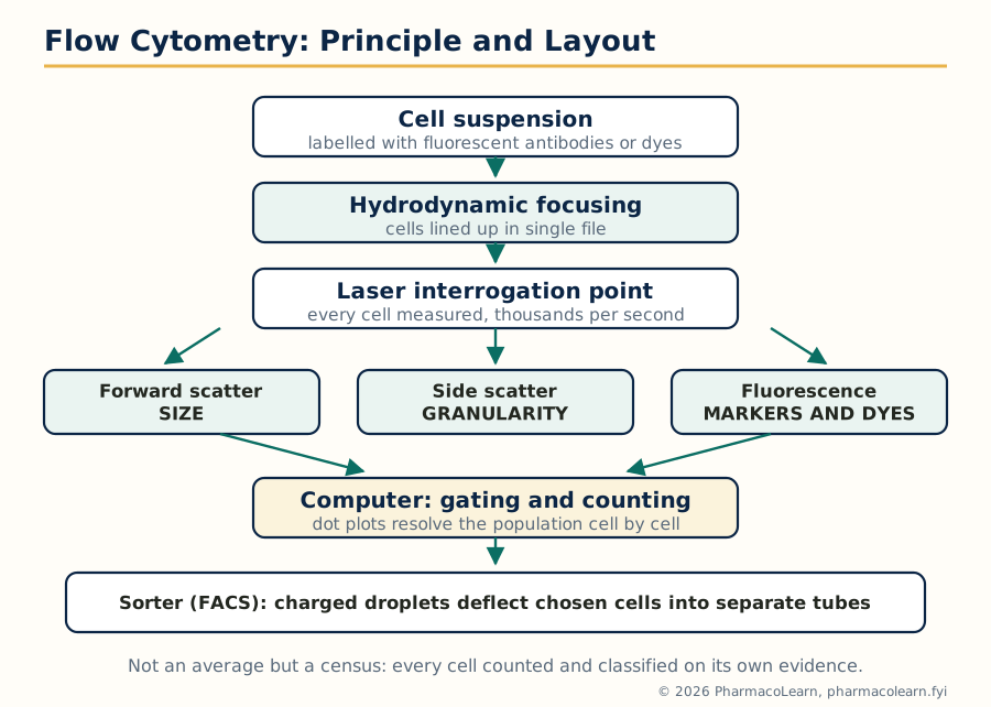
Figure: Flow cytometry in one pass. Single file cells, laser interrogation, scatter for physical character, fluorescence for identity, and, in the sorter, physical separation of the cells the gates select.
The applications in pharmacology and medicine are wide: immunophenotyping, counting CD4 T cells in HIV care and classifying leukaemias and lymphomas by their marker patterns; apoptosis measurement with Annexin V (which binds the phosphatidylserine that apoptotic cells expose, Unit 1.9) paired with a viability dye; cell cycle analysis from DNA content staining, reading out the phase distribution of Unit 1.7 and the effect of cycle active drugs; and the characterisation work of section 5.8. Its strength is statistical: it does not average a culture, it censuses it.
5.12 Biosimilars
Unit 5 closes with the products that the technologies of Units 3 and 4 created. A biologic medicine, an antibody, insulin, growth factor or vaccine, is made by living cells, is large and structurally complex, and carries the manufacturer's fingerprints in its glycosylation and folding: the process defines the product in a way that never applies to a small molecule. A biosimilar is a biologic shown to be highly similar to an already licensed reference product, with no clinically meaningful differences in quality, safety or efficacy. It is therefore not a generic: a generic small molecule is chemically identical to its reference and proves itself by bioequivalence, while a biosimilar can only be shown similar, through a stepwise comparability exercise that starts with extensive analytical and functional comparison (the tools of Unit 3) and confirms with comparative pharmacokinetic, pharmacodynamic and immunogenicity studies in patients. Regulation reflects this: the European agency built the first dedicated pathway, India regulates these products as similar biologics under guidelines issued by the CDSCO and the Department of Biotechnology (2012, revised 2016), and the United States reserves the additional label of interchangeable for products shown to support switching. Biosimilars of filgrastim, insulin glargine, trastuzumab, rituximab and adalimumab are now routine, several of them developed and manufactured in India. Their value is the value of competition: lower prices and wider access to the immunotherapeutics of Unit 4, guarded by pharmacovigilance and traceability, since a similar product is a distinct product and must be tracked as one.
Frequently Asked Questions
Q: What separates the intrinsic from the extrinsic pathway of apoptosis, and where do they meet? The intrinsic pathway is started from inside the cell, by damage, stress or withdrawal of growth signals, and runs through the mitochondria: Bax and Bak open the outer mitochondrial membrane, cytochrome c forms the apoptosome with Apaf-1, and caspase 9 is activated. The extrinsic pathway is started from outside, by death ligands such as Fas ligand and TNF binding their surface receptors, and activates caspase 8 at the death inducing signaling complex. They meet at the executioner caspase 3, and they are cross connected, because caspase 8 also cuts Bid to tBid, which engages the mitochondrial pathway and amplifies the signal.
Q: How do siRNA and microRNA differ, given that both silence genes? Both are short RNAs loaded into the RISC complex, but siRNA comes from long double stranded RNA (or is supplied synthetically), matches its single target messenger RNA perfectly, and directs Argonaute 2 to slice and destroy that message. microRNA is encoded by the cell's own genes, is processed from a hairpin precursor by Drosha and Dicer, matches its targets only partially, chiefly in the 3 prime untranslated region, and mainly represses translation and promotes decay; one microRNA therefore tunes many messages at once. siRNA is the precision tool and the drug (patisiran, inclisiran), microRNA is the cell's own fine tuning system.
Q: Why does the JAK/STAT pathway need no second messenger, and why does that matter for drugs? Cytokine receptors carry no enzyme of their own; the JAK kinases attached to their cytoplasmic tails do the work. Ligand binding activates the JAKs, which phosphorylate the receptor and then the STAT transcription factors, and the phosphorylated STATs dimerise and go straight to the nucleus. Receptor and gene are therefore only three proteins apart, the shortest signaling route in the subject. It matters because the borrowed kinase is a single druggable bottleneck for many cytokines at once: JAK inhibitors such as ruxolitinib and tofacitinib block a whole family of signals with one small molecule, which explains both their broad efficacy in immune disease and their broad adverse effect burden.
Q: RT-PCR and real time PCR sound like the same thing. Are they? No, and the similarity of the names is a standard examination trap. RT-PCR means reverse transcription PCR: a first step in which reverse transcriptase converts an RNA template into complementary DNA, without which RNA targets (RNA viruses, gene transcripts) could not be amplified at all. Real time PCR means the amplification is monitored as it happens by fluorescence, giving a Ct value that quantifies the starting template. The two combine freely: viral load tests for HIV and SARS-CoV-2 are reverse transcription real time PCR, doing both jobs in one run.
Q: Why can the same dose of codeine be harmless in one patient, useless in a second and fatal in a third? Codeine itself is nearly inactive; it must be converted to morphine by CYP2D6, a highly polymorphic enzyme. Poor metabolisers make little morphine, so the drug simply fails. Normal metabolisers make the expected amount. Ultrarapid metabolisers, who carry extra copies of the CYP2D6 gene, convert so fast that morphine levels spike into the toxic range, causing respiratory depression that has killed adults and breastfed infants of ultrarapid mothers. Identical prescription, three different drugs in practice: this is the founding clinical example of pharmacogenomics.
Q: What does it mean to call an antibody humanised, and why is it worth the engineering? A mouse monoclonal antibody is read as foreign protein by the human immune system, which raises anti mouse antibodies that neutralise the drug and cause reactions on repeat dosing. A chimeric antibody replaces the constant regions with human ones; a humanised antibody keeps only the mouse binding loops (the complementarity determining regions) grafted into a human framework, leaving about 5 to 10 percent mouse sequence; a fully human antibody has none. Each step cuts immunogenicity and extends usable life in the patient, which is why the stems in antibody names (-xi-, -zu-, -u-) record how far the engineering went.
Q: ELISA and western blotting both use antibodies. When is each the right tool? ELISA is the bulk measurement tool: fast, quantitative against standards, run in 96 well plates on hundreds of samples, but it reports only that something bound, in aggregate. Western blotting is the verification tool: the sample is first separated by size on SDS-PAGE, so the antibody signal comes with a molecular weight attached, confirming that the band is the right protein and revealing cut or modified forms. In practice ELISA screens and quantifies, western blotting confirms identity, and strong work uses each to police the other.
Q: How does the cell cycle explain the scheduling and toxicity of cancer chemotherapy? Most cytotoxic drugs kill cells in a particular phase: antimetabolites in S phase, vinca alkaloids and taxanes in M phase. A tumour's cells are not synchronised, so a single dose catches only the fraction currently in the sensitive phase, which is why chemotherapy is given in repeated cycles as more cells cycle through. The same logic predicts the toxicity pattern: the normal tissues that cycle fastest, bone marrow, gut lining and hair follicles, are hit hardest, producing myelosuppression, mucositis and alopecia. Checkpoint aware drugs such as the CDK4/6 inhibitors attack the control system itself, arresting cells in G1.
Q: The MTT assay is called a viability assay, but it does not count dead cells. What does it actually report? It reports metabolic activity. Living cells' dehydrogenases reduce the yellow MTT salt to purple formazan, so the colour intensity tracks active metabolism in the well, which under controlled conditions is proportional to viable cell number. The distinction matters: a drug that suppresses mitochondrial activity without killing cells reads as cytotoxic when it is not, and coloured or reducing compounds interfere directly. A viability claim from MTT alone is a proxy claim, and screening practice confirms hits with an assay on a different principle, such as ATP luminescence or membrane integrity.
Q: Why is a biosimilar not simply called a generic biologic? Because identity cannot be shown. A generic small molecule is the same chemical as its reference and needs only bioequivalence. A biologic is made by living cells, is far larger and more complex, and differs in fine details such as glycosylation between manufacturers and even between processes, so a follow on product can be demonstrated highly similar, through analytical, functional and clinical comparability studies, but never identical. The different name carries a different evidence standard, different regulation (similar biologics in India), and different clinical handling, including traceability of which product a patient received.
Complete MPL104T notes, all 5 units in one PDF, free to keep.
Other M.Pharm subjects: MPH101T · MPC101T · MPL101T · MPH102T · MPH103T · MPH104T · MPL102T · MPL103T · MPL104T · MPC102T · MPC103T · MPC104T · MPH201T · MPH202T · MPH203T · MPH204T · MPC201T · MPC202T · MPC203T · MPC204T · M.Pharm