
MPL103T — Pharmacological and Toxicological Screening Methods I
4 hrs/week, 4 credits, 60 hours, 100 marks. Notes follow the M.Pharm syllabus, unit by unit.
MPL103T Pharmacological and Toxicological Screening Methods I notes for M.Pharm Semester 1 Pharmacology: laboratory animals, CPCSEA and GLP, bioassay, and screening models for CNS, cardiovascular and metabolic drugs, with free unit wise PDF downloads on PharmacoLearn.
Units in this subject
- Unit 1 — Laboratory Animals
- Unit 2 — Preclinical Screening: General Principles, CNS and Autonomic Pharmacology
- Unit 3 — Preclinical Screening: Respiratory, Reproductive, Analgesic and Gastrointestinal Pharmacology
- Unit 4 — Preclinical Screening: Cardiovascular, Metabolic, Anticancer and Hepatoprotective Pharmacology
- Unit 5 — Preclinical Screening: Immunomodulators, Immunoassay, Alternatives and Extrapolation
Download MPL103T PDF Notes
Free PDFs for this subject, one per unit:
Pharmacological and Toxicological Screening Methods I, subject code MPL103T, is a core Semester 1 subject of M.Pharm Pharmacology. It carries 4 credits and 60 hours of theory, taught at 4 hours per week, for 100 marks.
Before any drug reaches a patient it must be screened: tested in living systems and in test systems that imitate living ones, to find out whether it works, how strongly it works, and where its dangers begin. This subject teaches that preclinical evaluation end to end. Unit 1 lays the foundation: the common laboratory animals and their strains, handling, transgenic animals, anaesthesia and euthanasia, animal house maintenance and breeding, the CPCSEA guidelines that govern animal experiments in India, good laboratory practice, and the principles and methods of bioassay. Units 2 to 5 then apply the idea of screening system by system. Unit 2 covers the general principles of preclinical screening and the screening of CNS and autonomic drugs, from behavioural tests to models of Parkinson's disease and Alzheimer's disease. Unit 3 covers respiratory, reproductive, analgesic, anti-inflammatory, antipyretic and gastrointestinal screening. Unit 4 covers cardiovascular screening, metabolic disorder screening, anticancer screening and hepatoprotective screening. Unit 5 covers immunomodulator screening, the principles and evaluation of immunoassay with the immunoassays of digoxin and insulin, the limitations of animal experimentation and its alternatives, and the extrapolation of in vitro and animal data towards humans. A student who completes this subject should be able to choose the right animal and the right model for a given drug class, name the endpoint and the standard drug of every classical screen, and judge what a preclinical result can and cannot promise for the clinic.
Unit 1: Laboratory Animals
1.1 Why animals stand at the start of drug discovery
A new chemical cannot be given to a human being on the strength of its structure or its behaviour in a test tube. The whole animal adds what no isolated system can: absorption, distribution, metabolism and excretion acting together, intact reflexes and feedback loops, and the integrated response of organs that talk to each other. Preclinical screening is the organised testing of new substances in animals and in animal derived systems to detect pharmacological activity, estimate potency, and give the first warning of toxicity before human exposure. Every screen in this subject follows the same grammar: a defined animal or preparation, a reproducible stimulus or disease like state, a measurable endpoint, and comparison against a standard drug whose behaviour in that model is already known.
The choice of species is itself a scientific decision. The animal must respond to the drug class in a way that resembles the human response, it must be economical enough to use in groups large enough for statistics, and its use must be ethically justified. The sections below take the common species one by one.
1.2 The mouse: the workhorse of screening
The mouse (Mus musculus) is the most widely used laboratory animal, accounting for the majority of animals used in pharmacology worldwide. It is small (adult weight about 20 to 40 g), cheap to house and feed, breeds rapidly with a gestation of about 19 to 21 days and large litters, and its small size means that only milligram quantities of a scarce new compound are needed per animal, a decisive advantage in early screening.
Applications follow from these virtues. Mice are the standard species for acute toxicity observation, for analgesic screening (writhing, hot plate), for CNS screening (locomotor activity, convulsions), for cancer chemotherapy screening using transplantable tumours, and for immunological work. Genetically defined mice, including transgenic and knockout lines (section 1.7), make the mouse the principal species for studying the role of a single gene or receptor in drug action. Its limitations are its very small blood volume, which restricts repeated sampling, and metabolic differences from humans that can mislead extrapolation (Unit 5).
1.3 The rat: the pharmacologist's standard animal
The rat (Rattus norvegicus) is the second great screening species and the default animal of experimental pharmacology teaching and research. Adults weigh about 200 to 400 g, large enough for surgery, repeated blood sampling and blood pressure recording, yet still economical. Two outbred stocks dominate: the Wistar rat, an albino stock valued for its docility, and the Sprague Dawley rat, a fast growing albino stock widely used in toxicology and reproduction studies.
Rats are the standard species for anti-inflammatory screening (carrageenan paw oedema), anti-ulcer studies (pylorus ligation), behavioural pharmacology (conditioned avoidance, maze learning), antihypertensive and diuretic screening, and most chronic toxicity work. The rat lacks a gall bladder and cannot vomit, two anatomical facts that shape its use: it is unsuitable for emetic studies, and bile collection is continuous rather than stored. Oestrous cycle monitoring by vaginal cytology makes the rat the standard species for reproductive screening (Unit 3).
1.4 Guinea pig, rabbit, hamster and the larger species
Each remaining species survives in the laboratory because it does one job better than the rodents.
| Species | Special features | Classic applications |
|---|---|---|
| Guinea pig (Cavia porcellus) | Bronchial smooth muscle very sensitive to histamine; needs dietary vitamin C; ileum rich in histamine and acetylcholine receptors | Isolated ileum bioassay, bronchospasm and anti-asthmatic screening, anaphylaxis and allergy models, hypoxia studies less so |
| Rabbit (Oryctolagus cuniculus) | Docile, large ear veins for intravenous dosing and sampling; eye accessible; some strains resistant to atropine | Pyrogen testing (now largely replaced), eye irritation and ocular pharmacology, isolated heart and ear preparations, pregnancy and teratology work |
| Syrian hamster | Cheek pouch accessible for study; hibernation physiology | Buccal pouch carcinogenesis, metabolic and infection models |
| Cat | Well developed vomiting centre; cardiovascular and CNS physiology close to classical teaching preparations | Emetic and anti-emetic work (historical), blood pressure and neurophysiology preparations |
| Dog (beagle) | Cardiovascular system and gastric physiology resemble humans; manageable size in the beagle | Cardiovascular safety and haemodynamic studies, emetic screening, chronic toxicology as the standard non-rodent species |
| Monkey (rhesus, cynomolgus) | Closest physiology and genetics to humans; reserved for questions rodents cannot answer | Reproductive pharmacology, CNS and behavioural studies, biologics and vaccine evaluation, used sparingly on ethical grounds |
The pattern to remember is that species selection is driven by the endpoint: the guinea pig ileum for spasmogens, the dog for emesis and haemodynamics, the rabbit for pyrogens and the eye, the primate only when no lower species can model the human system.
1.5 Strains: inbred, outbred and mutant
A strain is a population of animals with a defined genetic relationship, and the choice of strain decides the reproducibility of the whole experiment.
- Outbred stocks (Wistar, Sprague Dawley, Swiss albino mice) are bred to preserve genetic variation, so results generalise across genotypes much as a human population varies. They are cheaper and are the default for routine screening.
- Inbred strains are produced by brother to sister mating for at least 20 generations, making animals within the strain almost genetically identical, like identical twins. Examples are BALB/c and C57BL/6 mice and the Fischer 344 rat. Inbred strains remove genetic noise, accept tissue grafts from each other, and each strain has a characteristic profile (C57BL/6 mice, for example, are the background of most transgenic lines). Their weakness is that a result may be true only for that one genotype.
- Mutant and specialised strains carry a defined abnormality that is itself the model: the spontaneously hypertensive rat (SHR) for hypertension, the obese Zucker rat and the db/db mouse for metabolic disease, the nude (athymic) mouse that accepts human tumour xenografts, and the SCID mouse lacking functional immunity.
Sex, age and weight are part of the same discipline: a screening protocol states them for every group, because hormonal state, age and body weight all change drug response.
1.6 Handling and restraint
Correct handling is both an ethical duty and a scientific one, because a frightened, struggling animal releases catecholamines and corticosteroids that distort blood pressure, glucose, behaviour and almost every endpoint in this subject. The general rules are consistent across species: approach calmly, handle animals regularly before the experiment so they habituate, support the body weight fully, restrain for the shortest time needed, and never lift a rodent by the tip of the tail alone or squeeze the thorax.
In practice: mice are picked up by the base of the tail and restrained by scruffing the skin over the neck between finger and thumb, which also presents the abdomen for intraperitoneal injection. Rats are grasped firmly around the thorax from above with the head controlled, or scruffed for injections. Rabbits are lifted with one hand under the hindquarters and the ears and scruff controlled with the other, never by the ears alone. Guinea pigs are cupped with one hand under the chest and the other supporting the hindquarters. Restrainers (tubes and boxes sized to the animal) are used for dosing and sampling, and chemical restraint (section 1.9) replaces force whenever the procedure is more than brief.
Routes of administration taught with handling are oral (gavage tube in rodents), intraperitoneal, subcutaneous, intramuscular and intravenous (tail vein in rodents, ear vein in the rabbit, saphenous or jugular in larger animals).
1.7 Transgenic animals: production
A transgenic animal carries foreign DNA, or a deliberately altered version of its own DNA, in its genome, passed to its offspring through the germ line. Production follows three classical routes.
- Pronuclear microinjection: the DNA construct is injected into one pronucleus of a fertilised egg, which is then implanted into a pseudopregnant foster mother. Some pups integrate the construct and are screened for its presence; founders are bred to establish a line. Integration is random, so expression varies between lines.
- Embryonic stem cell targeting: the construct is introduced into embryonic stem cells in culture, where homologous recombination can place it at an exact locus. This precision allows knockout animals, in which a gene is disabled, and knock-in animals, in which a gene is replaced by a modified or human version. Selected stem cells are injected into a blastocyst, producing a chimeric animal whose germ line transmission gives the new strain.
- Viral and newer editing routes: retroviral and lentiviral vectors carry constructs into embryos, and nuclease based editing (the CRISPR Cas9 system) cuts DNA at a chosen sequence so that genes can be disrupted or corrected directly in the zygote, greatly speeding production.
1.8 Transgenic animals: maintenance and applications
Transgenic lines are maintained as breeding colonies with genotyping of each generation (usually from a tail or ear sample), because the inserted gene may be present in one or two copies and expression can drift. Cryopreservation of embryos and sperm protects valuable lines against disease and genetic drift.
Applications in pharmacology are direct. Target validation: if deleting a receptor gene abolishes the effect of a drug class, the receptor is confirmed as the target (the principle behind many knockout studies of opioid, dopamine and cannabinoid receptors). Disease models: transgenic mice expressing human amyloid precursor protein mutations model Alzheimer's disease for the screening of Unit 2; mice carrying human oncogenes model cancer. Humanised animals expressing human drug metabolising enzymes or human receptors predict human responses better than the wild type animal. Reporter animals, in which a gene of interest drives a visible marker, turn gene expression into a readable endpoint.
1.9 Anaesthesia of experimental animals
Anaesthesia in screening serves the animal (freedom from pain and distress) and the experiment (immobility for surgery and recording). The agent is chosen by species, duration and the endpoint at stake, because most anaesthetics themselves depress the CNS, respiration and cardiovascular system and can distort results.
| Agent or combination | Route and use | Notes |
|---|---|---|
| Ketamine with xylazine | Injectable, rodents and larger animals, short to medium procedures | Widely used combination; xylazine adds analgesia and muscle relaxation but depresses respiration and heart rate |
| Thiopentone and pentobarbitone | Injectable barbiturates, classical rodent and rabbit anaesthesia | Narrow margin between anaesthesia and respiratory death; pentobarbitone now more often an overdose agent for euthanasia |
| Isoflurane and sevoflurane | Inhalational, all species, precise control | Rapid induction and recovery, depth adjusted minute to minute; need a vaporiser and scavenging |
| Urethane | Injectable, terminal (non recovery) experiments only | Long lasting anaesthesia for acute cardiovascular and nerve recordings; carcinogenic, never used where the animal recovers |
| Local anaesthetics (lidocaine) | Infiltration or nerve block | For minor procedures, and combined with general agents to reduce their dose |
Stages of anaesthesia, depth monitoring (pedal reflex, respiratory rate, heart rate), body temperature support and fluid care are part of competent practice: a hypothermic anaesthetised rodent produces blood pressure and metabolic data that belong to the cold, not to the drug.
1.10 Euthanasia of experimental animals
Euthanasia is the humane killing of an animal at the end of an experiment, or earlier when a humane endpoint is reached (unrelieved pain, severe weight loss, moribund state). CPCSEA accepts methods that produce rapid unconsciousness and death with minimal pain and distress, and the method must not destroy the tissue or endpoint being studied.
Accepted routes include overdose of an anaesthetic agent (barbiturate overdose is the commonest physical chemical method), inhalation of carbon dioxide in rodents with a gradual fill of the chamber, and, in deeply anaesthetised animals, physical methods such as cervical dislocation in small rodents performed by trained personnel. Death must be confirmed (absence of heartbeat and respiration, fixed pupils) before disposal. Neonates, which resist hypoxia, need special care and longer exposure or an injectable agent. The scientific point is the mirror of anaesthesia: the method of death can alter blood chemistry, stress hormones and tissue enzymes, so it is standardised and recorded like any other part of the protocol.
1.11 Maintenance of the animal house: housing and environment
Maintenance begins with the building. A laboratory animal facility is barrier managed: clean and dirty corridors, controlled entry, quarantine for incoming animals, and separation of species and of infected from clean stock. Standard conditions in Indian guidelines for rodents are a temperature of about 22 plus or minus 3 degrees Celsius, relative humidity of 30 to 70 percent, 12 hour light and dark cycles, and 10 to 15 fresh air changes per hour, with noise and vibration controlled.
Cage level care decides data quality. Cages are sized to species norms so animals can stand, turn and rest normally; bedding is absorbent, dust free and changed on schedule; feed is a standardised pelleted diet (so that nutrition does not vary between batches of an experiment) with clean drinking water ad libitum; and environmental enrichment (nesting material, shelters) reduces stress behaviour. Health monitoring is continuous: sentinel animals, microbiological screening and post mortem checks keep specific pathogen free colonies genuinely free, because a silent infection changes immune and metabolic endpoints across an entire facility. Every animal carries records: species, strain, sex, source, date of birth and experimental allocation.
1.12 Breeding of laboratory animals
Breeding systems protect the genetics described in section 1.5. Monogamous pairing (one male with one female, the standard for mice) allows exact pedigree records. Polygamous or harem systems (one male with several females) save space for production colonies. Inbreeding by brother to sister mating maintains inbred strains and must be tracked generation by generation; outbreeding uses rotation systems that avoid mating close relatives, preserving the variation that outbred stocks exist to provide.
Practical numbers shape colony planning: mice gestate in about 19 to 21 days with litters commonly of 6 to 12 pups and can be rebred at the post partum oestrus; rats gestate in about 21 to 23 days; rabbits in about 30 to 32 days; guinea pigs in about 59 to 72 days with small precocious litters. Weaning is at about 3 weeks in mice and rats. A breeding nucleus of genetically monitored parents supplies production animals, so that experimental animals are never bred casually from the experimental stock itself.
1.13 CPCSEA guidelines and the ethics framework
In India, animal experimentation is regulated under the Prevention of Cruelty to Animals Act, 1960, through the Committee for the Purpose of Control and Supervision of Experiments on Animals (CPCSEA), a statutory body under the Ministry of Fisheries, Animal Husbandry and Dairying. No institution may conduct animal experiments unless it is registered with CPCSEA, and no experiment may begin without approval of its protocol by the institution's own Institutional Animal Ethics Committee (IAEC).
The framework, in sequence, is:
FROM IDEA TO APPROVED EXPERIMENT (CPCSEA / IAEC)

  Researcher drafts protocol
  (species, numbers, justification, humane endpoints)
              |
              v
  IAEC review: CPCSEA nominee must be present
  (welfare, 3Rs, statistics, alternatives considered)
              |
       approved? ---- no ----> revise and resubmit
              | yes
              v
  Registered establishment, CPCSEA norms for housing,
  breeding and transport; records kept and inspected
              |
              v
  Experiment runs; humane endpoints enforced;
  annual report and IAEC minutes to CPCSEA
The IAEC includes the CPCSEA nominee, a scientist from a different biological discipline, a veterinarian, and a socially aware member from outside the institution, so that welfare is judged by more than the experimenters themselves. CPCSEA norms also fix housing space, breeding and transport standards, and require that experiments use the minimum number of animals consistent with statistical validity, that alternatives be considered first (the 3Rs of Unit 5), and that anaesthesia and analgesia be used wherever pain is possible. Experiments on large animals and primates attract stricter scrutiny, and the CPCSEA main committee itself must clear certain categories.
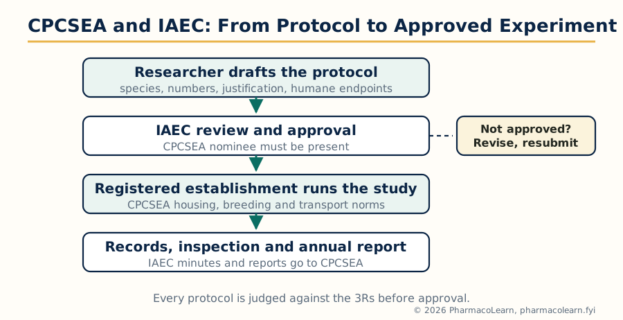
Figure: The CPCSEA and IAEC approval path. No animal experiment in India begins before the Institutional Animal Ethics Committee, with the CPCSEA nominee present, has approved the protocol.
1.14 Good laboratory practice
Good laboratory practice (GLP) is the quality system under which non clinical safety studies are planned, performed, monitored, recorded, archived and reported, so that a regulator can trust and reconstruct them years later. Its principles, harmonised through the OECD and monitored in India by the National GLP Compliance Monitoring Authority, include:
- A named study director responsible for the whole study, and an independent quality assurance unit that inspects it.
- Written standard operating procedures for every routine operation, and a study plan (protocol) approved before work begins.
- Characterised test items and test systems, calibrated instruments, and trained, documented personnel.
- Raw data recorded indelibly and contemporaneously; every correction traceable to a person and a reason.
- A final report that mirrors the raw data, with archives retained for the prescribed period.
In this subject GLP matters for two reasons: safety data submitted for drug approval must be GLP generated, and even academic screening borrows its disciplines (protocol first, raw data always, no unrecorded adjustments) because they are what make a result believable.
1.15 Bioassay: principle and scope
A bioassay (biological standardisation) measures the potency of a substance by the response it produces in a living system: an animal, an isolated tissue, a cell culture or an enzyme preparation. The principle is comparison: the unknown preparation is tested side by side with a standard preparation of the same substance (ideally an international or pharmacopoeial standard), and potency is expressed relative to that standard, never as an absolute property of the tissue.
Bioassay is the method of choice in defined circumstances, which form its scope:
- The active principle is unknown or cannot yet be measured chemically (historically insulin, posterior pituitary and many hormones and vitamins).
- No chemical method is sensitive or specific enough, or the substance exists as a mixture of related actives whose combined effect matters.
- The biological activity itself is the property of interest, as with vaccines, antisera and cytokines.
- A screening endpoint is needed for a new substance whose chemistry gives no clue to activity.
Its limitations are the reverse of its virtues: living systems vary, so bioassay is less precise than chemical assay and needs statistical design to reach acceptable error; it is slower, costlier and uses animals; and the response may not be specific, so interfering substances falsify results. Wherever a validated chemical or physical assay exists, pharmacopeias replace bioassay with it; bioassay survives where biology is the only measuring instrument available.
1.16 Bioassay: methods
Bioassay designs are classified by how the comparison with the standard is made.
| Design | How it works | Typical example |
|---|---|---|
| Matching (bracketing) | The dose of unknown producing the same response as a fixed standard dose is found by trial, bracketing the response between standard doses | Early insulin and pituitary assays; simple but imprecise |
| Interpolation | The response to the unknown is read off a previously plotted standard dose response curve | Reading an unknown histamine concentration from the guinea pig ileum curve |
| Multiple point (quantal and graded) assays | Several doses of standard and unknown are compared in a designed experiment: 2+2, 3+3 or 4+4 dose assays, with parallel line statistics | Pharmacopoeial assays of insulin, digitalis, hormones |
| Quantal (end point) methods | The response is all or none (dead or alive, convulsed or not); the dose effective in 50 percent of animals (ED50) or lethal in 50 percent (LD50) is estimated | Convulsion protection assays, toxicity endpoints |
| Microbiological assays | Growth or inhibition of a microorganism measures the substance | Antibiotic potency by cylinder plate or turbidimetric methods |
A 3 + 3 GRADED BIOASSAY, STEP BY STEP

  Plot log dose response curves for standard (S1 S2 S3)
  and test (T1 T2 T3) at three dose levels each
              |
              v
  Check validity: curves linear and parallel
  (non parallelism means the test is not behaving
   like the standard; the assay is invalid)
              |
              v
  Horizontal distance between the two lines
  = log potency ratio; convert to potency units
              |
              v
  Report potency with fiducial (confidence) limits
Preparation choice follows the substance: isolated guinea pig ileum for histamine and acetylcholine like activity, isolated frog rectus abdominis historically for acetylcholine, rabbit blood sugar or mouse convulsion methods historically for insulin, rat blood pressure for catecholamines. Every valid assay ends in statistics: potency is reported with fiducial limits, because a bioassay number without its error is not a result.
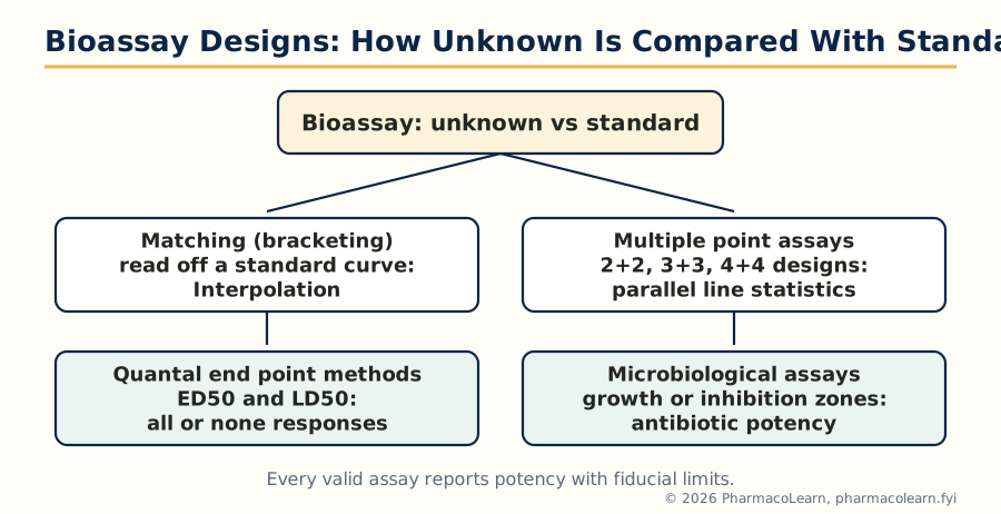
Figure: The four families of bioassay design. All compare the unknown against a standard; they differ in how the comparison is arranged and analysed.
Unit 2: Preclinical Screening: General Principles, CNS and Autonomic Pharmacology
2.1 General principles of preclinical screening
Screening is organised as a funnel. A new substance enters at the wide end, where cheap, fast, high capacity tests remove the inactive majority, and leaves at the narrow end, where expensive, slow, whole animal studies characterise the few survivors.
The principles that govern every unit of this subject are:
- Start simple, proceed to the complex. Primary screening in vitro (enzyme, receptor binding, cell systems) precedes secondary screening in vivo, which precedes detailed safety work.
- Always run a standard. Every experiment includes the reference drug of the class (diazepam in anxiety models, morphine in analgesia, haloperidol in psychosis models), so a failed day of testing is detected at once.
- Use more than one model. No single test defines a drug class; a profile across related models separates true activity from artefacts.
- Quantify. Dose response curves, ED50 values and potency relative to the standard replace impressions.
- Blind and randomise. The observer should not know which animal received which treatment, and animals are allocated randomly, because expectation moves subjective endpoints.
- Screen for the unwanted early. A compound active in the target model but toxic or sedating at the same dose is rejected early, using simple counter screens such as rotarod performance and general behavioural observation (the Irwin profile in mice, a systematic check of behaviour, autonomic signs and toxicity after a single dose).
THE SCREENING SEQUENCE

  Thousands of compounds
        |  in vitro binding, enzyme, cell assays
        v
  Primary in vivo screen (one or two models, few doses)
        |  inactive majority discarded
        v
  Secondary screens (model battery, dose response, ED50)
        |  counter screens: motor, autonomic, toxicity
        v
  Lead compound: pharmacokinetics, safety pharmacology
        |
        v
  Candidate for formal toxicology and clinical trials
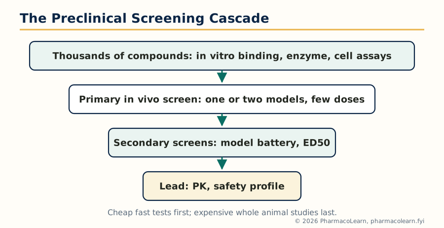
Figure: The screening funnel. Capacity falls and cost rises at every stage, so weak compounds must be removed as early as the science allows.
2.2 In vivo, in vitro and alternative models
The three families of test system trade realism against control.
| System | What it is | Strengths | Weaknesses |
|---|---|---|---|
| In vivo | Whole living animal | Integrated ADME and organ interaction; the only system that shows behaviour, blood pressure and disease course together | Slow, costly, ethically loaded, species differences |
| In vitro | Isolated organs, tissues, cells, enzymes, receptors outside the body | Precise dose control, mechanism visible, high throughput, no whole animal suffering | No ADME, no integrated physiology; activity may vanish or appear falsely |
| Alternative models | Non animal or lower organism systems: cell lines, computer (in silico) models, zebrafish embryos, invertebrates | Replace or reduce animal use (Unit 5), fast and cheap | Predictive value must be proven model by model |
Good programmes chain them: an in vitro hit earns an in vivo test; an in vivo profile earns comparison with human data before any clinical step.
2.3 Behavioural screening and locomotor activity
Behaviour is the readout of CNS screening, and the first question asked of any CNS active compound is whether it stimulates or depresses the intact animal.
- Actophotometer: the mouse or rat crosses beams of light in a cage; the count of crossings in a fixed time measures locomotor activity. Amphetamine raises the count, chlorpromazine and other sedatives lower it. It is the primary screen separating CNS stimulants from depressants.
- Open field test: the animal explores a novel arena; crossings, rearing and grooming are scored, and entries into the exposed centre reflect anxiety as much as movement.
- Irwin primary observation: a structured checklist (alertness, posture, gait, reflexes, autonomic signs, convulsions, death) after graded doses, giving the first behavioural and safety fingerprint of a new substance.
- Hole board and exploratory tests: head dipping into holes measures exploration, reduced by sedation and increased disinhibition by anxiolytics.
2.4 Muscle coordination and motor function
Motor tests serve twice: as screens in their own right (muscle relaxants, antiparkinsonian drugs) and as counter screens, because a drug that merely paralyses or sedates will falsely appear active in pain, anxiety and psychosis models.
- Rotarod: the animal walks on a rotating rod; the time it stays on measures coordination and balance. Neuroleptics, sedatives and muscle relaxants shorten the time; it defines the motor toxic dose (TD50) that is compared with the effective dose.
- Grip strength meter: measures forelimb grip force, reduced by muscle relaxants and neuromuscular blockers.
- Traction test: the mouse must pull itself up by the forepaws onto a wire; failure signals muscle relaxation.
- Chimney test: the mouse must climb backwards out of a vertical tube within a time limit; sedation and relaxation prolong it.
- Inclined plane and screen tests: the animal must hold position on a slope or mesh, a simple index of muscle tone and strength.
2.5 Screening of CNS stimulants
CNS stimulants are screened by reversal and enhancement designs. Increased locomotor counts in the actophotometer and increased open field activity are the primary signals. Analeptic activity (respiratory stimulation in barbiturate depressed animals) was the classical screen for agents like doxapram. Convulsant liability is measured by the dose lowering the threshold to electrically or chemically induced seizures, which is also a safety flag. Amphetamine serves as the standard stimulant; caffeine is the mild reference. A compound that increases activity only at doses that also cause stereotyped behaviour (repetitive sniffing, gnawing) is flagged as amphetamine like, with abuse potential implications.
2.6 Screening of CNS depressants
Depressants are detected by a coherent battery rather than one test: reduced actophotometer counts, potentiation of barbiturate sleeping time (the test drug prolongs the sleep produced by a fixed dose of pentobarbitone, the classical screen for sedative and hypnotic activity), loss of righting reflex at high doses, and reduced response to nociceptive and startle stimuli. Ethanol interaction and potentiation of ethanol induced sleep flag sedative load. Diazepam and pentobarbitone are the usual standards. The battery also grades depth: a compound that reduces activity but never abolishes the righting reflex is a sedative; one that abolishes it is a hypnotic or general depressant.
2.7 Screening of anxiolytics
Anxiety models exploit approach avoidance conflict: the animal wants to explore but fears exposure, light or punishment, and a true anxiolytic releases the suppressed behaviour without sedation.
| Model | Principle | Endpoint | Standard |
|---|---|---|---|
| Elevated plus maze | Rodent avoids open, raised arms | Increased time and entries in open arms | Diazepam |
| Light and dark box | Animal avoids the brightly lit compartment | More time and transitions into the light box | Diazepam |
| Vogel conflict test | Thirsty rat is punished by shock for drinking | Increased punished drinking (licks) | Diazepam |
| Four plate test | Mouse crossing between plates receives a shock | Increased punished crossings | Diazepam, buspirone |
| Social interaction test | Pairs of rats interact less under bright, unfamiliar conditions | Restored interaction time | Diazepam |
The discipline of the battery is the rotarod and actophotometer run alongside: an apparent anxiolytic effect at a dose that also impairs coordination is sedation, not anxiolysis.
2.8 Screening of antipsychotics
Antipsychotic screening rests on dopamine blockade signatures.
- Catalepsy test: the drug treated animal maintains an imposed abnormal posture (forepaws on a raised bar) for an abnormally long time. Haloperidol and other D2 blockers produce catalepsy; it models the extrapyramidal liability of typical agents as much as their efficacy.
- Blockade of amphetamine or apomorphine effects: antipsychotics antagonise amphetamine induced hyperactivity and stereotypy, and apomorphine induced climbing in mice and stereotyped behaviour in rats.
- Conditioned avoidance response: trained rats avoid a shock on a warning signal; antipsychotics selectively suppress avoidance while leaving escape intact, one of the most predictive classical screens.
- Pole climbing test: the animal climbs a pole to escape shock; blockade of climbing parallels antipsychotic potency.
Atypical agents (clozapine, risperidone) are distinguished by weak catalepsy at doses that still block apomorphine climbing, the signature of a better extrapyramidal profile.
2.9 Screening of antiepileptics
Two complementary seizure models divide the drug class between them, and every new antiepileptic is run through both.
- Maximal electroshock (MES): a high current through corneal or ear electrodes produces tonic hindlimb extension in mice or rats. Abolition of the extensor phase signals activity against generalised tonic clonic seizures; phenytoin and carbamazepine are the standards.
- Pentylenetetrazol (PTZ, metrazol) test: a convulsant dose of PTZ produces clonic seizures; protection signals activity against absence and myoclonic seizures; ethosuximide and valproate are active here, phenytoin is not, which is exactly the clinical pattern.
- Supporting models include subcutaneous PTZ threshold testing, kindling (repeated subthreshold stimulation producing a chronic epileptic focus, for disease modification), and genetic absence models such as the GAERS rat.
The MES to PTZ pattern is the classic teaching point: matching the animal profile to the clinical seizure type is how screening predicts use.
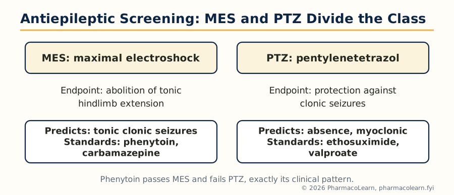
Figure: MES and PTZ divide the antiepileptic class between them; a new agent is always run through both.
2.10 Screening of nootropics and cognitive enhancers
Nootropics are screened in learning and memory models, usually against a pharmacologically induced amnesia.
- Morris water maze: the rat learns to find a hidden platform in a pool of opaque water using room cues; escape latency over training days measures spatial learning, and a probe trial without the platform measures memory.
- Passive avoidance: the animal learns to avoid a compartment where it received a shock; longer latency to re-enter on the test day means better memory. Scopolamine induced amnesia is the standard deficit, reversed by piracetam (the class standard) and by cholinesterase inhibitors.
- Radial arm maze and Y maze: working and reference memory are scored from arm choices, including spontaneous alternation in the Y maze, reduced by scopolamine and restored by nootropics.
- Elevated plus maze transfer latency and novel object recognition give simpler memory indices.
The trap of this area is stimulation masquerading as cognition: locomotor controls must accompany every memory claim.
2.11 Screening models of Parkinson's disease
Parkinson's models destroy or block the nigrostriatal dopamine pathway and ask whether the test drug restores movement.
- Reserpine model: reserpine depletes dopamine and produces akinesia, rigidity and hypothermia in rodents; levodopa and dopamine agonists reverse the syndrome.
- Neurotoxin models: 6-hydroxydopamine injected into the substantia nigra or striatum kills dopaminergic neurons on one side, producing rotational behaviour when the animal is challenged with apomorphine or amphetamine; the rotation count quantifies the lesion and its treatment. MPTP, which destroys nigral neurons through its metabolite MPP+, is used in mice and primates and is the closest toxin mimic of the human disease.
- Haloperidol catalepsy serves as a quick functional model of dopamine blockade.
- Tremor models (oxotremorine induced tremor) screen drugs for the tremor component, including anticholinergics such as trihexyphenidyl.
Levodopa with a peripheral decarboxylase inhibitor and bromocriptine are the reference standards.
2.12 Screening models of Alzheimer's disease and multiple sclerosis
Alzheimer's disease is modelled by deficit rather than by the full pathology. Scopolamine induced amnesia (section 2.10) screens symptomatic cholinergic drugs such as donepezil. Lesions of the basal forebrain cholinergic nuclei, intracerebral amyloid beta injection, and aged animals give deeper models. Transgenic mice overexpressing mutant human amyloid precursor protein develop amyloid plaques and memory loss and are the standard hosts for disease modifying candidates, including anti amyloid antibodies. Endpoints are maze performance plus amyloid and tau burden in brain tissue.
Multiple sclerosis is modelled by experimental autoimmune encephalomyelitis (EAE): rodents or primates are immunised with myelin proteins such as myelin basic protein or myelin oligodendrocyte glycoprotein in adjuvant, and develop ascending paralysis scored on a clinical scale. Fingolimod, glatiramer acetate and corticosteroids are active standards. Cuprizone feeding, which strips myelin chemically, models demyelination and remyelination. These are immunological models, linking forward to the immunomodulator screens of Unit 5.
2.13 Screening of drugs acting on the autonomic nervous system
Autonomic screening is the oldest branch of pharmacology and still runs on classical preparations.
- Isolated tissues: guinea pig ileum (acetylcholine, histamine), rat uterus, rabbit jejunum, rat vas deferens and the frog or rat rectus preparations quantify agonist potency and antagonism, with pA2 values from Schild analysis (Unit 1 bioassay methods applied to receptors).
- Whole animal haemodynamics: blood pressure and heart rate in anaesthetised or conscious rats and dogs reveal sympathomimetic and sympatholytic action; the classical dog and cat blood pressure preparations defined adrenaline reversal and ganglion blockade.
- Sympathomimetics are profiled by pressor responses, mydriasis and bronchodilation; sympatholytics by blockade of those responses and by ptosis and sedation signals in rodents.
- Parasympathomimetics and anticholinergics are read from salivation, pupil size, gut motility (charcoal meal transit in mice), and protection against acetylcholinesterase inhibitor toxicity; atropine is the universal anticholinergic standard.
- Neuromuscular blockers are screened on the rat phrenic nerve diaphragm and chick biventer cervicis preparations and in vivo by grip strength and head drop in rabbits, separating depolarising (succinylcholine) from non depolarising (tubocurarine) patterns.
Unit 3: Preclinical Screening: Respiratory, Reproductive, Analgesic and Gastrointestinal Pharmacology
3.1 Screening of anti-asthmatic drugs
Asthma screening asks two questions: does the drug relax bronchial smooth muscle, and does it prevent the inflammatory bronchospasm that allergens trigger?
- Isolated preparations: guinea pig tracheal chain and lung strips contract to histamine, acetylcholine and leukotrienes; relaxation or rightward shift of the curve measures bronchodilator action directly. Salbutamol is the standard beta2 agonist.
- Histamine and acetylcholine aerosol (bronchospasm in the conscious guinea pig): the animal is exposed to a nebulised spasmogen and the time to convulsions and collapse (preconvulsive time) is recorded; bronchodilators and antihistamines prolong it. This is the classical whole animal asthma screen.
- Allergic models: sensitised guinea pigs or rats challenged with ovalbumin develop early and late phase bronchoconstriction with eosinophil influx; bronchoalveolar lavage cell counts and lung histology measure the anti-inflammatory component that steroids (budesonide, dexamethasone) and leukotriene antagonists (montelukast) suppress.
- Supporting tests: inhibition of anaphylactic histamine release from mast cells (the basis of cromoglycate screening), and paw or ear anaphylaxis models in rats for systemic allergy.
3.2 Screening of drugs for COPD
Chronic obstructive pulmonary disease is modelled by producing chronic airway inflammation and emphysema rather than acute spasm. Rodents (usually mice or rats) are exposed to cigarette smoke for weeks to months, or receive intratracheal elastase or lipopolysaccharide, developing airspace enlargement, mucus hypersecretion and inflammatory cell influx. Endpoints are lung function (resistance and compliance), bronchoalveolar lavage counts, mean linear intercept as the emphysema measure, and cytokine levels. Roflumilast (phosphodiesterase 4 inhibitor) and long acting bronchodilators are the reference agents. The models are slow and expensive, which is why COPD screening leans heavily on the acute bronchospasm and inflammation models of section 3.1 for primary decisions.
3.3 Screening of anti-allergic drugs
Allergy screening targets the mast cell and the mediators it releases.
- Passive cutaneous anaphylaxis (PCA): antiserum containing IgE is injected into rat or mouse skin; intravenous antigen with a dye then produces a blue wheal where vessels leak; anti-allergic and antihistaminic drugs shrink the wheal. It is the standard in vivo allergy screen.
- Mast cell stabilisation in vitro: rat peritoneal mast cells are sensitised and challenged; histamine release into the medium is measured, with cromoglycate and ketotifen as standards.
- Systemic anaphylaxis: sensitised guinea pigs challenged intravenously with antigen develop fatal bronchospasm; protection measures anti-anaphylactic potency.
- Histamine based endpoints (wheal, ileum contraction, aerosol bronchospasm) run across this and the anti-asthmatic sections, with cetirizine and chlorpheniramine as the antihistamine standards.
3.4 Screening of aphrodisiacs
Aphrodisiac screening measures male sexual behaviour in rats under standardised conditions: a male is placed with a receptive (oestrous primed) female and trained observers record mount latency and frequency, intromission latency and frequency, and ejaculation latency. An active agent shortens latencies and raises frequencies without simply stimulating locomotion, so motor controls are run in parallel. Libido and potency components are separated by the penile erection index and by ex copula reflex tests. Sildenafil serves as a modern reference for erectile function (though its mechanism is vascular), while traditional claims for plant preparations are judged on the same behavioural endpoints. Hormonal assays (testosterone levels) accompany claims of androgenic action.
3.5 Screening of antifertility agents
Antifertility screening divides into female and male programmes.
Female agents are screened for: anti-ovulatory activity (blockade of spontaneous or gonadotropin induced ovulation in rats and rabbits), estrogenic and anti-estrogenic activity (uterine weight increase in immature rats, vaginal cornification), progestational activity (endometrial responses in primed animals), and anti-implantation activity (absence of implantation sites when dosing covers the implantation window after mating). The oestrous cycle is followed by vaginal smear cytology. Combined estrogen progestin preparations are the standards.
Male agents are screened by mating treated males with untreated females and counting pregnancies and litter size, supported by sperm count, motility and morphology, and testis and accessory gland weights. Gossypol is the historical reference compound. Every antifertility programme carries a reversibility arm: fertility must return after withdrawal for a contraceptive claim, and the same mating designs serve reproductive toxicology.
3.6 Screening of analgesics: central (narcotic) models
Analgesic screening begins by separating central from peripheral action, because the two classes treat different pain.
Central analgesics raise the threshold to thermal and other supraspinal stimuli:
- Hot plate test (Eddy and Leimbach): the mouse stands on a plate at about 55 degrees Celsius; the latency to paw licking or jumping is measured. Morphine prolongs it; NSAIDs do not. It is the primary screen for narcotic analgesics.
- Tail flick test: radiant heat is focused on the tail and the flick latency recorded; again morphine like drugs are active, and the test is spinally mediated, complementing the hot plate.
- Tail immersion and Haffner's tail clip test give variants of the same thermal and mechanical endpoints.
Morphine is the universal standard, and naloxone reversal is the confirmatory test: an effect abolished by naloxone is opioid mediated.
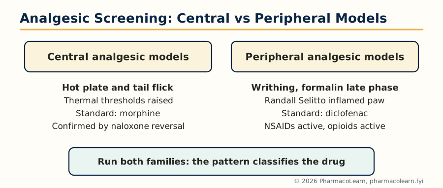
Figure: Central and peripheral analgesic models. The pattern of activity across the two families classifies a new analgesic.
3.7 Screening of analgesics: peripheral (non narcotic) models
Peripheral analgesics act where prostaglandins sensitise nociceptors, so their models are inflammatory and chemical.
- Acetic acid writhing (abdominal constriction): intraperitoneal dilute acetic acid makes the mouse writhe (stretching with abdominal contraction); both NSAIDs and opioids reduce the count, and the test is sensitive but not specific, so it is used as a primary screen only.
- Formalin test: formalin injected into the paw produces two phases of licking: an early neurogenic phase (blocked by opioids) and a late inflammatory phase (blocked by opioids and NSAIDs). The phase pattern classifies the agent.
- Randall Selitto test: increasing pressure is applied to an inflamed paw; the lowered pain threshold of inflamed tissue is raised by NSAIDs and opioids.
- Tail immersion in warm water and radiant heat on inflamed tissue give supporting thermal endpoints on a peripheral background.
Diclofenac and aspirin are the peripheral standards, morphine the central standard, and the hot plate to writhing comparison remains the quickest classroom demonstration that the two classes are genuinely different.
3.8 Screening of anti-inflammatory drugs
The carrageenan paw oedema of the rat is the most used screen in this whole subject. Carrageenan injected under the plantar skin produces a biphasic swelling: an early phase driven by histamine and serotonin, and a late phase driven by prostaglandins, which NSAIDs suppress. Paw volume is measured with a plethysmometer before and at intervals after injection, and percentage inhibition against the control group is the result. Indomethacin and diclofenac are the standards.
Chronic models complete the profile:
- Cotton pellet granuloma: sterile cotton pellets implanted under the skin gain dry weight from proliferating granulation tissue over days; agents that reduce the gain (steroids strongly, NSAIDs partly) act on the proliferative phase of inflammation.
- Adjuvant induced arthritis: Freund's complete adjuvant in the rat paw produces a polyarthritis over two to three weeks, the standard model for anti-rheumatic and disease modifying claims, read by paw volume, joint scores and radiology.
- Ultraviolet erythema in guinea pigs, croton oil ear oedema in mice and topical models screen agents intended for local use.
CARRAGEENAN PAW OEDEMA: ONE EXPERIMENT

  Fasted rats, groups: control, standard, test doses
              |
              v
  Dose orally 1 hour before challenge
              |
              v
  Inject 0.1 ml of 1 percent carrageenan
  under the plantar skin of the hind paw
              |
              v
  Measure paw volume at 0, 1, 2, 3, 4 hours
  (late phase, 3 to 4 hours, is prostaglandin driven)
              |
              v
  Percent inhibition vs control at 3 hours
  is the reportable endpoint
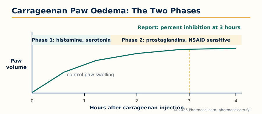
Figure: The two phases of carrageenan paw oedema. NSAIDs suppress the prostaglandin driven late phase, read as percent inhibition at 3 hours.
3.9 Screening of antipyretic agents
Fever is produced in rats or rabbits by injecting brewer's yeast suspension subcutaneously (the standard rat model; fever develops over about 18 hours) or bacterial endotoxin (lipopolysaccharide) and, historically, in rabbits for pyrogen work. Rectal temperature is recorded before and after dosing, and the fall in temperature over the following hours measures antipyretic action. Paracetamol is the standard. The model doubles as a discrimination test: antipyretics lower a raised temperature but do not lower normal body temperature, which separates them from hypothermic sedatives and anaesthetics.
3.10 Screening of anti-ulcer drugs
Anti-ulcer screening measures both aggression (acid, pepsin) and defence (mucus, mucosal blood flow, prostaglandins).
- Pylorus ligation (Shay rat): the pylorus is tied under anaesthesia; acid accumulates for several hours and ulcers form in the forestomach. Ulcer index, gastric volume, acidity and pH are measured. It screens antisecretory drugs; omeprazole and ranitidine are standards.
- Stress ulcer models: restraint and cold restraint stress produce mucosal erosions, screening cytoprotective and adaptogenic claims.
- Ethanol and NSAID induced ulcers: absolute ethanol or indomethacin/aspirin injures the mucosa directly; protection here signals mucosal defence (prostaglandin analogues such as misoprostol, sucralfate) rather than acid suppression.
- Acetic acid chronic ulcer: a serosal acetic acid application creates a chronic ulcer whose healing rate measures true ulcer healing, not just prevention.
- Supporting in vitro work uses isolated gastric glands and parietal cell preparations for H+/K+ ATPase and histamine H2 mechanisms.
3.11 Screening of anti-emetic drugs
The rat cannot vomit, so emesis screening uses species that can. The dog is the classical anti-emetic animal (apomorphine, a dopamine agonist acting on the chemoreceptor trigger zone, is the standard emetogen, and metoclopramide and haloperidol block it). The ferret is the modern standard for chemotherapy and radiation induced emesis (cisplatin challenge), the model in which ondansetron, a 5-HT3 antagonist, is the reference drug. The house musk shrew (Suncus murinus) offers a small animal alternative responding to cisplatin, motion and nicotine. In rodents, pica (eating non food material such as kaolin) after emetogenic stimuli serves as a nausea surrogate endpoint. Endpoints are the number of retches and vomits and the latency to the first episode.
3.12 Screening of antidiarrhoeal drugs and laxatives
The two classes are screened in opposite directions on the same physiology.
Antidiarrhoeals: castor oil induced diarrhoea in rats or mice is the primary screen (ricinoleic acid drives secretion and motility); the endpoints are onset time, number and weight of wet stools. The charcoal meal test measures gastrointestinal transit (the distance travelled by a charcoal marker as a fraction of intestinal length), slowed by loperamide, the standard, and by codeine. Enteropooling (fluid accumulation in the ligated intestine after castor oil or prostaglandin challenge) separates antisecretory from antimotility action.
Laxatives are screened by faecal output (number, weight and water content of pellets over a fixed period in rats) and by the same charcoal transit test run in the accelerating direction, with senna and bisacodyl as standards; isolated ileum and colon preparations characterise the stimulant mechanism.
Unit 4: Preclinical Screening: Cardiovascular, Metabolic, Anticancer and Hepatoprotective Pharmacology
4.1 Screening of antihypertensive drugs
Hypertension screening needs animals whose blood pressure is genuinely raised, plus acute preparations that reveal mechanism.
| Model | How hypertension is produced | What it screens best |
|---|---|---|
| Spontaneously hypertensive rat (SHR) | Genetic, pressure rises with age | Chronic oral therapy of all classes; the standard chronic model |
| Renal hypertension (Goldblatt) | Renal artery clipping (two kidney one clip, or one kidney one clip) raises renin dependent pressure | Renin angiotensin system drugs: ACE inhibitors, angiotensin receptor blockers |
| DOCA salt model | Mineralocorticoid plus salt and uninephrectomy, volume dependent, low renin | Diuretics and agents working in low renin states |
| L-NAME model | Chronic nitric oxide synthase inhibition | Endothelium targeted and vasodilator agents |
| Acute anaesthetised preparations | Direct arterial recording in rat, cat or dog | Immediate mechanism: ganglion blockade, alpha blockade, adrenaline reversal |
Endpoints are systolic and diastolic pressure and heart rate in conscious animals (tail cuff or, better, radiotelemetry) over days to weeks of dosing. Captopril or losartan are standard comparators in renin dependent models, amlodipine and hydrochlorothiazide across the battery.
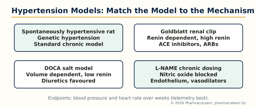
Figure: Four hypertension models, four different driving mechanisms. The model chosen decides which drug classes can show their effect.
4.2 Screening of antiarrhythmic drugs
Arrhythmias are induced by chemistry, electricity and ischaemia, and each inducer reads a different drug class.
- Chemically induced: aconitine (opens sodium channels; screens sodium channel blockers), ouabain or digoxin toxicity arrhythmias, adrenaline or isoprenaline arrhythmias in sensitised (halothane anaesthetised) animals, calcium chloride and chloroform adrenaline models, and barium chloride arrhythmias.
- Electrically induced: programmed stimulation and fibrillation threshold in isolated hearts (Langendorff) and in vivo.
- Ischaemia induced: coronary ligation in rats and dogs produces occlusion and reperfusion arrhythmias, the closest model of the clinical event.
- Isolated atrial and Purkinje fibre preparations classify the mechanism Vaughan Williams style (sodium, potassium, calcium channel or beta blockade) before whole animal work.
Lidocaine, amiodarone and verapamil serve as standards according to the model chosen.
4.3 Screening of antianginal drugs
Angina is a supply demand imbalance, so screening measures coronary flow and cardiac work. Isolated Langendorff hearts and working heart preparations quantify coronary flow, rate and contractility under controlled load. In vivo, isoprenaline induced myocardial necrosis and tachycardia in rats models demand ischaemia, and coronary ligation or ameroid constriction models supply failure; endpoints include ECG ST segment changes, infarct size (triphenyltetrazolium staining) and cardiac enzymes. Vasospastic components are read on isolated coronary artery rings contracted with ergonovine or potassium. Glyceryl trinitrate, propranolol and amlodipine are the standing standards for the nitrate, beta blocker and calcium channel mechanisms respectively.
4.4 Screening of antiatherosclerotic agents and diuretics
Antiatherosclerotic screening builds the lesion first: rabbits and, increasingly, transgenic mice (apoE knockout, LDL receptor knockout) are fed cholesterol rich diets for weeks, and serum lipids, aortic plaque area (en face staining) and arterial histology are the endpoints. The cholesterol fed rabbit made this field and remains the clearest lesion model.
Diuretic screening is one of the oldest quantitative screens in pharmacology. In the Lipschitz test, rats are given a standard water and salt load, urine is collected over 5 to 24 hours in metabolic cages, and volume plus sodium, potassium and chloride excretion are compared with control and with furosemide or hydrochlorothiazide standards. The sodium to potassium ratio separates potassium wasting loop and thiazide agents from potassium sparing ones; urine pH and bicarbonate excretion reveal carbonic anhydrase inhibition (acetazolamide). Isolated perfused kidney and clearance studies refine mechanism.
4.5 Screening of antidiabetic drugs
Diabetes models divide into insulin deficient and insulin resistant types, and the screen must match the claimed mechanism.
| Model | Induction | Suits |
|---|---|---|
| Streptozotocin (STZ) rat or mouse | Beta cell toxin, insulin deficient diabetes | Insulin, insulin secretagogues in residual function models |
| Alloxan model | Beta cell toxin (older, less selective) | Classical oral hypoglycaemic screening |
| Neonatal STZ, high fat diet plus low dose STZ | Partial deficiency with resistance | Type 2 agents broadly |
| db/db mouse, ob/ob mouse, Zucker fatty rat | Genetic obesity and insulin resistance | Insulin sensitisers, metformin, thiazolidinediones |
| Glucose tolerance tests (oral and intraperitoneal) | Glucose challenge in normal or diabetic animals | Any agent, first line screen |
Endpoints are fasting and post challenge glucose, HbA1c in chronic studies, insulin levels and tolerance tests. Alpha glucosidase inhibitors are screened in vitro against the enzyme and by sucrose loading tests in vivo (acarbose standard); DPP-4 inhibitors by enzyme assay plus glucose tolerance. Glibenclamide is the classical sulfonylurea standard and metformin the biguanide standard.
4.6 Screening of antidyslipidemic agents
Lipid screening begins in normal and diet loaded rats: a cholesterol and fat rich diet (often with cholic acid) raises serum cholesterol over days to weeks, and the test agent's effect on total cholesterol, LDL, HDL, triglycerides and the atherogenic index is compared with atorvastatin as the standing standard. The Triton WR-1339 model, in which a detergent blocks lipoprotein clearance and produces a sharp acute hyperlipidaemia, separates inhibitors of lipid synthesis from enhancers of clearance within hours. Transgenic apoE and LDL receptor knockout mice (section 4.4) carry the work into true atherosclerosis endpoints.
4.7 Screening of anticancer agents
Anticancer screening is the largest screening enterprise in pharmacology and runs in tiers.
- In vitro tier: panels of human tumour cell lines (the NCI 60 panel is the reference set) measure growth inhibition (GI50) across cancers of many organs; a distinctive pattern of sensitivity hints at mechanism. Normal cell lines run alongside to estimate selectivity.
- Transplantable animal tumours: rapidly growing syngeneic tumours in mice (Ehrlich ascites carcinoma, Sarcoma 180, Dalton's lymphoma, B16 melanoma, L1210 and P388 leukaemias) give the first in vivo read; endpoints are tumour weight and volume, and survival time.
- Human tumour xenografts: human cancer cells grown in nude or SCID mice (which cannot reject them, section 1.5) test the agent against actual human tumour biology.
- Chemically induced and transgenic models: agents such as DMBA and azoxymethane induce mammary and colon tumours for chemoprevention and slower growing tumour studies; oncogene transgenic mice model specific cancers.
Cyclophosphamide and 5-fluorouracil are among the standard comparators, chosen by tumour type. The toxicological shadow of this section is deliberate: anticancer screening always reports the therapeutic index, because these agents are given close to toxic doses.
4.8 Hepatoprotective screening methods
The liver is injured in a controlled way and protection is measured by enzymes, bilirubin and histology.
- Carbon tetrachloride (CCl4): converted by hepatic cytochrome P450 to a free radical that destroys hepatocytes; the classical acute hepatotoxicity model.
- Paracetamol overdose: glutathione depletion by the toxic metabolite NAPQI produces centrilobular necrosis; also the model where N-acetylcysteine is the mechanistic standard antidote.
- Thioacetamide and D-galactosamine: produce necrosis and a hepatitis like picture respectively; galactosamine injury is endotoxin sensitised and models viral like hepatitis.
- Anti-tubercular drug (isoniazid plus rifampicin) and alcohol models give clinically flavoured chronic injury.
Endpoints are serum SGOT (AST), SGPT (ALT), alkaline phosphatase and bilirubin, liver weight, and graded histopathology of sections. Silymarin, the milk thistle flavonolignan mixture, is the almost universal standard hepatoprotective comparator in this literature.
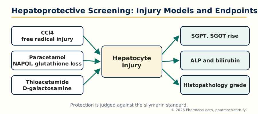
Figure: Hepatoprotective screening logic: controlled injury on the left, measurable liver endpoints on the right, silymarin as the standard.
Unit 5: Preclinical Screening: Immunomodulators, Immunoassay, Alternatives and Extrapolation
5.1 Screening of immunomodulators
An immunomodulator shifts immune function up or down, and screening must first say which direction, in which arm (cellular or humoral), and at what cost. The standard battery in rodents combines:
- Antibody response to sheep red blood cells (SRBC): animals are immunised with SRBC and the haemagglutination titre or plaque forming cell count in the spleen measures humoral immunity; suppression or enhancement against control is the primary endpoint.
- Delayed type hypersensitivity (DTH): SRBC or another antigen is given in the footpad after sensitisation; the swelling at 24 to 48 hours measures cell mediated immunity.
- Carbon clearance test: intravenous colloidal carbon is cleared by the reticuloendothelial system; the clearance rate (phagocytic index) measures macrophage function, raised by immunostimulants.
- Supporting counts: total and differential leucocyte counts, neutrophil adhesion, spleen and thymus weights, and resistance to a standard infection challenge.
Levamisole is the classical immunostimulant reference; cyclophosphamide induced immunosuppression provides the standard suppressed background against which restorative agents are proven.
5.2 Screening of immunosuppressants and immunostimulants
Immunosuppressants are screened for activity and for selectivity: the goal is suppression of the target response at doses that do not simply poison the animal. The SRBC antibody and DTH models of section 5.1, run in the suppressive direction, are the primary screens; skin graft survival prolongation is the classical transplant model; and mixed lymphocyte reactions and lymphocyte proliferation assays (mitogen stimulated) are the in vitro tier. Cyclosporine is the reference standard, with azathioprine and corticosteroids as supporting comparators. A selectivity index against general toxicity (weight loss, marrow counts) separates immunosuppression from cytotoxicity.
Immunostimulants are screened on the same models in the enhancing direction, and against cyclophosphamide suppressed animals for restorative claims: raised antibody titres, restored DTH, faster carbon clearance, and increased resistance in infection challenge models (for example survival after a standard bacterial inoculum). Levamisole is the standard; many plant derived preparations are examined in exactly this battery.
5.3 Immunoassay: theoretical basis
An immunoassay measures a substance using the binding of a specific antibody as the measuring event. Its theoretical basis is the law of mass action applied to a reversible antigen antibody equilibrium: antigen plus antibody form a complex whose amount, at equilibrium, depends on the concentrations and on the antibody's affinity constant. Because the complex itself is invisible, one partner carries a label (a radioisotope historically, now usually an enzyme, a fluorescent tag or a chemiluminescent marker) whose signal can be counted.
Two architectures follow from the equilibrium:
- Competitive (limited reagent) assays: labelled and unlabelled antigen compete for a fixed, limited number of antibody sites. The more analyte in the sample, the less label is bound, so signal falls as concentration rises. This design suits small molecules such as digoxin and hormones.
- Immunometric (excess reagent) assays: excess labelled antibody (or a capture antibody plus a labelled second antibody, the sandwich design) binds the analyte; signal rises with concentration. Sandwich designs need an analyte with two binding sites, so they suit large molecules such as insulin and protein hormones.
Sensitivity comes from the antibody's affinity and the label's detectability; specificity comes from the antibody's selectivity, which is why cross reactivity (section 5.6) is the standing weakness.
5.4 Optimisation of immunoassay
Optimisation means tuning the assay so that its dose response curve is steepest and most precise across the concentrations that matter clinically. The variables adjusted, one at a time and then together, are:
- Antibody concentration and affinity: lower antibody concentrations raise sensitivity in competitive designs, but too little antibody flattens the signal and worsens precision.
- Incubation time and temperature: long enough to approach equilibrium, short enough to be practical; non equilibrium (sequential) formats trade some equilibrium purity for speed.
- Label and tracer quality: specific activity, purity and stability of the labelled antigen or antibody.
- Separation conditions: in heterogeneous systems the bound free separation step (section 5.5) must be complete without disturbing the equilibrium being measured.
- Matrix controls: buffer composition, pH, ionic strength and protein content matched between standards and samples so that serum itself does not shift the curve (matrix effect).
- Standard curve design: enough calibrators, in the right range, fitted with the correct curve model, because every reported value is read from that curve.
OPTIMISING A COMPETITIVE IMMUNOASSAY

  Fix antibody, vary tracer amount
        |   choose the lowest tracer giving
        |   a countable, stable signal
        v
  Titrate antibody against fixed tracer
        |   select the dilution binding about
        |   50 percent of tracer with no analyte
        v
  Run full standard curve; check sensitivity
  (least detectable dose) and working range
        |
        v
  Confirm precision and recovery in real samples
  before accepting the assay for use
5.5 Heterogeneous and homogeneous immunoassay systems
Immunoassays are classified by whether bound and free label must be physically separated before measurement.
| Feature | Heterogeneous systems | Homogeneous systems |
|---|---|---|
| Separation step | Required: bound fraction separated from free (solid phase antibody, charcoal adsorption, precipitation, washing) | Not required: binding itself changes the label's behaviour, so the mixture is read directly |
| How binding is read | Signal of the separated bound (or free) fraction | Signal modulation in solution, for example enzyme activity switched by antibody binding |
| Classical examples | Radioimmunoassay (RIA), ELISA, immunoradiometric assay (IRMA) | EMIT (enzyme multiplied immunoassay technique), fluorescence polarisation immunoassay (FPIA), CEDIA |
| Strengths | High sensitivity, washing removes interference | Fast, simple, easily automated, no separation equipment |
| Weaknesses | More steps, longer, washing variability | More vulnerable to sample matrix interference; usually less sensitive |
ELISA, a heterogeneous enzyme labelled system on a solid phase, is the workhorse format of screening laboratories as well: the same architecture that measures digoxin in a patient sample also measures antibody titres in the immunomodulator screens of section 5.1.
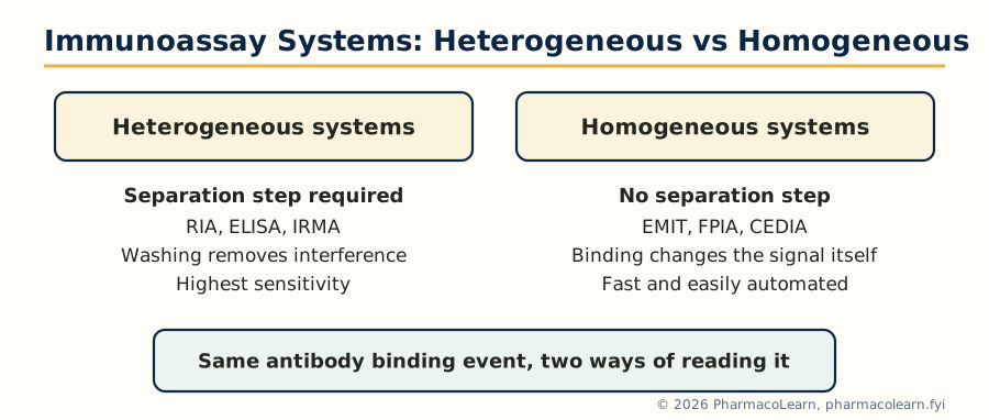
Figure: Heterogeneous and homogeneous immunoassays read the same antibody binding event in two different ways.
5.6 Evaluation of immunoassay methods: protocol outline, objectives and preparation
An immunoassay is only adopted after formal evaluation against a written protocol. The objectives are to prove that the method measures what it claims, accurately and reproducibly, in real samples. Preparation includes defining the reference method for comparison, collecting patient and control samples across the clinical range, setting acceptance criteria in advance, and training staff on the final written procedure.
The evaluation then measures:
- Accuracy and method comparison: results on the same samples by the new method and the reference method, assessed by correlation and bias across the range.
- Precision: within run and between run replication of control materials at low, medium and high levels, expressed as coefficient of variation.
- Sensitivity: the least detectable dose, from the variability of the zero standard.
- Recovery and dilution linearity (parallelism): known amounts added to samples are recovered quantitatively, and diluted samples read parallel to the standard curve; failure means matrix interference.
- Specificity and cross reactivity: related substances (for digoxin, its metabolites and other cardiac glycosides and steroids) are tested for false responses.
- Interference and stability: haemolysis, lipemia and bilirubin effects; reagent shelf life and calibration stability.
Only a method that passes its pre set criteria on all of these is released for routine use; that discipline, not the label technology, is what makes an immunoassay a measurement rather than a demonstration.
5.7 Immunoassay for digoxin
Digoxin is the model competitive immunoassay because it is a small molecule with a narrow therapeutic range (about 0.5 to 2 ng per ml in serum) that chemical methods cannot measure at clinical concentrations. The design is competitive: digoxin in the patient's serum competes with labelled digoxin (enzyme, fluorescent or chemiluminescent label in modern kits; iodine 125 in the classical RIA) for a limited amount of anti digoxin antibody, and the bound signal, read against the standard curve, falls as the sample concentration rises.
Evaluation points specific to digoxin illustrate section 5.6 in action: cross reactivity of digitoxin, digoxin metabolites and endogenous digoxin like immunoreactive substances (raised in renal failure, liver disease and pregnancy) can falsely elevate results; samples from patients given digoxin specific antibody fragments (Fab) after poisoning disturb the assay because much of the digoxin is antibody bound. The clinical use is therapeutic drug monitoring: dose adjustment in heart failure and atrial fibrillation, and confirmation of suspected digoxin toxicity.
5.8 Immunoassay for insulin
Insulin is the model immunometric assay because it is a peptide large enough to carry two antibody binding sites. The modern design is a two site sandwich: a capture antibody fixed to a solid phase binds one epitope of insulin, and a second, labelled antibody binds another epitope; the sandwich count is directly proportional to insulin concentration. The classical competitive radioimmunoassay of Berson and Yalow, the first immunoassay ever described, made insulin measurable at all and founded the whole field.
Specific insulin questions shape its evaluation: cross reaction with proinsulin (the precursor, raised in type 2 diabetes and insulinoma) falsely inflates older assays, so modern antibodies are chosen for true insulin specificity; insulin treated diabetic patients develop anti insulin antibodies that interfere; and paired measurement with C-peptide (secreted with insulin but not present in injected preparations) distinguishes endogenous secretion from injected insulin, a distinction used in evaluating hypoglycaemia and insulinoma. Fasting insulin and C-peptide values feed indices of insulin resistance and beta cell function in the metabolic screening of Unit 4.
5.9 Limitations of animal experimentation
Animal screening built pharmacology, but its limits must be stated as plainly as its successes.
- Species differences: metabolism, receptor distribution and sensitivity differ; thalidomide was quiet in rat tests yet teratogenic in humans, and penicillin is lethal to guinea pigs through gut flora disruption while being safe in humans. A clean animal result is evidence, not proof.
- Disease models are imitations: carrageenan oedema is not rheumatoid arthritis, and a scopolamine deficit is not Alzheimer's disease; models answer the question they embody and no other.
- Dose and exposure differences: animals often receive doses and durations far from clinical use, and routes differ.
- Statistical and design limits: small group sizes, strain peculiarities (section 1.5) and publication bias towards positive results all distort the record.
- Cost, time and ethics: whole animal work is slow and expensive, and every use must carry its ethical justification with it (Unit 1, CPCSEA framework).
5.10 Alternatives to animal experiments: the 3Rs
The organising idea is the 3Rs of Russell and Burch:
- Replacement: substitute animals with non animal systems where the science allows: cell and tissue culture, isolated organs, computer (in silico) models of binding and toxicity, lower organisms such as zebrafish embryos and invertebrates, and human volunteer and microdosing studies for defined questions.
- Reduction: use the fewest animals that keep the experiment statistically valid: better design, sharing control groups, in vitro pre screening so animals meet only promising compounds (the funnel of Unit 2), and full use of each animal's data.
- Refinement: reduce suffering in the animals still used: anaesthesia and analgesia, humane endpoints, better housing and handling, and non invasive measurements such as telemetry and imaging.
Validated alternatives already in routine use include reconstructed human skin models for irritation, cell based assays replacing some pyrogen and potency tests (the monocyte activation test replacing much rabbit pyrogen work), and in silico prediction in early toxicity screening. An alternative replaces an animal test only after validation shows it predicts as well, which is why the replacement programme itself is a screening science.
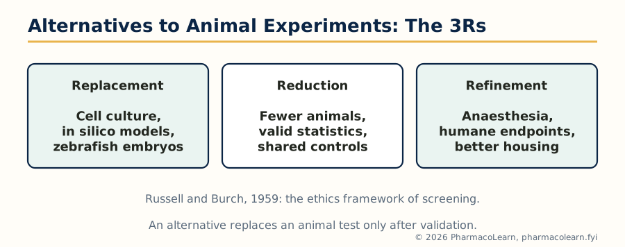
Figure: The 3Rs of Russell and Burch. Every CPCSEA protocol is expected to show how each of the three was considered.
5.11 Extrapolation of in vitro data to preclinical studies
Moving from the test system to the animal is the first extrapolation, and it succeeds or fails on concentration. The discipline is comparison: the concentration active in vitro must be reachable, as free drug, in the animal's plasma or tissue at tolerated doses. A compound potent at nanomolar levels in a receptor assay but never achieving even micromolar free levels in vivo will fail, and recognising that early is the point of the exercise. Binding to plasma proteins and tissue components lowers the free fraction in the animal in a way buffer systems do not reproduce; metabolism in the intact animal can destroy an in vitro winner or create the real active agent from an inactive looking parent (the prodrug pattern). Pharmacokinetic measurement in the screening species therefore accompanies the first in vivo efficacy tests, so that a negative result can be read correctly: no exposure, or exposure without effect. These are different verdicts and lead to different decisions.
5.12 Extrapolation from animals to humans
The final extrapolation crosses to the clinic, and its working tool is allometric scaling: physiological quantities such as clearance and dose relate to body weight by power laws across species, so a human equivalent dose can be estimated from animal doses by body surface area based conversion factors (the basis of first in human starting dose calculations, further discounted by safety factors from the no observed adverse effect level in toxicology). Beyond arithmetic, three judgement rules govern the crossing:
- Weight concordance: an effect seen in several species, at overlapping exposures, is more likely to appear in humans than a single species curiosity.
- Prefer mechanism over phenomenology: if the animal effect is explained by a receptor or enzyme that humans share, confidence rises; if it rides on a species specific pathway, confidence falls.
- Treat safety signals conservatively: toxicity in animals is assumed relevant to humans until shown otherwise, while efficacy is assumed provisional until the clinic confirms it.
Preclinical screening therefore does not predict the clinic; it prices the risk of entering it. That honest framing, from the CPCSEA protocol of Unit 1 to the first human dose, is the subject as a whole.
Frequently Asked Questions
Q: Which laboratory animals are most commonly used in pharmacological screening, and why? Mice and rats dominate because they are small, cheap, quick breeding and need only small amounts of a new compound. Guinea pigs serve bronchospasm and ileum work, rabbits pyrogen and eye studies, dogs cardiovascular and emetic work, and primates only where no lower species can answer the question.
Q: What is CPCSEA and what does an IAEC do? CPCSEA is the statutory committee under the Prevention of Cruelty to Animals Act, 1960 that registers establishments and sets norms for animal experiments in India. The Institutional Animal Ethics Committee of each establishment, which must include the CPCSEA nominee, approves every protocol before work begins and monitors welfare during it.
Q: What is the difference between inbred and outbred strains? Inbred strains come from at least 20 generations of brother to sister mating, so animals are almost genetically identical, which removes genetic noise but narrows generalisation. Outbred stocks are bred to keep genetic variation, so results generalise better across genotypes, and they are the routine screening default.
Q: What is bioassay and when is it preferred over chemical assay? Bioassay measures potency by the response of a living system, always relative to a standard preparation. It is preferred when the active principle is unknown, when no chemical method is sensitive or specific enough, or when biological activity itself is the property being measured, as with vaccines and hormones.
Q: How do the MES and PTZ tests divide antiepileptic drugs between them? The maximal electroshock test, judged by abolition of tonic hindlimb extension, detects drugs for generalised tonic clonic seizures such as phenytoin. The pentylenetetrazol test detects drugs for absence and myoclonic seizures such as ethosuximide and valproate. Phenytoin fails the PTZ test, exactly as it fails absence seizures clinically.
Q: Which models screen analgesics, and how are central and peripheral agents separated? The hot plate and tail flick tests respond to central, opioid analgesics such as morphine but not to NSAIDs. The acetic acid writhing and formalin late phase and Randall Selitto tests respond to peripheral agents such as diclofenac as well. Running both families, with naloxone reversal as confirmation, classifies a new analgesic.
Q: What is the carrageenan paw oedema model and what does its time course show? Carrageenan injected into the rat paw produces swelling in two phases: an early histamine and serotonin phase and a late prostaglandin phase. NSAIDs suppress the late phase, which is why percentage inhibition at about 3 hours, measured by plethysmometer against a diclofenac or indomethacin standard, is the reportable endpoint.
Q: Which animal models are used for screening antidiabetic drugs? Insulin deficient models (streptozotocin or alloxan treated rats) suit insulin and secretagogues; genetic models (db/db mice, Zucker fatty rats) and high fat diet models suit insulin sensitisers such as metformin. Glucose tolerance tests are the first line screen across classes.
Q: What is the difference between heterogeneous and homogeneous immunoassays? Heterogeneous assays such as RIA and ELISA must physically separate bound from free label before measuring, which gives high sensitivity at the cost of extra steps. Homogeneous assays such as EMIT need no separation because binding itself changes the label's signal, making them faster and easier to automate.
Q: What are the 3Rs in animal experimentation? Replacement of animals with non animal systems where possible, Reduction of animal numbers to the statistical minimum, and Refinement of procedures to reduce suffering, as framed by Russell and Burch. Indian CPCSEA review expects every protocol to show how the 3Rs were considered.
Complete MPL103T notes, all 5 units in one PDF, free to keep.
Other M.Pharm subjects: MPH101T · MPC101T · MPL101T · MPH102T · MPH103T · MPH104T · MPL102T · MPL103T · MPL104T · MPC102T · MPC103T · MPC104T · MPH201T · MPH202T · MPH203T · MPH204T · MPC201T · MPC202T · MPC203T · MPC204T · M.Pharm