
MPH201T — Molecular Pharmaceutics (Nano Tech and Targeted DDS)
4 hrs/week, 4 credits, 60 hours, 100 marks. Notes follow the M.Pharm syllabus, unit by unit.
MPH201T Molecular Pharmaceutics (Nano Tech and Targeted DDS) notes for M.Pharm Semester 2 Pharmaceutics: drug targeting, nanoparticles and liposomes, microspheres, monoclonal antibodies, pulmonary and nasal delivery, gene therapy, with free unit wise PDF downloads on PharmacoLearn.
Units in this subject
- Unit 1 — Targeted Drug Delivery Systems
- Unit 2 — Targeting Methods, Nanoparticles and Liposomes
- Unit 3 — Microcapsules, Microspheres and Vesicular and Related Carriers
- Unit 4 — Pulmonary and Intra Nasal Drug Delivery Systems
- Unit 5 — Nucleic Acid Based Therapeutic Delivery Systems
Download MPH201T PDF Notes
Free PDFs for this subject, one per unit:
Molecular Pharmaceutics (Nano Tech and Targeted DDS), subject code MPH201T, is a core Semester 2 subject of M.Pharm Pharmaceutics. It carries 4 credits and 60 hours of theory, taught at 4 hours per week, for 100 marks.
This subject is about delivering a drug to the place in the body where it is needed, in the amount needed, instead of distributing it everywhere and accepting toxicity in healthy tissues. The first unit builds the concept of targeted drug delivery, the biological events a carrier must survive, and the two model problems of tumour targeting and brain specific delivery. The second unit studies the methods of targeting and the two carrier families examined in most depth, nanoparticles and liposomes, with their types, preparation and evaluation. The third unit covers microcapsules and microspheres, monoclonal antibodies as targeting agents, and the vesicular and related carriers niosomes, aquasomes, phytosomes and electrosomes. The fourth unit turns to site specific routes in their own right: pulmonary delivery by aerosols and intra nasal delivery. The last unit deals with nucleic acid based therapeutics: gene therapy, viral and nonviral gene transfer, liposomal gene delivery, and the antisense molecules and aptamers that are expected to become the drugs of the future. A student who completes this subject should be able to explain how a targeted system is designed, prepare and evaluate the main carrier systems in the laboratory, and judge which carrier suits a given drug and target.
Unit 1: Targeted Drug Delivery Systems
1.1 The concept of drug targeting
Conventional dosage forms distribute the drug throughout the body according to the physicochemical properties of the molecule. Only a small fraction of the dose reaches the tissue that needs treatment, while the rest is taken up by healthy organs, where it causes side effects, and is cleared by the liver and kidneys. A targeted drug delivery system is designed to reverse this distribution: the drug, alone or loaded in a carrier, is transported preferentially to the target tissue, organ, cell or even an intracellular compartment, so that the concentration at the site of action rises and the exposure of the rest of the body falls.
The idea is old. Paul Ehrlich proposed the concept of a magic bullet (Zauberkugel) at the beginning of the twentieth century: a carrier that would seek out the diseased site and deliver its toxic payload only there. Targeted delivery is the practical attempt to build that bullet, usually by attaching the drug to a carrier (a nanoparticle, liposome, microsphere or antibody) whose size, surface and ligands decide where it goes.
The advantages expected from targeting are:
- Higher efficacy at a lower dose: more of the administered drug reaches the receptor site, so the same effect is obtained with a smaller total dose.
- Lower toxicity: healthy tissues are exposed to less drug, which matters most for cytotoxic anticancer drugs with a narrow therapeutic index.
- Protection of the drug: the carrier shields labile molecules such as peptides, enzymes and nucleic acids from enzymes and pH during transit.
- Control of release: the carrier can be designed to release the drug slowly, or in response to a local stimulus such as pH, temperature or an enzyme.
- Delivery to difficult sites: carriers can cross or bypass barriers that the free drug cannot, for example the blood brain barrier or the interior of a solid tumour.
- Improved patient acceptance: less frequent dosing and fewer side effects improve compliance.
Targeting is not free of problems. The carrier itself must be non toxic, non immunogenic, biodegradable or safely eliminable, stable on storage, and manufacturable at scale and at acceptable cost. The body also treats most carriers as foreign particles and removes them through the reticuloendothelial system (section 1.2), so a targeting design is always a negotiation between the carrier and the body's defences.
1.2 Events and biological processes involved in drug targeting
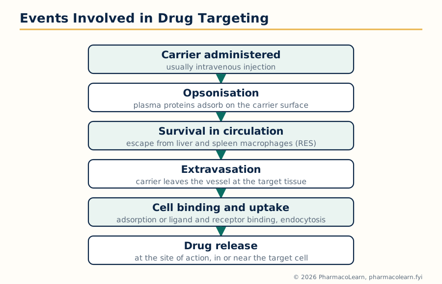 Figure: The sequence of events a targeted carrier must pass through: it can fail at any step, and carrier design is the attempt to survive each one.
Between injection or administration and arrival at the target, a carrier passes through a sequence of events, and it can fail at every step. Understanding this sequence is the basis of all carrier design.
Sequence of events in drug targeting
Carrier administered (usually intravenous)
        |
        v
Circulation: plasma proteins adsorb (opsonisation)
        |
        v
Survival against reticuloendothelial clearance
        |
        v
Extravasation: carrier leaves the blood vessel at the target tissue
        |
        v
Interstitial transport to the target cell
        |
        v
Binding to the cell surface (ligand and receptor, if active targeting)
        |
        v
Internalisation (endocytosis) and intracellular trafficking
        |
        v
Drug release at the site of action
The main biological processes in this sequence are:
- Opsonisation. As soon as a particle enters the blood, plasma proteins called opsonins (immunoglobulins, complement components such as C3b, fibronectin) adsorb onto its surface. This protein layer, often called the protein corona, marks the particle as foreign.
- Reticuloendothelial clearance. Opsonised particles are recognised and phagocytosed by the fixed macrophages of the reticuloendothelial system (RES), also called the mononuclear phagocyte system, mainly the Kupffer cells of the liver and the macrophages of the spleen and bone marrow. Conventional particles are cleared within minutes. This is the single biggest obstacle to targeting any site other than the liver and spleen themselves, and it is why surface modification with hydrophilic polymers such as polyethylene glycol (PEG), which reduces opsonisation, is used to make long circulating or stealth carriers.
- Extravasation. To reach tissue cells, the carrier must leave the blood vessel. Normal continuous endothelium allows only small molecules through, but the endothelium in inflamed tissue and solid tumours is fenestrated and leaky, and the liver and spleen sinusoids are discontinuous, so particle size decides which tissues a carrier can enter.
- Interstitial transport. After leaving the vessel, the carrier must diffuse through the extracellular matrix to reach the cells. High interstitial pressure inside tumours opposes this movement.
- Cell binding and uptake. The carrier binds to the cell, by simple adsorption in passive systems or by a ligand binding its receptor in active systems, and is then usually taken in by endocytosis (phagocytosis for large particles, pinocytosis and receptor mediated endocytosis for smaller ones).
- Intracellular fate and release. Endocytosed carriers enter endosomes and then lysosomes, where enzymes and low pH can destroy the drug. A successful carrier either releases the drug before this happens, escapes the endosome, or is designed so that the lysosomal enzymes themselves trigger release.
Targeting is also described by orders or levels, according to how precise the destination is. In first order targeting the carrier reaches a whole organ or tissue compartment (for example the liver). In second order targeting it reaches a particular cell type within that organ (for example the hepatocyte rather than the Kupffer cell). In third order targeting it reaches an intracellular destination, such as the nucleus or a lysosome. The higher the order, the more sophisticated the carrier design must be.
1.3 Why most carriers accumulate in liver and spleen
The pattern of RES clearance explains the default biodistribution of almost every particulate carrier: the majority of an intravenous dose of conventional nanoparticles, liposomes or microspheres ends in the liver and spleen. This is a failure when the target is elsewhere, but it can be used deliberately. Liver targeting is exploited for drugs against hepatic infections and for lysosomal storage disorders, and splenic and macrophage targeting is useful against infections that hide inside macrophages, such as leishmaniasis, where liposomal amphotericin B reaches the infected macrophages of the liver and spleen directly. The lesson for design is that biodistribution is decided mainly by size, surface charge and surface hydrophilicity, and only secondarily by the drug inside the carrier.
| Property of carrier | Effect on fate in the body |
|---|---|
| Size above about 200 nm | Rapid uptake by spleen and liver macrophages |
| Strong positive or negative surface charge | More opsonisation, faster RES clearance |
| Hydrophobic surface | More protein adsorption, faster clearance |
| PEG coated (stealth) surface | Reduced opsonisation, long circulation, more time to reach the target |
| Ligand on the surface | Binding to cells carrying the matching receptor, if the carrier reaches them |
1.4 Tumour targeting
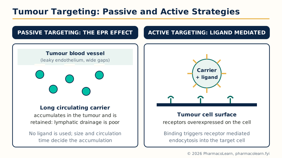 Figure: Passive targeting accumulates long circulating carriers in tumours through the EPR effect; active targeting adds a ligand so the carrier binds and enters receptor bearing cells.
Solid tumours are the most studied target in this subject, because anticancer drugs are potent, toxic and needed in high local concentration. Tumour targeting rests on the unusual biology of the tumour tissue:
- Angiogenesis and leaky vasculature. A growing tumour secretes angiogenic factors such as vascular endothelial growth factor (VEGF) and builds new blood vessels in haste. These vessels are poorly organised, with wide fenestrations and gaps in the endothelium (openings of the order of 100 to 800 nm are reported between tumour endothelial cells, much wider than in normal vessels), and an incomplete basement membrane.
- Poor lymphatic drainage. Tumours lack a functional lymphatic system, so material that enters the tumour interstitium is not drained away efficiently and is retained.
- Enhanced permeability and retention (EPR) effect. The combination of leaky vessels and poor drainage, described by Maeda and co-workers, means that macromolecules and nanoparticles in a suitable size range (roughly 10 to 200 nm) extravasate preferentially into tumours and accumulate there to concentrations many times higher than in normal tissue. This is the basis of passive tumour targeting.
- A distinctive microenvironment. Tumour tissue is often slightly acidic, hypoxic in its core, and rich in certain enzymes, which can be used as triggers for stimulus responsive release.
Two strategies are used. In passive targeting, no ligand is used; the carrier is made long circulating (for example PEGylated) and of a size that exploits the EPR effect, so accumulation follows from the tumour's own anatomy. The first approved nanodrug, Doxil (PEGylated liposomal doxorubicin, approved in 1995), works in exactly this way and reduces the cardiac toxicity of free doxorubicin. In active targeting, a ligand such as an antibody, a peptide, folic acid or transferrin is attached to the carrier surface so that it binds a receptor overexpressed on tumour cells (for example HER2, folate receptor or transferrin receptor) and is internalised by receptor mediated endocytosis. Active targeting mainly improves cell uptake after the carrier has already arrived in the tumour; it does not by itself bring more carrier out of the blood stream, which is why it is combined with a long circulating design. A third approach, stimulus responsive targeting, uses carriers that release the drug when they meet the tumour's low pH, raised temperature or specific enzymes.
Limitations must be stated honestly: the EPR effect varies between tumour types and between patients, tumour interstitial pressure is high and opposes carrier entry, and necrotic tumour cores are poorly perfused. Even so, tumour targeting remains the model application in which nearly every carrier in Units 2 and 3 is tested.
1.5 Brain specific delivery
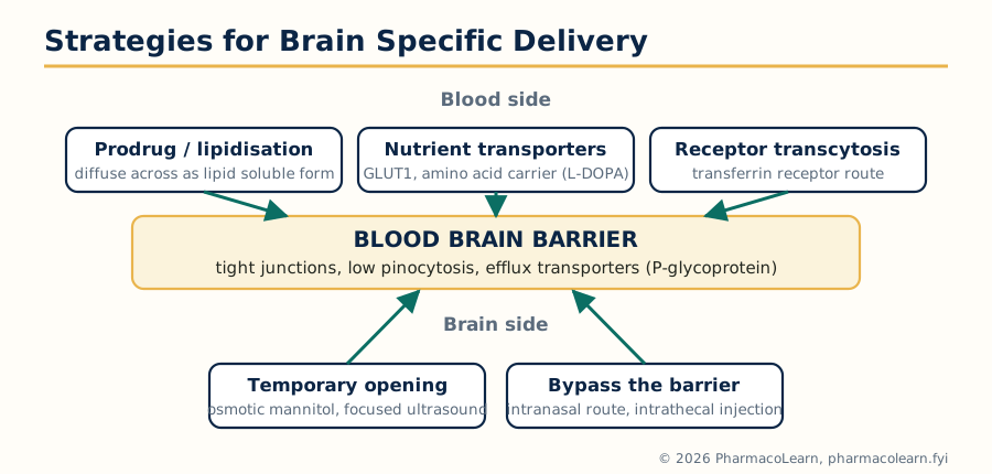 Figure: The blood brain barrier and the five families of strategy used to cross it or bypass it.
The brain is protected by the blood brain barrier (BBB), a continuous layer of endothelial cells joined by tight junctions, supported by pericytes and astrocyte end feet, with very low pinocytotic activity and with efflux transporters such as P-glycoprotein that pump foreign molecules back into the blood. The barrier keeps out almost all large molecules and the great majority of small molecules too, which protects the brain but also makes brain tumours, infections and neurodegenerative diseases very difficult to treat with drugs given by ordinary routes.
The main strategies for brain specific delivery are:
- Lipidisation and prodrugs. Making the molecule more lipid soluble, or masking polar groups in a prodrug that is converted after crossing, improves passive diffusion across the endothelium. The classic example is heroin (diamorphine), the lipid soluble prodrug of morphine, which crosses far faster than morphine itself.
- Carrier mediated transport. The BBB carries nutrient transporters, for example the glucose transporter GLUT1 and the large neutral amino acid transporter. L-DOPA is the standard example of a drug that crosses on the amino acid transporter and is then converted to dopamine inside the brain.
- Receptor mediated transcytosis. Receptors such as the transferrin receptor and the insulin receptor carry their ligands across the endothelium by transcytosis. Attaching a drug or a carrier to transferrin, or to an antibody against the transferrin receptor, hijacks this route; this is one of the main approaches for delivering nanoparticles and biologics to the brain.
- Adsorptive mediated transcytosis. Cationic molecules and cationised proteins bind the negatively charged endothelial surface and are carried across; the route is effective but not brain specific.
- Temporary disruption of the barrier. Osmotic opening with hypertonic mannitol, or focused ultrasound with microbubbles, opens the tight junctions for a short time so that drugs can enter; the risk is that unwanted substances enter too.
- Bypassing the barrier. Intranasal delivery along the olfactory and trigeminal pathways (Unit 4), intrathecal injection and direct implants avoid the BBB altogether.
- Nanocarriers. Lipid nanoparticles and polymeric nanoparticles, especially when surface modified with ligands such as transferrin or with polysorbate coatings that adsorb apolipoproteins from plasma, show improved brain uptake in experimental work and are an active research direction.
The design lesson of this unit is that a targeted system is defined by three things together: the biology of the target, the properties of the carrier, and the events in between. Units 2 and 3 now take up the carriers one by one.
Unit 2: Targeting Methods, Nanoparticles and Liposomes
2.1 Targeting methods: a general introduction
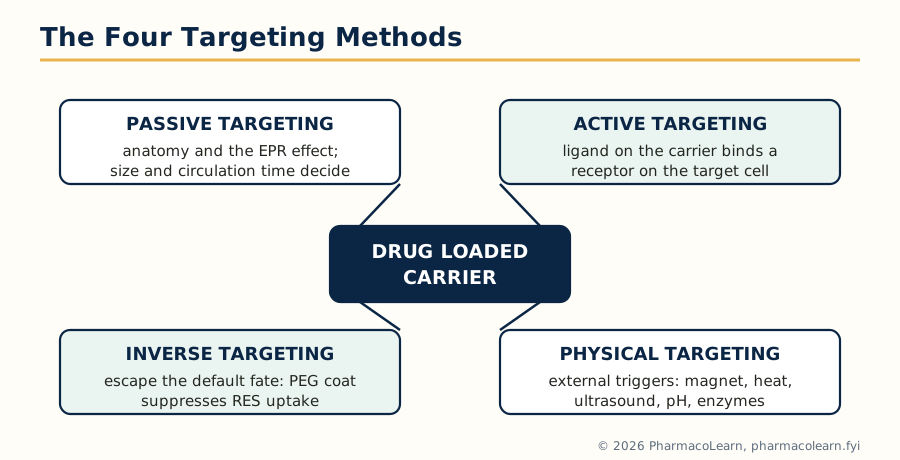 Figure: The four general targeting methods. Every carrier in this subject is used in one or more of these ways.
The carriers in this subject are used in four general ways, and every preparation and evaluation method in the unit is a means of making one of these ways work.
- Passive targeting uses the natural distribution of the carrier. It exploits anatomy and physiology: the fenestrations of the liver and spleen sinusoids, the leaky vasculature of tumours and inflamed tissue (the EPR effect, Unit 1), and the uptake of particles by phagocytic cells. No ligand is involved. The design variables are size, surface charge and circulation time.
- Active targeting attaches a ligand to the carrier surface so that it binds a specific receptor or antigen on the target cell and is taken up by receptor mediated endocytosis. Ligands used include monoclonal antibodies and their fragments, peptides, aptamers, and small molecules such as folic acid and galactose (for the asialoglycoprotein receptor of hepatocytes).
- Inverse targeting is the deliberate avoidance of the usual fate: instead of accepting RES uptake, the carrier surface is made hydrophilic (for example by PEGylation or poloxamer coating) so that opsonisation and macrophage uptake are suppressed and the carrier can reach non-RES targets. It is targeting by escaping the default destination.
- Physical (and chemical) targeting uses an external or local physical trigger: a magnet held over the site for magnetic carriers, local heating, ultrasound, or a stimulus at the site itself such as low pH or a specific enzyme that triggers release from a responsive carrier.
Whichever method is chosen, an ideal carrier has the same set of properties: it should carry enough drug, protect it during transit, be non toxic, non immunogenic and biodegradable or safely excreted, be stable during storage, release the drug at the required rate at the site, and be simple and cheap enough to manufacture reproducibly at scale. Preparation methods are judged by how close they come to this list, and evaluation methods are the tests that measure it.
2.2 Nanoparticles: introduction and types
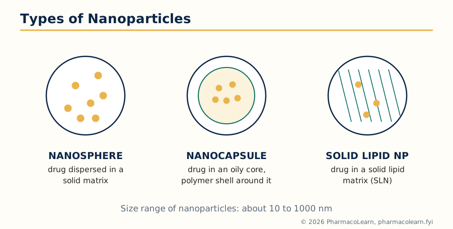 Figure: The three structural types of nanoparticle: matrix (nanosphere), reservoir (nanocapsule) and solid lipid nanoparticle.
Nanoparticles are solid colloidal particles in the approximate size range of 10 to 1000 nm that carry the drug dissolved, entrapped, encapsulated or adsorbed in or on a polymer or lipid matrix. Because of their small size they can pass through fenestrated endothelium, are taken up by cells, and can be given by several routes including intravenous, oral and ocular. Their large surface area to volume ratio also allows extensive surface modification with ligands and stealth coatings.
The main types are:
- Nanospheres: matrix systems in which the drug is dispersed uniformly through a solid polymer or lipid matrix and is released by diffusion and by erosion of the matrix.
- Nanocapsules: reservoir systems with a liquid or oily core containing the drug, surrounded by a thin polymer shell; release is controlled mainly by the shell.
- Polymeric nanoparticles: made from biodegradable polymers such as poly(lactic acid) (PLA), poly(lactic-co-glycolic acid) (PLGA), poly(caprolactone) and poly(alkyl cyanoacrylates), or from natural polymers such as albumin, gelatin and chitosan.
- Solid lipid nanoparticles (SLN): the liquid oil of an emulsion is replaced by a lipid that is solid at room and body temperature (for example triglycerides, waxes or fatty acids), stabilised by surfactants. They combine the tolerability of lipids with solid matrix release control, and are used for drugs such as vaccines, peptides and poorly soluble compounds, and in cosmetics and brain delivery research.
- Nanocrystals: pure drug in crystalline nanoparticle form, stabilised by surfactants or polymers, used to raise the dissolution rate and bioavailability of poorly soluble drugs.
| Feature | Nanosphere | Nanocapsule |
|---|---|---|
| Structure | Solid matrix throughout | Core surrounded by a shell |
| Drug location | Dispersed in the matrix | Dissolved in the core (often oily) |
| Release control | Diffusion plus matrix erosion | Membrane (shell) controlled diffusion |
| Drug loading | Limited by matrix capacity | Often higher for lipophilic drugs in an oily core |
2.3 Preparation of nanoparticles
The methods fall into two families: building particles from monomers (polymerisation) and forming particles from a preformed polymer or lipid.
Polymerisation methods
- Emulsion polymerisation: the monomer (for example an alkyl cyanoacrylate) is dispersed in an aqueous phase containing surfactant; polymerisation starts in the micelles or droplets and nanoparticles form directly in the dispersion. The drug may be added before polymerisation (incorporated) or afterwards (adsorbed).
- Interfacial polymerisation: monomer and drug are dissolved in an organic phase that is emulsified into water; polymerisation occurs at the oil water interface and produces nanocapsules with an oily core.
Methods from preformed polymers
- Nanoprecipitation (solvent displacement): the polymer and drug are dissolved in a water miscible organic solvent (acetone or ethanol); this phase is poured into water containing a stabiliser, the solvent diffuses out rapidly, and the polymer precipitates as nanoparticles. The method is simple and reproducible, works best for lipophilic drugs, and is the most common laboratory method for PLA and PLGA particles.
- Emulsification and solvent evaporation: polymer and drug are dissolved in a water immiscible volatile solvent (dichloromethane or ethyl acetate), emulsified into an aqueous phase (oil in water, or water in oil in water double emulsion for hydrophilic drugs), and the solvent is removed by evaporation under stirring or reduced pressure, hardening the droplets into particles.
- Emulsification and solvent diffusion: a partially water miscible solvent is used; on dilution with excess water the solvent diffuses out of the droplets and the polymer precipitates.
- Salting out: the polymer solution in a water miscible solvent is emulsified into an aqueous phase rich in salts (for example magnesium chloride) and stabiliser that temporarily prevents solvent diffusion; dilution with water then allows diffusion and particle formation. No chlorinated solvent is needed and no heating is used, which suits labile drugs.
- Supercritical fluid methods: a supercritical fluid (usually carbon dioxide) is used as solvent or antisolvent to produce fine, solvent free particles; equipment cost limits routine use.
- High pressure homogenisation (for SLN): the drug is melted or dissolved in molten lipid and dispersed in hot surfactant solution; the pre emulsion is passed through a high pressure homogeniser and, on cooling, the droplets solidify into solid lipid nanoparticles (hot homogenisation). A cold variant homogenises solid drug lipid microparticles for heat sensitive drugs.
After preparation, the particles are separated and purified (centrifugation, ultrafiltration or dialysis), and are often freeze dried with a cryoprotectant such as trehalose or mannitol for storage.
2.4 Evaluation of nanoparticles
Evaluation answers four questions: what size and surface the particles have, how much drug they carry, how they release it, and whether they stay that way.
- Particle size and size distribution: measured by photon correlation spectroscopy (dynamic light scattering), which gives the mean size and the polydispersity index; a narrow distribution (low polydispersity index) is wanted for reproducible biodistribution. Laser diffraction and Coulter counting cover larger particles.
- Morphology and surface: scanning and transmission electron microscopy show shape and confirm size; atomic force microscopy gives surface detail.
- Surface charge (zeta potential): measured from electrophoretic mobility. A zeta potential magnitude of about 30 mV or more (positive or negative) generally gives good electrostatic stability against aggregation; near zero charge, particles aggregate quickly unless sterically stabilised.
- Drug content and encapsulation efficiency: the drug is extracted from a known quantity of particles and assayed; encapsulation efficiency is the percentage of the drug taken that is actually entrapped, and drug loading is the drug mass as a percentage of the particle mass.
- In vitro release: studied in a release medium at body temperature, usually by dialysis bag or sample and separate methods, and fitted to release models (zero order, first order, Higuchi, Korsmeyer-Peppas) to identify the mechanism.
- Stability: size, charge, drug content and release are followed on storage, including after freeze drying and reconstitution.
- Biological evaluation: sterility and pyrogen testing for injectables, haemolysis and cell culture studies in development, and animal biodistribution studies to prove targeting.
2.5 Liposomes: introduction and structure
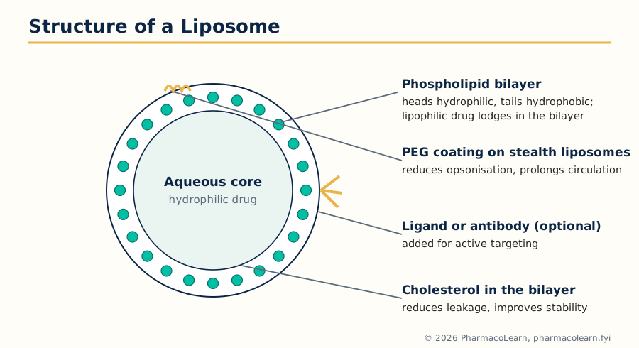 Figure: Structure of a liposome: the bilayer holds lipophilic drugs, the aqueous core holds hydrophilic drugs, and the surface can be PEGylated or fitted with a targeting ligand.
Liposomes are microscopic vesicles in which an aqueous compartment is enclosed by one or more bilayers of phospholipid. They were described by Bangham in the 1960s and remain the most successful carrier family in clinical use. The structure explains their versatility: hydrophilic drugs are held in the aqueous core, lipophilic drugs lodge in the bilayer, and amphiphilic drugs sit at the interface, so one carrier suits all three classes. Liposomes are made of the same phospholipids as cell membranes (phosphatidylcholine, phosphatidylethanolamine and related lipids, usually with cholesterol), so they are biocompatible and biodegradable, and their surface can be modified with PEG (stealth liposomes) or with ligands and antibodies (immunoliposomes). Their limitations are physical and chemical instability (leakage, fusion, oxidation and hydrolysis of the lipids), rapid RES clearance of conventional vesicles, and cost of manufacture.
2.6 Types of liposomes
Liposomes are classified in two main ways.
By size and lamellarity (number of bilayers)
- Multilamellar vesicles (MLV): several concentric bilayers like an onion, typically about 0.5 to 5 micrometres and heterogeneous; high lipid content, suited to lipophilic drugs, but cleared rapidly.
- Large unilamellar vesicles (LUV): a single bilayer enclosing a large aqueous volume, about 100 to 1000 nm; high aqueous trapped volume, good for hydrophilic drugs.
- Small unilamellar vesicles (SUV): a single bilayer, about 20 to 100 nm; small and uniform, longer circulating than MLV, but low trapped volume per unit lipid.
By composition and behaviour
- Conventional liposomes: neutral or negatively charged phospholipid with cholesterol; rapidly taken up by the RES.
- Stealth (long circulating) liposomes: surface coated with PEG, which forms a hydrated steric barrier, reduces opsonisation and greatly prolongs circulation, allowing EPR based tumour accumulation (Doxil is the model product).
- Targeted liposomes and immunoliposomes: carry a ligand or antibody on the surface for active targeting (Unit 1).
- Cationic liposomes: made with positively charged lipids; they bind negatively charged nucleic acids and cell surfaces and are the standard nonviral gene delivery carrier (Unit 5).
- Stimulus sensitive liposomes: pH sensitive or temperature sensitive compositions that destabilise and release their contents at the target site.
Cholesterol deserves a note of its own: inserted into the bilayer, it reduces membrane fluidity and permeability, decreases leakage of water soluble drugs, and improves stability in plasma; most practical formulations contain it.
2.7 Preparation of liposomes
- Thin film hydration (Bangham method): the lipids are dissolved in an organic solvent (chloroform or chloroform methanol) in a round bottom flask, and the solvent is removed under vacuum to leave a thin dry lipid film on the flask wall. The aqueous phase (buffer, with hydrophilic drug dissolved in it) is added and the flask is shaken or rotated above the lipid transition temperature; the film hydrates and peels off as MLVs. Lipophilic drugs are dissolved with the lipids in the organic phase. This is the standard laboratory method; the MLVs are then sized down.
Thin film hydration (Bangham) method
Lipids and cholesterol dissolved in chloroform
        |
        v
Solvent removed under vacuum: thin lipid film on flask wall
        |
        v
Aqueous phase added, hydrate above transition temperature
        |
        v
Multilamellar vesicles (MLV) formed
        |
        v
Size reduction: sonication, extrusion or homogenisation
        |
        v
Separation of unentrapped drug; sterilisation; storage
- Reverse phase evaporation: lipids are dissolved in ether and the aqueous phase is added; the mixture is sonicated to a water in oil emulsion and the ether is evaporated under vacuum, so the emulsion collapses into LUVs with a large trapped volume and high encapsulation of hydrophilic drugs.
- Solvent injection: in the ether injection method, the lipid solution in ether is injected slowly into warm aqueous phase and the ether evaporates, forming vesicles; in the ethanol injection method, the ethanolic lipid solution is injected into water and the ethanol is later removed, a method that is simple and suitable for scale up.
- Detergent removal (dialysis): lipids and detergent are mixed into micelles; as the detergent is dialysed away, the micelles grow and close into uniform unilamellar vesicles.
- Size reduction methods: probe or bath sonication (gives SUVs but risks drug and lipid degradation and metal contamination), membrane extrusion through polycarbonate filters of defined pore size (gives reproducible LUVs of the filter size and is the standard sizing step), the French pressure cell, and microfluidisation or high pressure homogenisation for larger scale.
- Active (remote) loading: for weak base drugs such as doxorubicin, an ammonium sulphate or pH gradient across the bilayer of preformed liposomes is used to draw the drug in and trap it, giving very high encapsulation; this is how the commercial liposomal anticancer products are loaded.
Unentrapped drug is removed by dialysis, gel filtration or centrifugation, and injectable liposomes are sterilised by filtration where size permits or prepared aseptically.
2.8 Evaluation of liposomes
- Vesicle size and distribution: dynamic light scattering for the mean size and polydispersity; microscopy for the larger MLVs.
- Lamellarity and morphology: electron microscopy (freeze fracture and negative staining), and phosphorus NMR methods in research settings.
- Trapped (encapsulated) volume and encapsulation efficiency: the liposomes are separated from free drug and the entrapped drug is assayed after disrupting the vesicles with a detergent or solvent; the trapped volume (aqueous volume per unit lipid) is measured with an entrapped marker.
- Surface charge: zeta potential, as for nanoparticles.
- Bilayer properties: the phase transition temperature by differential scanning calorimetry, and membrane fluidity in research studies.
- Drug leakage and stability: leakage of an entrapped marker or drug on storage and in plasma, lipid oxidation and hydrolysis, and size growth by fusion or aggregation.
- In vitro release and in vivo performance: release in buffer and plasma, then pharmacokinetic and biodistribution studies comparing free and liposomal drug.
- Quality tests for the finished product: sterility, pyrogen (endotoxin) content, and residual solvent, because production uses organic solvents.
2.9 Applications and marketed products
Nanoparticle and liposome systems are applied mainly in cancer chemotherapy, fungal and intracellular infections, vaccine delivery, and the delivery of peptides, proteins and nucleic acids. Products that reached the market show what the methods of this unit can achieve: Doxil (PEGylated liposomal doxorubicin, the first approved nanodrug, 1995) and DaunoXome (liposomal daunorubicin) in oncology, AmBisome (liposomal amphotericin B) for systemic fungal infection and visceral leishmaniasis, DepoCyt (liposomal cytarabine) for lymphomatous meningitis, and Abraxane (albumin bound paclitaxel nanoparticles, about 130 nm) in oncology. Each succeeds because the carrier changes the biodistribution or the toxicity profile of a drug whose free form was limited, which is the practical test of every design in this unit.
Unit 3: Microcapsules, Microspheres and Vesicular and Related Carriers
3.1 Microcapsules and microspheres: definitions
Microencapsulation packages solid, liquid or even gaseous material inside a polymeric coating, producing microparticles roughly in the range of 1 to 1000 micrometres. Two structures result:
- Microcapsules are reservoir systems: a distinct core of drug is surrounded by a continuous wall or shell of polymer. The wall controls release, protects the core and masks taste and odour.
- Microspheres are matrix systems: the drug is dissolved or dispersed throughout a solid polymer matrix, often in a spherical particle, and release occurs by diffusion through the matrix and by its erosion.
The two are used for sustained and controlled release, for protecting labile drugs from moisture, oxygen and gastric fluid, for masking the taste of bitter drugs, for converting liquids into free flowing solids, and for targeted and site specific delivery, since microparticles of a chosen size can be lodged in a chosen capillary bed or taken up by a chosen tissue. Biodegradable microspheres based on PLGA are a standard long acting injectable platform; the first such product, Lupron Depot (leuprolide acetate microspheres, 1989), releases the peptide for a month after one intramuscular injection, and Risperdal Consta (risperidone microspheres) applies the same idea in psychiatry.
3.2 Types of microcapsules and microspheres
- By structure: mononuclear (single core) and polynuclear (many cores in one shell) capsules, matrix type microspheres, and coated or double walled systems.
- By polymer origin: natural polymers (gelatin, gum acacia, albumin, chitosan, sodium alginate, starch) and synthetic polymers (PLGA, PLA, polycaprolactone, polymethyl methacrylate, ethyl cellulose, cellulose acetate phthalate, Eudragit grades).
- By degradability: biodegradable systems (PLGA, PLA, albumin, gelatin, chitosan), which release drug as the matrix erodes and need no removal, and non biodegradable systems (ethyl cellulose, PMMA), used where the device stays outside the body or is excreted intact, such as oral products.
- Magnetic microspheres, containing magnetite, belong to physical targeting (Unit 2) and are localised by an external magnet in research and clinical studies.
The choice of polymer decides the release mechanism (diffusion, erosion, or both), the route the product can be given by, and the preparation method that can be used.
3.3 Preparation of microcapsules and microspheres
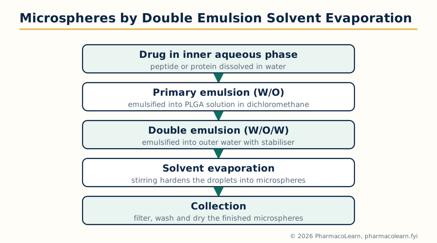 Figure: The water in oil in water double emulsion solvent evaporation method, the standard preparation for peptide loaded PLGA microspheres.
- Phase separation (coacervation): the coating polymer is made to separate from solution as a polymer rich phase (coacervate) that deposits around the dispersed core. In simple coacervation a single polymer (for example gelatin) is desolvated by adding a competing substance or changing temperature; in complex coacervation two oppositely charged polymers (classically gelatin and gum acacia) are mixed and the pH is adjusted so that they separate together around the core. The soft coating is then hardened, for example with glutaraldehyde.
- Interfacial polymerisation: two reactive monomers, one in each of two immiscible phases, meet and polymerise at the interface around the droplets, forming a thin strong shell; used for both microcapsules and nanocapsules.
- Emulsion and solvent evaporation: the workhorse method for microspheres. The drug and polymer are dissolved or dispersed in a volatile organic solvent and emulsified into an aqueous phase (oil in water single emulsion for lipophilic drugs). For water soluble drugs and peptides a water in oil in water double emulsion is used, with the drug solution as the inner water phase. The solvent then evaporates under stirring, the droplets harden into microspheres, and the product is filtered, washed and dried.
Double emulsion solvent evaporation (for peptides and proteins)
Drug dissolved in inner aqueous phase
        |
        v
Emulsify into polymer solution in dichloromethane (W/O)
        |
        v
Emulsify into outer water with stabiliser (W/O/W)
        |
        v
Stir: solvent evaporates, droplets harden
        |
        v
Filter, wash, dry: PLGA microspheres
- Emulsion crosslinking: droplets of a natural polymer solution containing the drug (for example albumin or chitosan) are hardened by chemical crosslinking (glutaraldehyde) or by heat, giving matrix microspheres.
- Ionotropic gelation: sodium alginate solution containing the drug is dropped into calcium chloride solution; calcium ions gel the alginate instantly and form beads, a gentle solvent free method suited to labile drugs and cells.
- Spray drying and spray congealing: a drug polymer solution or suspension is atomised into hot air (spray drying) and the solvent flashes off to give microparticles in one step; in spray congealing the drug is dispersed in a molten wax or polymer that is atomised into cold air and solidifies.
- Coating methods (pan coating and air suspension): used for larger cores, where a coating is applied in a rotating pan or in a fluidised bed (Wurster) coater; these are coating rather than particle forming processes but are part of classical microencapsulation practice.
3.4 Evaluation of microcapsules and microspheres
- Particle size and distribution: sieving, microscopy, laser diffraction and Coulter counting; size decides injectability, deposition site and release rate.
- Shape and surface morphology: scanning electron microscopy shows sphericity, surface smoothness, pores and cracks, all of which affect release.
- Drug content and encapsulation efficiency: drug is extracted from a weighed sample and assayed; high efficiency matters most for expensive peptide drugs.
- Yield and flow properties: percentage yield of the process; angle of repose, bulk density and flow for products to be filled into capsules or sachets.
- Swelling and density: swelling behaviour of hydrophilic matrices in release medium; density for floating or sinking behaviour where relevant.
- In vitro drug release: release in a suitable apparatus or dialysis system over the full intended duration (weeks for PLGA depots), fitted to kinetic models; an initial burst release is looked for and controlled, because a burst of a potent drug can be toxic.
- Stability and (for injectables) sterility, endotoxin and syringeability through the intended needle.
3.5 Monoclonal antibodies: preparation and application
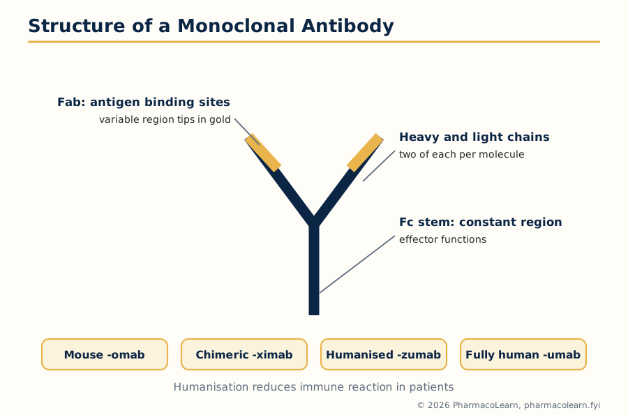 Figure: Structure of a monoclonal antibody and the naming suffixes that show how human the molecule is.
A monoclonal antibody is a homogeneous population of identical antibodies, all produced by one clone of cells and all directed against one epitope of one antigen. Antibodies are Y shaped immunoglobulin molecules: two heavy and two light chains, with antigen binding sites in the variable regions at the tips of the arms (Fab) and effector functions in the stem (Fc).
Preparation. The classical method is the hybridoma technique of Kohler and Milstein (1975). An animal (usually a mouse) is immunised with the antigen; its spleen B lymphocytes, which make the wanted antibody but cannot be cultured indefinitely, are fused with immortal myeloma cells that do not make antibody. The fused hybridoma cells are selected in HAT medium (in which unfused cells die), screened for the wanted antibody, and cloned so that one clone producing one antibody is expanded. The clone is then cultured at scale, and the antibody is purified from the culture medium (protein A affinity chromatography is the standard capture step), characterised and formulated. Because mouse antibodies provoke a human anti mouse antibody response in patients, therapeutic antibodies are progressively humanised: chimeric antibodies carry mouse variable regions on human constant regions, humanised antibodies carry only the mouse antigen binding loops (CDRs) in a human framework, and fully human antibodies are produced by phage display or transgenic animals. The suffix of the generic name shows the origin: -omab (mouse), -ximab (chimeric), -zumab (humanised), -umab (fully human). Trastuzumab (HER2 positive breast cancer) and rituximab (CD20, B cell lymphomas) are standard examples.
Applications in drug delivery. Monoclonal antibodies are the most specific targeting ligands available:
- Immunoliposomes and immunonanoparticles: the antibody is attached to the carrier surface so that the whole carrier binds antigen bearing cells (active targeting, Unit 2).
- Antibody drug conjugates (ADC): the cytotoxic drug is chemically linked to the antibody, which carries it to the antigen positive cell, where the conjugate is internalised and the drug is released. Ado-trastuzumab emtansine (Kadcyla) and brentuximab vedotin (Adcetris) are approved examples.
- Direct therapy: the antibody itself is the drug (blocking a receptor, or marking a cell for immune destruction), which is therapy rather than delivery but uses the same products.
- Diagnostics and targeting support: labelled antibodies localise tumours for imaging and guide the choice of targeted therapy.
The limits are cost, the need for the target antigen to be accessible and fairly specific, possible immune reactions, and poor penetration of large antibody molecules into solid tissue.
3.6 Niosomes
Niosomes are vesicular carriers built like liposomes but from nonionic surfactants instead of phospholipids. Hydration of a film of a nonionic surfactant (commonly Span 20, 40 or 60, or Tween grades) with cholesterol and a small amount of a charge inducer such as dicetyl phosphate gives closed bilayer vesicles with an aqueous core. Cholesterol stiffens the bilayer and reduces leakage, as it does in liposomes. Preparation follows the liposome methods of Unit 2: thin film hydration is standard, with ether injection, sonication and other variants also used. Niosomes were developed to overcome the weaknesses of liposomes: the surfactants are cheaper and chemically more stable than phospholipids (no oxidative degradation of unsaturated chains), the vesicles are more stable on storage, and the surfactants themselves can improve skin penetration. They are used experimentally and in marketed cosmetic and some pharmaceutical preparations for antifungal, anti inflammatory, anticancer and vaccine delivery, and systemically they behave like other vesicles (RES uptake unless surface modified).
3.7 Aquasomes
Aquasomes are three layered nanoparticulate carriers, typically 60 to 300 nm. The three layers are: a solid ceramic core (calcium phosphate is the most used; diamond and tin oxide cores are also described), a coating of a polyhydroxy oligomer (a carbohydrate such as cellobiose, sucrose, trehalose or lactose) adsorbed on the core, and the drug, which is adsorbed on the coated core surface without covalent bonding. Preparation is equally stepwise: the core is prepared and cleaned, the carbohydrate is adsorbed onto it, and the drug solution is added and the drug adsorbs, followed by freeze drying. Because the drug sits on the surface held by weak forces rather than being encapsulated or chemically bound, it keeps its native conformation and activity, and the carbohydrate layer acts as a protective, water like environment; the design is meant to mimic the way molecules sit in the aqueous environment of the body. Aquasomes (developed by Kossovsky and co-workers) are applied to the delivery of proteins and peptides such as insulin, enzymes, antigens for vaccines, and haemoglobin as an oxygen carrier in experimental work.
3.8 Phytosomes
Many plant constituents with good pharmacological activity (flavonoids such as silymarin, curcumin, ginkgo flavonoids, catechins) are poorly absorbed because they are either too water soluble to cross lipid membranes or too poorly soluble to dissolve. A phytosome is a molecular complex of such a phytoconstituent with a phospholipid, usually phosphatidylcholine, in a defined ratio (commonly 1:1 or 1:2). Preparation is a reaction, not just a mixing: the constituent and phosphatidylcholine are reacted in an aprotic organic solvent (such as dioxane, acetone or ethanol in some processes), and the solvent is removed to give the complex, in which the polar head of the phospholipid forms hydrogen bonds with the polar groups of the constituent. The resulting complex is lipid compatible as a whole, so it crosses membranes and is absorbed far better than the free constituent, while the constituent keeps its activity. Characterisation uses spectroscopy (NMR, IR) to confirm complex formation, solubility and partition studies, and dissolution and pharmacokinetic comparison with the free extract. Marketed examples include silymarin phytosome (Siliphos) and Ginkgo biloba phytosome (Ginkgoselect), developed by Indena, for liver protection and vascular indications.
3.9 Electrosomes
Electrosomes are soft bilayer vesicular carriers in the phospholipid family, described in the prescribed texts as electrically responsive delivery systems: the vesicles are designed to carry drug across biological barriers, particularly the skin, when an electric field is applied, for example by iontophoresis or electroporation assisted delivery. The applied field promotes the movement of the charged vesicles and the transient permeabilisation of the barrier, so that molecules which otherwise cross poorly (including some large and hydrophilic molecules) can be delivered. They remain an experimental system, taught here as the PCI syllabus names them: a vesicular carrier related to liposomes whose application depends on electrical stimulation of the site of administration.
| Carrier | Building material | Drug location | Key advantage |
|---|---|---|---|
| Liposome | Phospholipid bilayer | Aqueous core and bilayer | Versatility, clinical record |
| Niosome | Nonionic surfactant + cholesterol | Aqueous core and bilayer | Stability and low cost |
| Aquasome | Ceramic core + carbohydrate coat | Adsorbed on the surface | Native conformation of proteins preserved |
| Phytosome | Phytoconstituent phospholipid complex | Part of the complex itself | Much improved absorption of plant actives |
Unit 4: Pulmonary and Intra Nasal Drug Delivery Systems
4.1 Pulmonary delivery: rationale and deposition
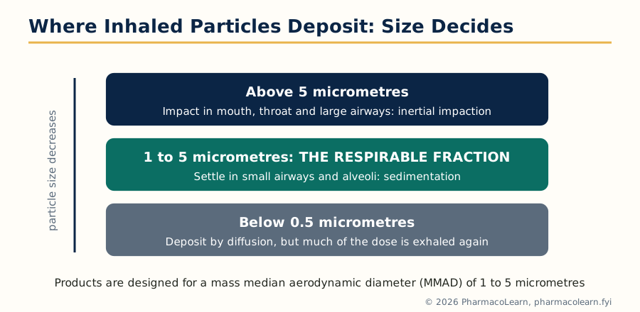 Figure: Aerodynamic particle size decides where an inhaled dose deposits; the 1 to 5 micrometre band is the respirable fraction products are designed for.
The lungs offer a very large absorptive surface (of the order of 100 square metres in an adult), a thin alveolar barrier (a fraction of a micrometre), rich blood supply and relatively low enzyme activity compared with the gut and liver. Pulmonary delivery is therefore used in two ways: for local treatment of airway disease (asthma, chronic obstructive pulmonary disease, infections, cystic fibrosis), where a small dose acts directly on the diseased tissue with little systemic exposure, and for systemic delivery of drugs that are poorly absorbed orally or destroyed in the gut, including peptides, although local therapy dominates in practice. The same routes and devices are studied in Unit 4 of MPH102T's companion teaching; here the emphasis is on the aerosol science and the evaluation of the systems.
Where an inhaled particle deposits is decided mainly by its aerodynamic diameter:
- Inertial impaction: particles larger than about 5 micrometres cannot follow the airstream around bends and impact in the mouth, throat and large airways; this dominates for large particles and fast inhalation.
- Gravitational sedimentation: particles of about 1 to 5 micrometres settle out by gravity in the smaller airways and alveoli, where the air moves slowly; a breath hold increases this deposition, which is why patients are told to hold the breath after inhaling.
- Brownian diffusion: particles smaller than about 0.5 micrometres deposit by diffusion, but a large fraction of very fine particles is simply exhaled again.
The practical result is that the respirable fraction, the mass fraction of the dose with an aerodynamic diameter of about 1 to 5 micrometres, is the part that reaches the lower airways; the mass median aerodynamic diameter (MMAD) and its spread (geometric standard deviation) are therefore core quality attributes of every inhalation product. Against delivery stand the lung's defences: the mucus layer and mucociliary escalator clear deposited material upwards, alveolar macrophages phagocytose particles, and enzymes and the tight alveolar epithelium limit absorption of large molecules.
4.2 Pharmaceutical aerosols and propellants
A pharmaceutical aerosol is a pressurised dosage form in which the drug, in solution or suspension, is expelled from its container by the pressure of a compressed or liquefied gas propellant through a valve, as a fine spray or mist. A formulation contains the drug concentrate (with solvents such as ethanol and co-solvents and, for suspensions, surfactants such as oleic acid or sorbitan esters to keep particles dispersed and lubricate the valve), the propellant, and sometimes antioxidants and flavouring agents.
Propellants provide the pressure and also act as a solvent and the dispersing medium. The requirements are that they be non toxic, non flammable as far as possible, inert, compatible with the drug and container, and have suitable vapour pressure and density.
- Chlorofluorocarbons (CFCs): the original propellants (CFC 11, 12 and 114 blends) were withdrawn from inhalers because they destroy stratospheric ozone; their use was phased out under the Montreal Protocol.
- Hydrofluoroalkanes (HFAs): the current standard for metered dose inhalers, mainly HFA 134a (norflurane) and HFA 227 (apaflurane). They do not damage ozone, but they are different solvents from CFCs, so CFC formulations could not simply be refilled with HFA: surfactant solubility, valve elastomers and dose characteristics all changed and products had to be reformulated.
- Hydrocarbons (propane, butane, isobutane): used in topical and some oral aerosols; flammable, so limited in inhalation products.
- Compressed gases (nitrogen, carbon dioxide, nitrous oxide): used where a liquefied propellant is not wanted; pressure falls as the product is used, which limits their use in metered inhalers.
4.3 Containers and valves
The container must withstand the propellant pressure, protect the contents and not react with them. Materials are tinplate (tin coated steel, often with an additional lacquer coating), aluminium (light, seamless monobloc cans, often anodised or coated), stainless steel and glass (plastic coated glass for low pressure products). Coatings of epoxy or phenolic resins are applied where the formulation attacks the metal. The valve is the functional heart of the aerosol: it seals the container, and for a metered dose inhaler its metering chamber delivers a fixed volume (commonly 25 to 100 microlitres) at each actuation, so that the patient receives a reproducible dose. The actuator (the plastic mouthpiece) contains the orifice that shapes the spray, and its design affects droplet size and spray pattern. Gaskets and valve elastomers must be compatible with modern HFA propellants.
4.4 Types of pulmonary delivery systems and their preparation
- Metered dose inhalers (MDI): the pressurised aerosol described above. Preparation involves compounding the drug concentrate, filling it into cans, crimping the metering valve and pressure filling with propellant through the valve (or cold filling with chilled concentrate and propellant at low temperature for suitable products). The dose is reproducible, the device is small and portable, but it demands coordination between pressing and inhaling, and only a modest fraction of the dose reaches the lungs; spacers (holding chambers) are used to reduce throat deposition and coordination problems.
- Dry powder inhalers (DPI): breath actuated devices holding the drug as a micronised powder, alone or blended with a coarse lactose carrier or in engineered particles. Inspiration provides the energy to deaggregate the powder, so lung deposition depends on the patient's inspiratory effort. DPIs contain no propellant, are stable and simple, and include capsule based single dose devices and multi dose reservoir and blister devices (for example Rotahaler, Diskus and Turbuhaler type designs). Preparation centres on producing respirable sized drug particles (micronisation, spray drying) and a blend or particle design that disperses well but flows and fills reproducibly.
- Nebulisers: devices that convert a drug solution or suspension into a continuous mist for tidal breathing over several minutes, used for patients who cannot use inhalers (children, the elderly, the very ill) and for large doses. Jet nebulisers use compressed gas through a venturi, ultrasonic nebulisers use a piezoelectric crystal, and vibrating mesh nebulisers push the liquid through a fine vibrating mesh to give a controlled mist with little residual volume.
| Feature | MDI | DPI | Nebuliser |
|---|---|---|---|
| Driving energy | Propellant pressure | Patient's inhalation | External (gas, electricity) |
| Dose per use | Small, metered | Small, metered | Larger, continuous over minutes |
| Patient coordination | Needed (or use a spacer) | Not needed; needs adequate inspiratory flow | Not needed |
| Propellant | Yes (HFA) | No | No |
| Portability | High | High | Low (bench devices) |
4.5 Evaluation of aerosol and inhalation products
- Container and valve tests: pressure testing and leak testing of cans, valve delivery (shot weight) and dose uniformity through the life of the can, including the number of actuations per container.
- Spray characteristics: spray pattern and plume geometry, and for topical products the rate of discharge.
- Aerodynamic particle size distribution: measured with cascade impactors (such as the Andersen cascade impactor) or liquid impingers, giving the MMAD, geometric standard deviation and the fine particle (respirable) fraction; this is the central performance test of an inhalation product.
- Formulation tests: moisture content (water promotes particle growth and valve problems), flammability of the spray, net contents, and microbial limits.
- For DPIs: delivered dose uniformity, fine particle dose at defined flow rates, and performance of the powder blend (flow, blend uniformity, moisture sensitivity).
- For nebulisers: output rate, total output, droplet size distribution and residual volume.
4.6 Intra nasal delivery: rationale and physiology
The nasal route is studied as a site specific system in its own right. The nasal cavity is lined by a highly vascular respiratory mucosa with a surface area of about 150 square centimetres, underlain by a rich blood supply that drains towards the systemic circulation. Drugs absorbed across it enter the blood quickly and avoid both gastrointestinal degradation and hepatic first pass metabolism, so the route suits potent drugs that are poorly absorbed orally, including many peptides at least in principle. Its limits are the small volume that can be given (a metered spray delivers about 50 to 140 microlitres per actuation, and volumes much above about 200 microlitres per nostril are swallowed or run out), the mucociliary clearance that sweeps the mucus blanket towards the throat with a transit time of roughly 15 to 20 minutes, nasal enzymes, and irritation with chronic use. Nasal absorption is mainly by passive diffusion across the respiratory epithelium for lipophilic small molecules, with carrier and paracellular pathways contributing for some compounds, and absorption enhancers and mucoadhesive polymers (chitosan, cellulose derivatives, carbomers) are used to prolong residence and open the pathway. Licensed nasal products include corticosteroid sprays for rhinitis, calcitonin and desmopressin sprays, sumatriptan nasal spray for migraine and naloxone nasal spray for opioid overdose, which shows the range from local to rapid systemic action.
A second, specialised objective is nose to brain delivery: drug deposited on the olfactory region can reach the brain and cerebrospinal fluid along the olfactory and trigeminal nerve pathways, bypassing the blood brain barrier (Unit 1). This is an active research area, using solutions, gels and particulate carriers (mucoadhesive microspheres and nanoparticles) designed to deposit high in the cavity and resist clearance.
4.7 Types of intra nasal systems: preparation and evaluation
- Nasal drops and simple solutions: the simplest systems, instilled with a dropper; dose is imprecise and much of the dose is swallowed, but they are cheap and familiar.
- Metered spray pumps: the standard modern system. A mechanical pump delivers a fixed volume per actuation as a spray of controlled droplet size; the drug is formulated as a solution or suspension with preservative, viscosity modifier and tonicity agents, and filled into the pump bottle. Droplet size is kept coarse enough (mostly above 10 micrometres) to deposit in the nose rather than be inhaled into the lungs.
- Nasal gels and mucoadhesive systems: viscous or in situ gelling formulations (including thermosensitive poloxamer gels) that resist mucociliary clearance and prolong contact; prepared by dispersing the drug in the gel base under controlled conditions.
- Nasal powders and particulate carriers: dry powders, and microspheres or nanoparticles of mucoadhesive polymers such as chitosan and starch, prepared by the methods of Units 2 and 3 and administered by powder insufflators; particulates prolong residence and are the main platform studied for nose to brain delivery.
- Pressurised nasal aerosols: historically used, now largely replaced by pump sprays.
Evaluation covers: delivered dose and dose uniformity per actuation and through the container life, spray pattern and plume geometry, droplet or particle size distribution, pH, viscosity and tonicity of the formulation, mucoadhesion and residence time studies, in vitro permeation across excised nasal mucosa, ciliary toxicity screening (because preservatives and enhancers can damage the clearance mechanism the product depends on), and stability and microbial quality.
Unit 5: Nucleic Acid Based Therapeutic Delivery Systems
5.1 Gene therapy: introduction
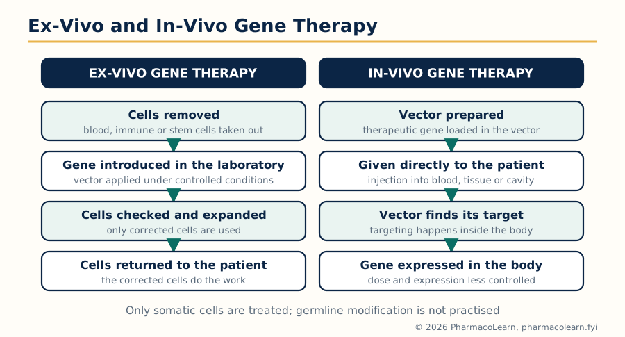 Figure: Ex-vivo gene therapy corrects the patient's cells in the laboratory; in-vivo gene therapy delivers the vector directly to the patient.
Gene therapy treats disease by delivering nucleic acid: it introduces a functional copy of a gene to replace a defective one, adds a gene whose product fights the disease, or silences or edits a harmful gene. The delivered DNA must survive transport, enter the target cell, reach the nucleus and be expressed at the right level for long enough; each of those steps is a delivery problem, which is why this unit sits in a drug delivery subject. Two features distinguish it from ordinary drug therapy: the effect can persist for years after one administration because the cell keeps expressing the gene, and the delivery system (the vector) is as much a part of the product as the gene itself.
Two approaches are distinguished:
- Ex-vivo gene therapy: cells are removed from the patient, the gene is introduced into them in the laboratory, the corrected cells are selected, checked and expanded, and they are then returned to the patient. The first clinical trial of gene therapy (1990, for adenosine deaminase deficiency, ADA-SCID) used this approach with the patient's own T lymphocytes. Quality control is possible before the cells go back, and no vector is given to the patient directly, but the method is individual, costly and limited to cells that can be removed and returned (blood and immune cells, skin, stem cells).
- In-vivo gene therapy: the gene, in its delivery system, is given directly to the patient, by injection into the blood, a tissue, or a body cavity, and must find its target cells in the body. It is simpler in concept and can reach internal organs, but targeting, dose control and safety monitoring are all harder.
Somatic cell gene therapy (treating the patient's body cells, with effects confined to that patient) is the accepted field; germline modification, which would pass changes to children, is not practised.
5.2 Potential target diseases for gene therapy
- Inherited (monogenic) disorders: diseases caused by one defective gene are the natural first targets, because one working copy can in principle correct them. Examples studied include ADA-SCID and other severe combined immunodeficiencies, cystic fibrosis (the CFTR gene, delivered to the airways), haemophilia A and B (factor VIII and IX genes, delivered to the liver), Duchenne muscular dystrophy, and inherited retinal disease. The commercially successful products reflect this list: Luxturna (voretigene neparvovec, an AAV vector carrying the RPE65 gene, 2017) for inherited retinal blindness and Zolgensma (onasemnogene abeparvovec, AAV carrying the SMN1 gene, 2019) for spinal muscular atrophy are in-vivo products, and Strimvelis (2016) is an ex-vivo stem cell product for ADA-SCID.
- Cancer: gene therapy attacks cancer in several ways: replacing a defective tumour suppressor gene (delivery of normal p53), suicide gene therapy (introducing an enzyme gene such as herpes simplex thymidine kinase that converts a prodrug, ganciclovir, into a toxic product inside the tumour cell), immunotherapy (engineering the patient's T cells to carry a chimeric antigen receptor, the CAR-T products such as Kymriah, 2017, which is ex-vivo gene therapy), and oncolytic viruses engineered to multiply in and destroy tumour cells.
- Other fields under study: infectious disease (for example engineering resistance to HIV), cardiovascular disease (angiogenic growth factor genes) and neurological disease.
5.3 Gene expression systems
A gene delivery product is more than a gene. The expression cassette carried by the vector contains the therapeutic gene (coding sequence) together with the control elements that decide where, when and how strongly it is expressed: a promoter (constitutive promoters such as the CMV promoter for strong general expression, or tissue specific promoters to restrict expression to, for example, liver or muscle), enhancer sequences, a polyadenylation signal, and in some designs inducible elements that allow expression to be switched by a drug. The choice of expression system is a formulation decision: too little expression gives no benefit, too much or expression in the wrong tissue can be toxic, and permanent expression is desirable for a missing enzyme but unnecessary or unwanted for a temporary effect such as a growth factor pulse. Gene transfer systems are judged not only by how much DNA they deliver but by the expression profile they produce.
5.4 Viral gene transfer systems
Viruses are nature's gene delivery vehicles: they are built to enter cells and place their genetic material where it will be expressed. Gene therapy vectors are viruses from which the disease causing and replication genes have been removed and replaced by the therapeutic gene.
| Vector | Carried form | Main advantages | Main limitations |
|---|---|---|---|
| Retrovirus (gamma retrovirus) | RNA, integrated into host DNA | Stable integration, lifelong expression in dividing cells | Enters dividing cells only; insertional mutagenesis risk (leukaemia occurred in early SCID trials) |
| Lentivirus | RNA, integrated | Infects dividing and non dividing cells; used for ex-vivo stem cell and CAR-T products | Integration risk remains; complex manufacture |
| Adenovirus | DNA, stays episomal | High expression, infects many cell types, large capacity | Strong immune response, expression is temporary, cannot be redosed easily |
| Adeno associated virus (AAV) | DNA, mainly episomal | Low pathogenicity and immunogenicity, long expression in non dividing tissues (eye, liver, muscle, nerve) | Small packaging capacity (about 4.7 kb); pre existing immunity to natural AAV in many patients |
| Herpes simplex virus | DNA, episomal | Very large capacity, natural nerve tropism | Cytotoxicity and latency need careful engineering |
5.5 Nonviral gene transfer systems
Nonviral systems avoid the safety and manufacturing problems of viruses but are less efficient, because they must overcome every barrier (serum nucleases, cell entry, endosomal escape, nuclear entry) without viral machinery.
- Naked DNA: plasmid DNA injected directly, taken up poorly except in muscle and skin; used in DNA vaccine research and with physical assistance.
- Physical methods: electroporation (electric pulses open transient pores in the cell membrane), the gene gun (DNA coated gold particles shot into tissue), ultrasound and magnet assisted delivery; effective locally but hard to apply to deep tissues.
- Chemical carriers: calcium phosphate coprecipitation (the classical laboratory method), DEAE-dextran, cationic polymers such as polyethyleneimine (which buffers the endosome and promotes escape, the proton sponge idea), dendrimers, and cationic lipids and liposomes (next section). These condense the negatively charged DNA into small complexes (polyplexes or lipoplexes) that cells take up by endocytosis.
5.6 Liposomal gene delivery systems
Cationic liposomes and lipid nanoparticles are the most developed nonviral systems and connect this unit back to Unit 2. A cationic lipid (for example DOTAP or DOTMA in research systems) mixed with a helper lipid such as DOPE forms liposomes with a positive surface; the anionic DNA binds electrostatically and is condensed into a lipoplex, which is protected from nucleases, binds the negatively charged cell surface, enters by endocytosis, and with the help of the fusogenic helper lipid escapes the endosome so that the DNA can reach the nucleus. The design variables are the lipid structure, the lipid to DNA charge ratio, particle size and the route of administration. Lipid systems are chemically defined, can be manufactured reproducibly, can carry large DNA, and provoke less immune response than viruses, but efficiency is lower and cationic lipids are dose limitingly toxic, so they are used where transient expression is acceptable. Their clinical coming of age is the lipid nanoparticle delivery of small interfering RNA in Onpattro (patisiran, 2018), the first approved siRNA medicine, in which an ionisable lipid nanoparticle carries the siRNA to hepatocytes after intravenous infusion. siRNA is not gene therapy in the strict gene addition sense, but the delivery system is the liposomal platform of this section.
5.7 Biodistribution and pharmacokinetics of gene delivery systems
Nucleic acids behave unlike small molecule drugs, and their delivery systems have their own pharmacokinetics. Unprotected DNA and RNA are degraded by serum and tissue nucleases within minutes, so protection by the carrier is a precondition of any effect. After intravenous administration, complexes and vectors distribute like other nanoparticles: larger and charged complexes are cleared rapidly by the RES to liver and spleen, lung can trap larger aggregates at first pass, and only a small fraction reaches other targets, which is why local administration (into the eye, muscle, tumour or airway) is common in the approved products. Clearance also has an immune component: viral vectors provoke antibody and T cell responses that limit repeat dosing, and innate sensing of nucleic acids (for example through Toll like receptors) causes dose limiting inflammation. Biodistribution studies (where the vector and the expressed gene are found, using PCR based assays) and shedding studies (whether the patient excretes vector) are therefore formal parts of gene therapy development and regulation, and the pharmacokinetics of the expressed protein, not of the DNA alone, decides the duration of treatment effect.
5.8 Antisense molecules as drugs
Antisense oligonucleotides are short synthetic single stranded nucleic acid sequences (typically about 15 to 25 nucleotides) designed to bind a specific messenger RNA by base pairing and so prevent production of one chosen protein. The main mechanisms are: recruiting the enzyme RNase H to cut the bound mRNA; sterically blocking the ribosome so translation stops; and modulating splicing, by blocking splice sites so that an exon is skipped or included. Because naked oligonucleotides are destroyed by nucleases and cross membranes poorly, they are chemically modified: phosphorothioate backbones resist nucleases and bind plasma proteins (improving distribution), and 2 prime sugar modifications (2'-O-methyl, 2'-O-methoxyethyl) and locked nucleic acids increase binding strength and stability. Approved examples show the range: fomivirsen (Vitravene, 1998), the first antisense drug, given into the eye for cytomegalovirus retinitis; mipomersen (Kynamro), against apolipoprotein B mRNA for homozygous familial hypercholesterolaemia; and nusinersen (Spinraza), which corrects the splicing of the SMN2 gene in spinal muscular atrophy and is given intrathecally. Delivery remains the limiting step for this class, which is why antisense chemistry and delivery are developed together.
5.9 Aptamers: drugs of the future
Aptamers are short single stranded DNA or RNA molecules that fold into defined three dimensional shapes and bind a target molecule (a protein, or occasionally a small molecule) with antibody like specificity and affinity, but by shape recognition rather than by base pairing with nucleic acid. They are selected in vitro by SELEX (systematic evolution of ligands by exponential enrichment): a huge random library (of the order of 10^15 sequences) is exposed to the target, the binding sequences are recovered and amplified by PCR, and the cycle is repeated until binders of very high affinity dominate. Aptamers are then chemically stabilised (2 prime modifications, capping, and PEGylation to slow renal clearance). Their advantages over antibodies are that they are made by chemical synthesis (cheap, reproducible, no animals or cells), are small (better tissue penetration), and are generally of low immunogenicity; their disadvantages are nuclease sensitivity without modification and rapid renal clearance of small molecules, both addressed by the modifications above. The first approved aptamer drug was pegaptanib (Macugen, 2004), a PEGylated anti VEGF aptamer injected into the eye for wet age related macular degeneration. Together with antisense molecules and siRNA, aptamers represent the class the syllabus calls the drugs of future: information based medicines whose specificity is designed from a sequence or a shape rather than discovered by screening, and whose success depends on the delivery and stability solutions developed in this unit.
Frequently Asked Questions
Q: What is targeted drug delivery and how does it differ from conventional drug delivery? Targeted drug delivery transports the drug preferentially to the tissue, cell or compartment where it is needed, usually by loading it in a carrier whose size, surface and ligands control its distribution. Conventional delivery distributes the drug throughout the body according to its own physicochemical properties, so most of the dose reaches healthy tissue, causing side effects, while the target may receive too little. Targeting aims to raise the concentration at the site of action and lower it everywhere else, improving efficacy and reducing toxicity.
Q: What is the EPR effect and why does it matter in tumour targeting? The enhanced permeability and retention effect is the tendency of macromolecules and nanoparticles of a suitable size to accumulate in solid tumours. Tumour blood vessels are leaky, with wide endothelial gaps, and the tumour lacks working lymphatic drainage, so material that leaves the blood in a tumour stays there. It is the basis of passive tumour targeting and of PEGylated products such as Doxil, which circulate long enough to exploit it.
Q: What is the role of the reticuloendothelial system in drug targeting? The reticuloendothelial system (mononuclear phagocyte system), mainly the macrophages of the liver and spleen, recognises opsonised particles and removes them from the blood within minutes. It is the main obstacle to targeting sites outside the liver and spleen, and carriers are made long circulating by hydrophilic surface coating (PEGylation) to reduce opsonisation. The same uptake is used deliberately when the target is the liver, spleen or macrophages themselves, as in liposomal amphotericin B for visceral leishmaniasis.
Q: Compare nanospheres and nanocapsules. Nanospheres are matrix systems in which the drug is dispersed uniformly through a solid polymer or lipid matrix and is released by diffusion and erosion. Nanocapsules are reservoir systems with a drug containing core, often oily, surrounded by a thin polymer shell that controls release. Nanocapsules often achieve higher loading of lipophilic drugs, while nanospheres are simpler to prepare and their release is governed by the matrix properties.
Q: How are liposomes classified? Give the preparation of liposomes by thin film hydration. By size and lamellarity, liposomes are multilamellar vesicles (several bilayers, about 0.5 to 5 micrometres), large unilamellar vesicles (one bilayer, about 100 to 1000 nm) and small unilamellar vesicles (one bilayer, about 20 to 100 nm); by composition they are conventional, stealth (PEGylated), targeted or immunoliposomes, cationic and stimulus sensitive. In thin film hydration, the lipids and cholesterol are dissolved in chloroform and the solvent is removed under vacuum to leave a thin film; aqueous phase is added and the film is hydrated above the lipid transition temperature to form multilamellar vesicles, which are then sized down by sonication or extrusion and separated from unentrapped drug.
Q: What are the main methods of preparing microspheres and how are they evaluated? The main methods are phase separation (coacervation), interfacial polymerisation, emulsion solvent evaporation (single emulsion for lipophilic drugs, double emulsion for peptides and proteins), emulsion crosslinking, ionotropic gelation, spray drying and spray congealing. Evaluation covers particle size and distribution, shape and surface morphology by SEM, drug content and encapsulation efficiency, yield and flow, in vitro release over the full intended duration with attention to burst release, and stability, sterility and endotoxin for injectable depots.
Q: How are monoclonal antibodies prepared, and how are they used in drug delivery? Monoclonal antibodies are prepared by the hybridoma technique: an immunised animal's spleen B cells are fused with myeloma cells, the hybridomas are selected in HAT medium, screened and cloned, and the single clone's antibody is produced in culture and purified; humanisation reduces immune reactions in patients. In delivery they act as highly specific ligands, as the targeting part of immunoliposomes and antibody coated nanoparticles, and in antibody drug conjugates such as ado-trastuzumab emtansine, where the antibody carries a cytotoxic drug to antigen positive cells.
Q: What is the difference between an aerosol, a metered dose inhaler and a nebuliser, and what decides where an inhaled particle deposits? An aerosol is the pressurised dosage form itself, drug in solution or suspension expelled by a propellant through a valve. A metered dose inhaler is the patient device that delivers a fixed metered volume of that aerosol per actuation. A nebuliser is a separate device that converts a drug solution into a continuous mist for tidal breathing, needing no propellant or coordination. Deposition is decided mainly by aerodynamic particle size: above about 5 micrometres particles impact in the throat and large airways, 1 to 5 micrometres sediment in the small airways and alveoli (the respirable fraction), and below about 0.5 micrometres particles deposit by diffusion or are exhaled.
Q: Compare ex-vivo and in-vivo gene therapy, and viral and nonviral gene transfer. In ex-vivo gene therapy, cells are removed from the patient, corrected in the laboratory, checked and returned; it gives quality control and no direct vector exposure but is individual, costly and limited to removable cells. In in-vivo therapy the vector is given directly to the patient; it can reach internal organs but targeting, dose control and safety are harder. Viral vectors (retrovirus, lentivirus, adenovirus, AAV) transfer genes efficiently but carry immunogenicity and, for integrating vectors, mutagenesis risks. Nonviral systems (naked DNA, physical methods, cationic polymers and liposomes) are safer and easier to manufacture but less efficient, largely because of endosomal escape and nuclear entry barriers.
Q: What are antisense molecules and aptamers, and why are they called drugs of the future? Antisense molecules are short synthetic oligonucleotides that bind a chosen mRNA by base pairing and block production of one protein, by RNase H cleavage, steric blocking or splicing modulation; examples are fomivirsen, mipomersen and nusinersen. Aptamers are short DNA or RNA molecules selected by SELEX that fold into shapes binding a target with antibody like specificity; pegaptanib was the first approved aptamer. They are called drugs of the future because their specificity is designed from sequence or shape rather than discovered by screening, offering precise, programmable medicines, provided the delivery and stability problems that this unit discusses are solved.
Complete MPH201T notes, all 5 units in one PDF, free to keep.
Other M.Pharm subjects: MPH101T · MPC101T · MPL101T · MPH102T · MPH103T · MPH104T · MPL102T · MPL103T · MPL104T · MPC102T · MPC103T · MPC104T · MPH201T · MPH202T · MPH203T · MPH204T · MPC201T · MPC202T · MPC203T · MPC204T · M.Pharm