
MPH102T — Drug Delivery System
4 hrs/week, 4 credits, 60 hours, 100 marks. Notes follow the M.Pharm syllabus, unit by unit.
MPH102T Drug Delivery System notes for M.Pharm Semester 1 Pharmaceutics: SR and CR formulations, polymers, rate controlled, gastro-retentive, buccal, ocular and transdermal systems, protein and vaccine delivery, with free unit wise PDF downloads on PharmacoLearn.
Units in this subject
- Unit 1 — Sustained Release and Controlled Release Formulations, Polymers and Dosage Forms for Personalized Medicine
- Unit 2 — Rate Controlled Drug Delivery Systems
- Unit 3 — Gastro-Retentive and Buccal Drug Delivery Systems
- Unit 4 — Ocular Drug Delivery Systems
- Unit 5 — Transdermal Drug Delivery Systems
- Unit 6 — Protein and Peptide Delivery
- Unit 7 — Vaccine Delivery Systems
Download MPH102T PDF Notes
Free PDFs for this subject, one per unit:
Unit 1 PDF Unit 2 PDF Unit 3 PDF Unit 4 PDF Unit 5 PDF Unit 6 PDF Unit 7 PDF
Drug Delivery System, subject code MPH102T, is a core Semester 1 subject of M.Pharm Pharmaceutics. It carries 4 credits and 60 hours of theory, taught at 4 hours per week, for 100 marks.
This subject teaches how a drug delivery system is designed so that the drug reaches the right site, at the right rate, for the right length of time. A conventional tablet releases its drug quickly and the blood level rises and falls with every dose. The systems in this subject change that behaviour: they sustain or control the release rate, hold the dosage form in the stomach or on a mucosal surface, carry the drug across the skin or the eye, protect fragile protein drugs, and deliver vaccines in new ways. A student who completes this subject should be able to select a suitable drug and polymer for a delivery system, explain the mechanism by which the system releases the drug, and describe how such a system is formulated and evaluated. The seven units move from the basic concepts of sustained and controlled release, through rate controlled, gastro-retentive, buccal, ocular and transdermal systems, to protein, peptide and vaccine delivery.
Unit 1: Sustained Release and Controlled Release Formulations, Polymers and Dosage Forms for Personalized Medicine
1.1 Introduction and basic concepts
When a conventional immediate release dosage form is given repeatedly, the plasma concentration rises after each dose and falls before the next one. If the interval between doses is long compared with the half life of the drug, the concentration spends part of the time below the minimum effective concentration (MEC) and, with a narrow therapeutic index drug, part of the time near or above the minimum toxic concentration (MTC). The aim of a modified release system is to flatten this curve.
Some basic terms must be clear before any design work:
- Sustained release (SR): a dosage form that releases the drug slowly over an extended period, so that the dosing frequency is reduced, usually from three or four times a day to once or twice a day. The release rate is prolonged but is not necessarily constant, and the system does not target a specific site.
- Controlled release (CR): a system that releases the drug at a constant, predetermined rate, ideally zero order, so that the plasma concentration stays nearly constant for the period of therapy. Controlled release is the stricter term: rate control is designed into the system and is largely independent of the biological environment.
- Extended release, prolonged release and delayed release are related labels. Delayed release means release begins after a lag, for example an enteric coated tablet that releases only in the intestine. Extended and prolonged release are used loosely for systems that release over a longer time than a conventional form.
- Loading dose and maintenance dose: many SR/CR designs contain an immediate release portion that produces the therapeutic level quickly (the loading part), followed by a slow release portion that maintains it. The total dose is therefore higher than a single conventional dose, which is one reason dose dumping is a safety concern.
The rationale for SR/CR is strongest for drugs with a half life of about 2 to 8 hours, a wide enough therapeutic index, uniform absorption from the gastrointestinal tract, and a dose that is not too large to fit into one unit.
1.2 Advantages and disadvantages of SR/CR formulations
Advantages:
- Reduced frequency of dosing, which improves patient compliance, especially in chronic therapy such as hypertension, diabetes and arthritis.
- More constant plasma levels: fewer peaks and troughs, so fewer peak related side effects and fewer periods of lost effect, including better night time control.
- Reduced total drug usage in some cases, because the drug is used more efficiently and first pass loss per dose event is spread out.
- Better safety margin in therapy where fluctuation itself is harmful, for example antianginal and antihypertensive therapy.
- Reduced gastrointestinal irritation for some drugs, because the local concentration released at one spot at one time is lower, though this depends on the design.
- Economic and nursing time savings in hospitals because fewer administrations are needed.
Disadvantages and limitations:
- Higher cost per unit than conventional forms, because of polymers, coating and process technology.
- Dose dumping: if the system fails (a cracked coat, chewing of a matrix tablet, alcohol induced failure of some coatings), the whole large dose can be released at once and cause toxicity.
- The dosage form cannot be crushed, split or chewed in most cases, which is a problem for patients who cannot swallow whole units and for feeding tube administration.
- Poor correlation sometimes between in vitro release and in vivo performance, because gut motility, food and pH vary between patients.
- Retrieval of the drug is difficult once the dose is taken: therapy cannot be stopped quickly if an adverse reaction occurs.
- Not every drug is suitable. Very short half life drugs need too large a dose, very long half life drugs do not need the technology, poorly soluble or site specific absorption drugs behave unpredictably, and large dose drugs cannot fit.
1.3 Factors influencing SR/CR design
Physicochemical factors of the drug:
- Aqueous solubility: very soluble drugs are hard to slow down because they dissolve and diffuse quickly; very insoluble drugs are dissolution rate limited already, and release becomes erratic. A moderate solubility is easiest to work with.
- Partition coefficient: the drug must cross lipid membranes after release. Very high lipid solubility can cause the drug to be held in fatty tissue; very low lipid solubility gives poor absorption.
- Molecular size and diffusivity: large molecules diffuse slowly through polymer matrices and membranes, which limits some designs and is a central problem for proteins (Unit 6).
- Drug stability: the drug must survive the pH range and enzymes of the gut for the whole transit period, because the form releases over many hours.
- Ionisation (pKa): only the unionised form crosses membranes well, and solubility changes with pH along the gut, so release and absorption both shift with site.
- Dose size: as a working rule, single doses above about 500 to 600 mg are difficult to make into a swallowable SR unit.
Biological factors:
- Biological half life: the ideal range is roughly 2 to 8 hours. Drugs below about 1 hour need an impractically large dose in the system; drugs beyond about 12 hours are already sustained by the body itself.
- Absorption window: some drugs absorb only in the upper gut (for example many weak bases and drugs carried by transporters). Once the form passes that window, the remaining dose is wasted. This is the exact problem gastro-retentive systems in Unit 3 are built to solve.
- Absorption rate: release must be slower than absorption, otherwise the release step cannot control the blood level (the flip flop situation is intended here: the slowest step, release, should govern the profile).
- Metabolism: drugs with heavy, variable first pass metabolism give inconsistent levels from any oral system; enzyme induction or saturation also changes the picture during long therapy.
- Distribution and protein binding: highly bound or widely distributed drugs have their own buffering, which changes how much benefit release control adds.
- Disease state, food and motility: gastric emptying time varies with food, posture, age and disease, so the residence of the form is never fully in the formulator's hands.
1.4 Physicochemical and biological approaches for SR/CR formulation
The practical methods used to build SR/CR products fall into a few repeatable approaches:
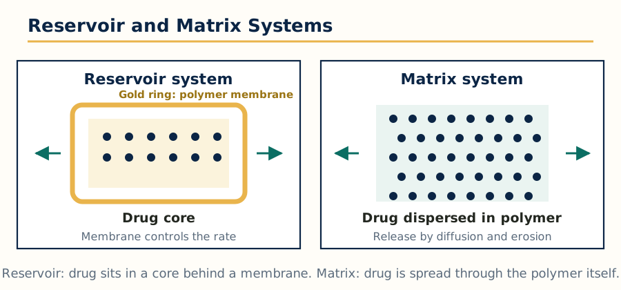 Figure: Reservoir and matrix systems, the two basic diffusion controlled designs for SR and CR formulations.
Physicochemical approaches:
- Matrix (monolithic) systems: the drug is dispersed in a polymer matrix and release happens as the medium penetrates, dissolves the drug and lets it diffuse out, or as the matrix itself erodes. Hydrophilic matrices (for example HPMC) swell and form a gel layer; inert plastic matrices (for example ethyl cellulose, polyethylene) keep their shape and release by diffusion through pores; wax and fatty matrices erode slowly.
- Reservoir systems: the drug core is enclosed by a polymer membrane and release is by diffusion across that membrane. Coated pellets, microcapsules and membrane coated tablets belong here. A pinhole or crack in the coat can dump the dose, so coat quality is critical.
- Ion exchange resin complexes: the drug is bound to an ion exchange resin and is released in the gut by exchange with sodium, potassium or hydrogen ions. Useful for taste masking plus sustained action.
- Osmotic systems: water enters through a semipermeable membrane and pushes the drug out through a laser drilled hole at a constant rate. Covered in detail in Unit 2.
- Prodrugs and drug modification: chemical change of the drug itself, for example an insoluble salt or ester, slows dissolution and prolongs action. This is a drug level approach rather than a dosage form approach.
- pH dependent coatings and solubility modifiers: release is tied to the pH of the region, or the microenvironment pH inside the form is adjusted with buffers to hold solubility steady.
Biological approaches:
- Using the body's own slow step: designing for a drug whose elimination is naturally slow is not formulation, so biological approaches mainly mean enzyme based or site based ideas: prodrugs activated by enzymes at the target region (for example colon targeted prodrugs split by colonic bacteria), antibody or ligand directed carriers studied in targeted delivery, and systems timed with biological rhythms (chronotherapy), where release is matched to the time of day the disease is worst, such as early morning asthma or blood pressure surge.
1.5 Mechanism of drug delivery from SR/CR formulations
Three mechanisms explain nearly every SR/CR product:
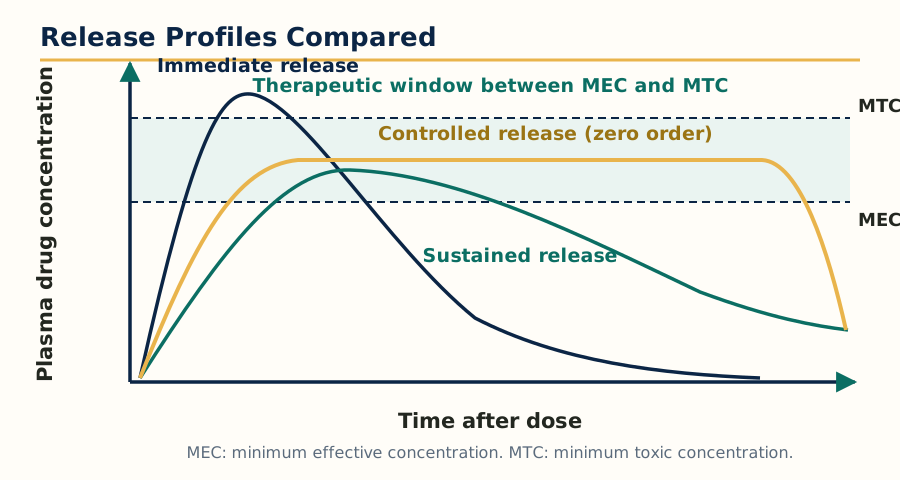 Figure: Plasma profiles compared: immediate release peaks and falls fast, sustained release is lower and broader, controlled release holds a flat zero order plateau inside the therapeutic window.
- Dissolution controlled release: the rate limiting step is the dissolution of the drug or of a coating around it. Encapsulation dissolution control coats drug particles with a slowly dissolving material of graded thickness; matrix dissolution control embeds the drug in a slowly dissolving wax matrix. Release follows the Noyes Whitney relationship: the slower the surface dissolves, the slower the supply of dissolved drug.
- Diffusion controlled release: the drug diffuses through a polymer. In reservoir systems the membrane gives near zero order release while solid drug remains inside, because the concentration gradient across the membrane stays constant. In matrix systems the amount released grows with the square root of time (the Higuchi relationship for a planar matrix), because the diffusion path length increases as the drug depletes from the outer layer inward. Students should remember: reservoir membrane systems are the classic route to zero order; homogeneous matrices are not, they slow down with time.
- Combined diffusion and erosion, and osmotic release: many real systems mix mechanisms, for example a swelling HPMC matrix releases by diffusion through the gel plus erosion of the gel surface. Fitting release data to models (zero order, first order, Higuchi, Hixson Crowell for erosion, and Korsmeyer Peppas, whose release exponent n indicates Fickian diffusion, non Fickian transport or case II erosion controlled release) is the standard way the mechanism is identified in evaluation work.
Drug release mechanisms from SR/CR systems
Dissolution controlled  -> slow dissolving coat or matrix limits supply of drug
Diffusion controlled    -> drug diffuses through membrane (reservoir) or matrix
Erosion controlled      -> polymer surface wears away and exposes fresh drug
Osmotic controlled      -> water inflow pushes drug out at a constant rate
Most real products      -> a combination, most often diffusion plus erosion
1.6 Polymers: introduction, definition, classification, properties and application
A polymer is a very large molecule built by joining many small repeating units called monomers. In drug delivery, polymers are the working material of almost every system in this subject: they form matrices, membranes, coatings, gels, implants and microparticles, and they decide the release rate more than any other excipient.
Classification:
- By source: natural (for example gelatin, alginate, chitosan, xanthan gum, guar gum), semi synthetic (cellulose derivatives such as HPMC, ethyl cellulose, cellulose acetate phthalate) and synthetic (for example polylactic acid, PLGA, polymethacrylates sold as Eudragit grades, polyethylene oxide, silicones, carbomers).
- By biodegradability: biodegradable polymers break down in the body into safe products (PLA, PLGA, polycaprolactone, albumin, gelatin) and suit implants and injectable depots; non biodegradable polymers stay intact (ethyl cellulose, silicone, ethylene vinyl acetate) and must be removable if implanted, or pass harmlessly through the gut if oral.
- By response to water: hydrophilic polymers swell or dissolve in water and make gel barriers (HPMC, PEO, carbopol); hydrophobic polymers resist water and slow penetration (ethyl cellulose, waxes, Eudragit RS and RL, which are water insoluble but permeable).
- By function in the system: matrix formers, film formers and coating materials, mucoadhesive polymers (Unit 3), enteric polymers that dissolve only above a set pH (cellulose acetate phthalate, Eudragit L and S), and smart or stimulus responsive polymers that change with pH or temperature.
Key properties a formulator checks: molecular weight and viscosity grade (higher viscosity grade, slower release from a gel matrix), glass transition temperature (decides film flexibility and whether a plasticiser is needed), solubility and swelling behaviour, permeability to the drug, mechanical strength, stability, safety and regulatory acceptance, and the degradation products for biodegradable grades.
Applications: rate controlling membranes of reservoir systems, hydrophilic matrix tablets, film coating, microencapsulation, osmotic pump membranes (cellulose acetate), implants and microspheres (PLGA), transdermal adhesives and matrices (Unit 5), mucoadhesive buccal films (Unit 3), and hydrogels for protein delivery (Unit 6). Polymer selection is therefore not one decision but a matching exercise between drug properties (Section 1.3), target profile and route.
| Feature | Hydrophilic matrix polymer (example HPMC) | Hydrophobic polymer (example ethyl cellulose) |
|---|---|---|
| Behaviour in water | Swells and forms a gel layer | Does not swell, stays intact |
| Main release mechanism | Diffusion through gel plus surface erosion | Diffusion through pores and channels |
| Effect of higher polymer level | Slower, more gel controlled release | Slower, more tortuous diffusion path |
| Typical use | Once daily matrix tablets | Coating of pellets, inert matrices |
| Failure risk | Gel can be disrupted by alcohol or strong agitation | Film cracks can cause dose dumping |
1.7 Dosage forms for personalized medicine
Personalized medicine means selecting or designing the treatment for the individual patient instead of giving every patient the same drug, dose and form. The syllabus topics under this head are:
- Pharmacogenetics: the study of how genetic differences change drug response. Variants in metabolising enzymes (for example CYP2C9, CYP2D6, CYP2C19), transporters and drug targets explain why one patient needs a low dose, another a standard dose and a third gets no effect or toxicity at the same dose. Testing before therapy (as with clopidogrel and CYP2C19, or fluorouracil and DPYD) is the applied form of this idea.
- Categories of patients for personalized medicines: broadly, patients can be grouped by genotype (poor, intermediate, extensive and ultra rapid metabolisers), by disease subtype and biomarkers (for example HER2 positive breast cancer treated with trastuzumab), by age and organ function (children, elderly, renal or hepatic impairment), and by lifestyle and adherence pattern. Each group is a candidate for a different strength, release profile or dosage form.
- Customized drug delivery systems: delivery designed around one patient or one narrow group: dose flexible forms that allow titration (mini tablets counted per dose, oral films cut or printed to strength), systems matched to a patient's swallowing ability or daily routine, and combination products built for a specific comorbidity pattern.
- Bioelectronic medicines: small implantable or wearable devices that deliver electrical stimulation to nerves to treat disease, instead of or alongside a drug (for example vagus nerve stimulation). The delivery thinking is the same as for drugs: dose (stimulus strength), site (the nerve) and duration are controlled by the device.
- 3D printing of pharmaceuticals: layer by layer fabrication of dosage forms from a digital design. Fused deposition modelling, binder jetting and semi solid extrusion can print tablets with exact individual doses, multiple drugs in one polypill with separate release profiles, and shapes that change surface area and release. The first regulatory approved printed drug product (levetiracetam, Spritam, made by binder jetting) shows the route is practical; the exam points are personalisation of dose, complex internal geometry, and on demand manufacture close to the patient.
- Telepharmacy: delivery of pharmacy services at a distance through telecommunications: remote prescription review, counselling by video, remote supervision of dispensing and follow up of adherence. It supports personalized care where a pharmacist is not physically present, and pairs naturally with customized dispensing.
The common thread of this unit: release control starts with the drug and the polymer, but the end point is a system fitted to a patient, not only to a molecule.
Unit 2: Rate Controlled Drug Delivery Systems
2.1 Principles and fundamentals
A rate controlled drug delivery system is designed so that the release rate, not the body's absorption or elimination, decides the plasma profile. The fundamental idea is simple: if the drug input rate is known and constant, and the body handles the drug by first order elimination, the plasma level settles to a predictable steady state, Css = input rate divided by clearance. The formulator therefore designs the input, and physiology does the rest.
Rate control can be engineered at three levels:
- Rate preprogrammed systems: the release rate pattern is fixed during manufacture (membrane reservoir systems, osmotic pumps). The system cannot change its rate in the body, but it delivers that rate faithfully.
- Activation modulated systems: release stays off or minimal until an activation force switches it on or speeds it: mechanical force, pH change, enzymes or osmotic pressure. These are the modulated systems of this unit.
- Feedback regulated systems: a sensor detects a body signal (for example glucose) and the system responds by releasing drug in proportion to the signal. This is the closed loop concept, the most advanced level.
The general design requirements repeat across all types: a rate controlling element (membrane, matrix, osmotic core), a drug reservoir protected from the environment until release, reproducibility between units, and stability of the rate over the whole dosing period.
2.2 Types of rate controlled systems
By mechanism, the working classification used in the standard texts is:
Rate controlled drug delivery systems
1. Rate preprogrammed
   - Polymer membrane permeation controlled (reservoir with membrane)
   - Polymer matrix diffusion controlled (monolithic matrix)
   - Microreservoir partition controlled (drug in microscopic reservoirs in a matrix)
2. Activation modulated
   - Mechanically activated    - pH activated
   - Enzyme activated          - Osmotic activated
   - Also listed: magnetic, ultrasound, iontophoresis and hydration activated
3. Feedback regulated
   - Bioerosion regulated, bioresponsive, self regulated systems
- Membrane permeation controlled: saturated drug reservoir behind a rate controlling polymer membrane; while solid drug remains, the activity inside is constant and release is near zero order.
- Matrix diffusion controlled: drug dispersed in a polymer slab, cylinder or sphere; release falls with the square root of time (Higuchi) as the depleted zone thickens.
- Microreservoir systems: the drug is first dissolved or dispersed in microscopic compartments (for example an aqueous drug solution emulsified into a polymer matrix, as in the Nitrodisc type design), combining reservoir behaviour at micro scale with a matrix at macro scale.
2.3 Mechanically activated drug delivery systems
In these systems a mechanical force applied by the patient or by a device drives delivery, which gives on demand dosing.
- Infusion pumps: external electromechanical pumps deliver drug (classically insulin) through a catheter at programmed basal rates with patient triggered bolus doses. Implantable pumps do the same inside the body for long term therapy.
- Metered dose inhalers and nasal sprays: each actuation (a mechanical press) releases a fixed metered volume through a valve. The metering valve is the rate and dose controlling element.
- Working principle to state in exams: energy for delivery comes from an external mechanical action, not from the formulation, so dose accuracy depends on device engineering and patient technique; the formulation mainly needs stability and compatibility with the device.
2.4 pH activated drug delivery systems
These systems exploit the pH gradient of the body, above all the gastrointestinal tract: stomach pH about 1 to 3, small intestine about 6 to 7.5, colon near neutral, and specific sites such as the vagina or inflamed tissue having their own values.
- Enteric systems: polymers such as cellulose acetate phthalate and methacrylic acid copolymers (Eudragit L, dissolves above pH 6; Eudragit S, above pH 7) stay intact in acid and dissolve in the intestine. Used to protect acid labile drugs (for example erythromycin, pancreatic enzymes, proton pump inhibitors) and to protect the stomach from irritant drugs (for example enteric coated aspirin and NSAIDs).
- Colon targeted systems: pH alone is unreliable for the colon, so pH polymers are combined with time dependent or enzyme degradable layers (see also 2.5) to release drugs for ulcerative colitis and Crohn disease (mesalamine products are the standard example).
- pH sensitive hydrogels and particles: polymers carrying acidic or basic groups swell or shrink as pH changes, squeezing out or releasing the drug; studied for oral insulin protection through the stomach with release in the intestine.
- Limitation to remember: gastrointestinal pH varies with food, disease and co-medication (a patient on a proton pump inhibitor has a raised gastric pH), so pH triggering is a probability, not a switch, and good designs add a second safeguard.
2.5 Enzyme activated drug delivery systems
Here an enzyme performs the activation step, so release is linked to a biochemical event at the site.
- Enzyme triggered release in the colon: coatings and matrices of polysaccharides (pectin, guar gum, chitosan, dextran) and azo polymer systems survive the upper gut but are degraded by colonic bacterial enzymes (azoreductase, glycosidases). Azo prodrugs are the drug level version: sulfasalazine is split by colonic azoreductase into sulfapyridine and 5-aminosalicylic acid, the active part for bowel disease.
- Enzyme responsive hydrogels and carriers: a carrier cross linked by peptide bonds is cleaved by enzymes that are raised at a diseased site (for example matrix metalloproteinases in tumours and inflamed tissue), releasing the drug where the enzyme activity is highest.
- Design checks: enzyme levels differ between patients and with diet and antibiotics (which can wipe out the colonic flora the system depends on), so the formulator must test release with and without the enzyme and treat the system as site assisted rather than perfectly site specific.
2.6 Osmotic activated drug delivery systems
Osmotic systems are the most reliable route to true zero order oral delivery, so this section deserves careful study.
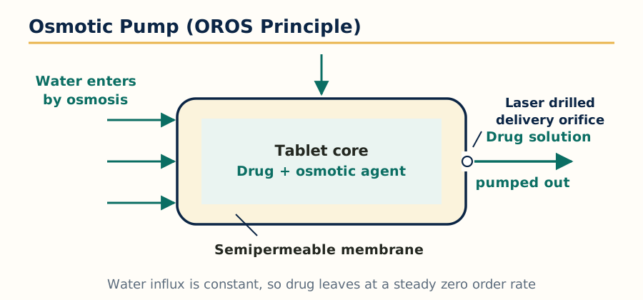 Figure: The elementary osmotic pump: water drawn in through the semipermeable membrane pushes drug solution out of the orifice at a constant rate.
Principle: water crosses a semipermeable membrane (classically cellulose acetate) into a core containing an osmotically active substance. The incoming water creates hydrostatic pressure that pushes the drug solution or suspension out through a small delivery orifice drilled in the membrane. The release rate depends on the membrane area, thickness and permeability, the osmotic pressure difference and the orifice size, and stays constant as long as solid osmotic agent remains to keep the incoming fluid saturated. In simple words: constant water inflow, constant drug outflow, largely independent of pH, food and agitation, which is why in vitro and in vivo rates match unusually well for these systems.
The elementary osmotic pump (EOP): a core of drug with or without added osmogen, coated with the semipermeable membrane, with one laser drilled hole. Rose and Nelson's early pump and Theeuwes' elementary osmotic pump are the textbook line of development. It works best for moderately soluble drugs; a very insoluble drug leaves as a solution only in small amount, and a very soluble drug needs membrane and osmogen balancing.
Push pull osmotic pump (PPOP): two layer core. The push layer contains a swellable polymer and osmogen; the drug layer contains the drug, often poorly soluble, in a suspendable form. Water enters both layers, the push layer expands and squeezes the drug suspension out of the orifice at a constant rate. Most modern once daily products (for example nifedipine GITS, glipizide and paliperidone systems) use this design. GITS (gastrointestinal therapeutic system) is the trade name family for these.
Elementary osmotic pump, working sequence
Tablet swallowed
  -> water enters through semipermeable cellulose acetate coat
  -> core dissolves, osmotic pressure builds inside
  -> pressure pushes drug solution out of the laser drilled orifice
  -> constant rate while solid core material remains (zero order)
  -> exhausted shell is finally excreted in the faeces
Other osmotic designs: push pull pump (two layer core for insoluble drugs),
osmotic bursting pump (no orifice, coat bursts after a lag),
sandwiched and controlled porosity pumps (pore formers in the coat)
Advantages: zero order release, rate set by the device not the patient, reduced food and pH effects, good IVIVC, one daily dosing for many chronic drugs. Limitations: cost and laser drilling technology, the spent shell is excreted intact (patients must be told, the "ghost tablet" in stool is expected), the rigid non deformable shell is a caution in patients with severe gastrointestinal narrowing, and a coat defect can release the whole dose.
2.7 Feedback regulated drug delivery systems: principles and fundamentals
A feedback regulated system closes the loop: it senses a biochemical signal, decides, and releases. Three parts are always present:
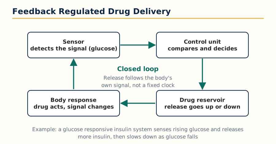 Figure: A feedback regulated system is a closed loop: sense, decide, release, respond, and sense again.
- A sensor or sensing chemistry that responds to the signal substance.
- A rate controlling mechanism whose permeability or erosion changes with the signal.
- The drug reservoir, released in proportion to the signal, and stopping or slowing when the signal falls.
The classic research example is self regulated insulin delivery: glucose oxidase inside a hydrogel converts glucose to gluconic acid, the local pH falls, the pH sensitive gel swells and lets insulin out; when glucose falls, acid production slows, the gel shrinks and release stops. Other studied versions use concanavalin A competitive binding (glucose competes with glycosylated insulin for binding sites, freeing insulin in proportion to glucose) and bioerosion regulated designs where the signal substance triggers surface erosion of the carrier.
Exam level judgement expected: the concept is proven in the laboratory and in animal work, and modern sensor based insulin pumps with electronic feedback (closed loop or artificial pancreas systems) achieve the same goal with devices; a purely chemical self regulated formulation is still not a routine marketed product because sensor response is slow, materials fatigue, and fail safe behaviour is hard to guarantee. That gap between principle and product is exactly why this topic sits at the end of the unit.
Unit 3: Gastro-Retentive and Buccal Drug Delivery Systems
3.1 Gastro-retentive systems: principle and concepts
A gastro-retentive drug delivery system (GRDDS) is designed to stay in the stomach for a prolonged, predictable time instead of emptying with the normal gastric contents. Normal gastric residence of a dosage form is short and variable, often under 2 to 3 hours, and is decided by the migrating myoelectric complex, the housekeeper waves that sweep the fasted stomach roughly every 90 to 120 minutes.
Why retain a form in the stomach at all? Four clear reasons:
- The drug has an absorption window in the stomach or upper small intestine (for example levodopa, furosemide, riboflavin, many weak bases) and is lost if the form travels beyond it.
- The drug acts locally in the stomach (antacids, and antibiotics against Helicobacter pylori such as amoxicillin and clarithromycin combinations).
- The drug is more soluble or more stable in acid and dissolves better if released slowly into gastric fluid (weak bases that precipitate at intestinal pH).
- Sustained local supply to the upper gut smooths the plasma profile for short half life drugs absorbed mainly from that region.
Basic physiological facts the design must respect: gastric fluid volume is small at rest (about 25 to 50 mL) and rises with food and fluid intake; a fed stomach empties solids slowly over hours, while the fasted stomach clears even large objects in the next housekeeper wave; objects larger than the pyloric opening (about 12 to 15 mm in the fed state) are retained longer; posture, age, sex and disease (diabetic gastroparesis, for example) all shift emptying.
Advantages: better bioavailability for window absorbed drugs, reduced dosing frequency, local action at a controlled rate, and less drug wasted in the colon. Disadvantages: retention is never guaranteed in the fasted state, large single units can be uncomfortable and carry a small risk of obstruction in narrowed stomachs, drugs that irritate the gastric mucosa or are unstable in acid are excluded, and food dependence makes performance differ between patients and days.
3.2 Modulation of GI transit time: approaches to extend GI transit
The recognised approaches, which are also the formulation methods for GRDDS:
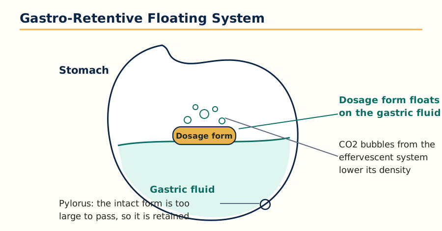 Figure: A floating gastro-retentive system stays buoyant on the gastric contents and is too large to pass the pylorus while intact.
Approaches to extend gastric residence time
Floating systems        -> density below gastric fluid, form floats on contents
  - effervescent (gas generating) and non effervescent (swelling gel) types
High density systems   -> density about 2.5 g per mL or more, form sinks
                          and is held in the antrum folds
Expandable / swelling  -> form swells or unfolds larger than the pylorus
  systems                 after swallowing, shrinks later for exit
Mucoadhesive systems   -> form sticks to the gastric mucosa
Magnetic systems       -> internal magnet in the form, external magnet on skin
Raft forming systems   -> viscous gel raft floats on gastric contents
                          (the alginate raft principle of antacid products)
- Floating systems: the dosage form has an overall density below that of gastric fluid (1.004 to 1.01 g per mL). Effervescent systems generate carbon dioxide inside a polymer matrix (sodium bicarbonate plus citric or tartaric acid with a hydrocolloid such as HPMC); the gas is trapped and the form floats, releasing drug by diffusion and erosion. Non effervescent systems swell into a gel that traps air. Evaluation must report floating lag time and total floating duration.
- High density systems: pellets or tablets made dense with inert heavy materials (barium sulphate, iron powder, zinc oxide, titanium dioxide) sink to the bottom of the stomach and resist the housekeeper waves in the antral folds.
- Expandable and unfoldable systems: a capsule sized form that, once in the stomach, swells (superporous hydrogels) or unfolds (spring like or geometric shapes of biodegradable polymer film) to a size the pylorus cannot pass, then degrades or softens after the release period so it can leave safely. The safety rule is absolute: the exit mechanism must be as reliable as the retention mechanism.
- Mucoadhesive systems: polymers such as carbopol, chitosan and HPMC hydrate and bond to gastric mucin (mechanism in Section 3.4). Gastric mucus turns over quickly, which limits how long adhesion alone can retain the form.
- Magnetic systems: a small magnet inside the form and an external magnet worn over the stomach hold the form in place; effective in studies, limited in practice by patient acceptance.
- Evaluation of GRDDS in general: buoyancy tests for floating forms (lag time, duration), swelling index for expandable forms, in vitro release in simulated gastric fluid, mucoadhesive strength where relevant, and in vivo confirmation by gamma scintigraphy or X ray with radio opaque markers, because in vitro retention claims are meaningless without imaging in humans.
3.3 Buccal drug delivery: the route and its logic
The buccal route delivers drug across the lining of the cheek into the systemic circulation. The buccal mucosa is relatively immobile, well supplied with blood, heals quickly after minor injury, and drains into the jugular vein, so the drug avoids first pass metabolism and avoids the acid and enzymes of the stomach. It suits potent drugs with heavy first pass loss, drugs destroyed in the gut, and situations where a patient cannot swallow or oral dosing is impractical (nausea, unconsciousness risk groups under supervision). Marketed logic examples: buccal and sublingual nitroglycerin for rapid angina relief, fentanyl buccal products for breakthrough pain, buccal midazolam for acute seizures, prochlorperazine buccal tablets for nausea.
Advantages: rapid onset, bypass of first pass and gut degradation, easy termination (the form can be removed), good patient acceptance compared with injections, and usefulness in emergencies. Disadvantages: small surface area (the buccal surface is only about 50 cm2) and low total fluid volume limit the dose and dissolution; saliva flow washes the form and the drug away and causes swallowing of part of the dose; taste, mouth feel and irritation restrict candidates; eating, drinking and talking must be managed while the form is in place; only small, potent, moderately lipophilic drugs qualify.
3.4 Principle of mucoadhesion
Mucoadhesion is the attachment of a polymer based dosage form to a mucosal surface. It happens in two stages: first the contact stage, in which the polymer wets, swells and makes intimate contact with the mucus layer; then the consolidation stage, in which polymer chains interpenetrate the mucin chains and weak bonds (hydrogen bonds, van der Waals forces, electrostatic attraction) build a durable joint.
The theories used to explain it, any two of which may be asked:
- Wetting theory: the polymer spreads over the mucus like a liquid adhesive; good spreading (low contact angle) means good adhesion.
- Diffusion or interpenetration theory: polymer and mucin chains diffuse into each other to a depth of about 0.2 to 0.5 micrometres and entangle; needs similar solubility character and flexible chains.
- Adsorption theory: surface forces, mainly hydrogen bonding and van der Waals attraction, hold the joint; the most widely used explanation for dry polymer systems.
- Electronic theory: electron transfer between polymer and mucus creates an electrical double layer that attracts; applies to charged polymers such as cationic chitosan against negatively charged mucin.
- Fracture theory: adhesion is judged by the force needed to break the joint, which is the basis of detachment force measurement in evaluation.
Polymers used: carbopol (polycarbophil family) is the benchmark anionic mucoadhesive; chitosan is the main cationic one and also opens tight junctions slightly; HPMC, hydroxypropyl cellulose, sodium carboxymethyl cellulose, sodium alginate, pectin and xanthan are common partners, often blended to balance adhesion, release rate and comfort. Second generation ideas (thiolated polymers forming disulphide bonds with mucin, and lectin based targeting) increase bond strength but belong to advanced discussion, one line is enough at this level.
3.5 Mechanism of drug permeation across the buccal mucosa
The buccal epithelium is a stratified squamous, non keratinised lining about 500 to 800 micrometres thick. Drug crosses by:
- Transcellular route: through the cells; favoured by lipophilic drugs that partition into cell membranes, crossing many lipid bilayers in series.
- Paracellular route: between the cells through the intercellular spaces; favoured by small hydrophilic drugs, limited by the lipid material extruded between the upper cell layers, which is the main permeability barrier of the buccal mucosa.
- The barrier is therefore lipid in nature but the tissue is far more permeable than skin (no stratum corneum) and far less enzymatic than the gut, though saliva and mucosal enzymes (esterases, peptidases) still degrade sensitive drugs, which matters for peptides.
- Permeation enhancers used in research and some products: bile salts, fatty acids (oleic acid), surfactants, chelators and chitosan; each raises permeability by disturbing intercellular lipids or tight junctions, and each must pass irritation and reversibility checks.
Only the unionised, moderately lipophilic fraction crosses well, so buccal pH (about 6.2 to 7.4 in saliva) and drug pKa decide the absorbed fraction, the same pH partition logic as the gut.
3.6 Methods of formulation of buccal systems
- Buccal tablets: small flat tablets, often bilayer, with drug and mucoadhesive polymer, and an impermeable backing layer (for example ethyl cellulose) so the drug is released towards the mucosa only, not into saliva. Made by direct compression.
- Buccal films and patches: thin flexible films cast or hot melt extruded from film forming polymers with plasticiser; they conform to the cheek, are discreet, and are the most comfortable modern form. Multilayer films add a backing and a separate drug layer for unidirectional release.
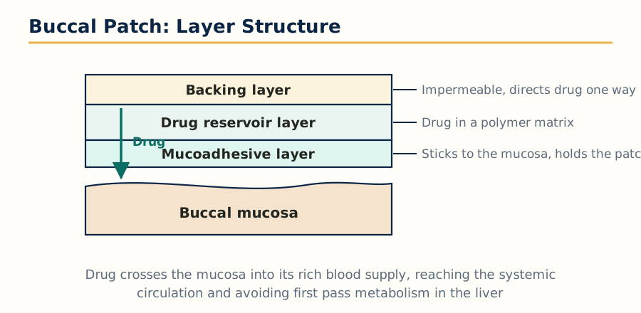 Figure: Layers of a buccal patch, from the impermeable backing to the mucoadhesive layer that holds it on the mucosa.
- Buccal gels, ointments and pastes: semi solids with mucoadhesive gel bases; easier to apply over a larger area but shorter residence and less dose accuracy.
- Sprays and liquids: metered sprays coat the mucosa for rapid absorption of potent drugs; residence is short, so they suit rapid onset more than sustained action.
- Wafers and lyophilised forms: fast dissolving porous wafers for rapid release on the mucosa.
3.7 Evaluation of buccal systems
The standard evaluation set for a buccal formulation in practical and exam work:
- Physical tests: weight and thickness uniformity, surface pH (must sit near saliva pH to avoid irritation), folding endurance for films, swelling index.
- Mucoadhesive strength: measured as the force needed to detach the form from a mucosal surface (modified balance or texture analyser, using excised animal mucosa such as porcine or sheep buccal mucosa), and residence time in vitro and in volunteers.
- In vitro release and permeation: release into simulated saliva in a dissolution setup, and permeation across excised buccal mucosa in a Franz diffusion cell to get flux and permeability coefficient.
- In vivo checks: residence time, irritation and acceptability in volunteers, and pharmacokinetic comparison against the reference route to prove the first pass advantage.
- Stability: drug and adhesive performance after storage, since a polymer that loses tack or a film that turns brittle fails the whole concept.
Unit 4: Ocular Drug Delivery Systems
4.1 Why ocular delivery is difficult
The eye is protected so well that most of an eye drop never reaches its target. Less than about 5 percent of an instilled dose typically enters the eye; the rest is drained or absorbed into the blood through the nose. Understanding the barriers is the whole unit, because every ocular delivery technology is an answer to one of them.
4.2 Barriers of drug permeation
- Tear film and tear turnover: the precorneal tear film is only a few microlitres, the cul de sac holds about 30 microlitres at most while a commercial drop is 25 to 50 microlitres, so much of the drop spills or drains at once. Tear turnover (about 1 microlitre per minute, faster with reflex tearing from irritation) washes the drug away within minutes.
- Nasolacrimal drainage: solution drains through the puncta into the nose, where the drug is absorbed into the systemic circulation across the nasal mucosa without first pass protection. This is why eye drops can cause systemic side effects: timolol eye drops can slow the heart and constrict airways in sensitive patients.
- The cornea: the main route into the anterior chamber and a three layer barrier in series: the lipophilic epithelium (with tight junctions), the hydrophilic stroma (about 90 percent of corneal thickness), and the lipophilic endothelium. A drug must therefore be both water soluble enough to cross the stroma and lipid soluble enough to cross the epithelium: an intermediate partition coefficient, log P roughly 1 to 3, performs best. Ionised drug crosses poorly.
- The conjunctiva and sclera: the conjunctiva is leakier than the cornea and rich in blood vessels, so drug absorbed there is largely lost to the blood (non productive absorption), though the conjunctival and scleral route is genuinely useful for larger molecules aiming at tissues behind the eye.
- Blood ocular barriers: the blood aqueous barrier (tight junctions of the iris and ciliary vessels) and the blood retinal barrier (retinal pigment epithelium and retinal vessel endothelium) block entry from the blood into the eye, which is why systemic dosing barely treats intraocular disease and why local delivery dominates eye therapy.
- Efflux and metabolism: transporters and enzymes in the cornea and iris ciliary body remove or break down part of what does enter.
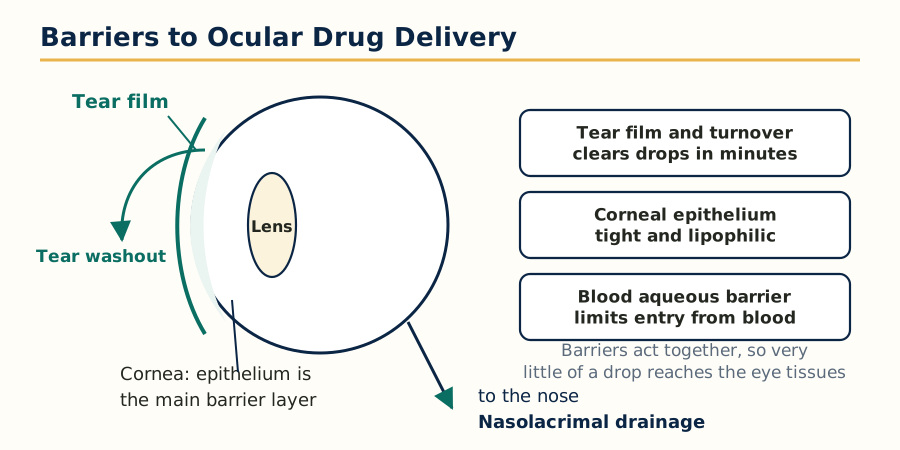 Figure: The barriers of the eye act together: tear washout, the corneal epithelium, drainage and the blood aqueous barrier.
4.3 Methods to overcome the barriers
- Increasing residence time: viscosity enhancers (HPMC, polyvinyl alcohol, carbomers) slow drainage; in situ gelling systems are solutions that gel on the eye with temperature (poloxamers), pH (carbopol) or ions in tears (gellan gum, alginate); mucoadhesive polymers grip the mucin of the tear film.
- Particulate and carrier systems: liposomes, nanoparticles, nanosuspensions and dendrimers prolong contact, protect the drug and can carry it across the cornea; cyclodextrins keep poorly soluble drugs in solution at the corneal surface without irritation.
- Ocular inserts and devices: soluble and insoluble inserts placed in the cul de sac release drug over hours to days. The classic example is the Ocusert, a membrane controlled pilocarpine reservoir that released a set amount per hour for a week, replacing four times daily drops. Collagen shields, drug eluting contact lenses and punctal plugs (which also block the drainage route physically) are the modern relatives.
- Prodrugs: a lipophilic ester form crosses the cornea and enzymes inside convert it to the active drug: dipivefrin (a prodrug of adrenaline) and the prostaglandin analogues such as latanoprost (an isopropyl ester prodrug) are the standard teaching examples.
- Permeation enhancers and physical methods: preservatives such as benzalkonium chloride loosen epithelial junctions (an effect balanced against their toxicity); research methods include iontophoresis (small electric current drives ionised drug), microneedles and ultrasound for delivery to the back of the eye.
- Invasive routes for the posterior segment: intravitreal injections (anti VEGF agents for wet macular degeneration are the biggest example), periocular and subretinal routes, and sustained implants (biodegradable and non biodegradable implants releasing steroid or antiviral drug over months) bypass every surface barrier at the cost of procedure risk.
4.4 Formulation and evaluation of ophthalmic dosage forms (key concepts)
Formulation musts for eye preparations: sterility (a non negotiable requirement; aseptic manufacture or sterilisation, single dose units where possible), isotonicity close to tears (about 0.9 percent sodium chloride equivalent; the eye tolerates a small range), pH near tear pH 7.4 within comfort limits (about 6.5 to 7.8 preferred; buffers are kept weak so tears can restore pH), freedom from particulate matter, controlled viscosity (high enough to stay, low enough to not blur vision or feel sticky), and a preservative system for multidose containers chosen to balance antimicrobial cover against corneal toxicity.
Evaluation: sterility testing, clarity and particulate inspection, pH, osmolality and viscosity measurement, drug content and in vitro release for inserts and gels, preservative efficacy testing for multidose packs, and irritation assessment historically by the Draize test and now increasingly by in vitro alternatives. For delivery systems specifically, residence and pharmacokinetic studies (drug levels in aqueous humour or tears over time) prove that the barrier strategy actually worked.
| Barrier | What it does to the dose | Main formulation answers |
|---|---|---|
| Tear turnover and drainage | Removes most of the dose in minutes | Viscosity enhancers, in situ gels, inserts, punctal occlusion |
| Corneal epithelium and stroma | Blocks very lipophilic and very hydrophilic drugs | Balanced log P design, prodrugs, carriers, cyclodextrins |
| Conjunctival blood flow | Steals drug into systemic circulation | Target corneal route, reduce dose volume, controlled release |
| Blood retinal barrier | Stops systemic drugs reaching the retina | Intravitreal injection and implants, periocular routes |
Unit 5: Transdermal Drug Delivery Systems
5.1 Structure of skin and the barrier
Skin is the largest organ of the body, about 2 square metres in an adult, and it is built to keep things out. Its layers, from the surface:
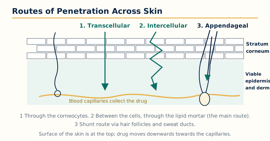 Figure: The three routes across the stratum corneum: transcellular, intercellular and appendageal.
- Stratum corneum: the outer 10 to 20 micrometres, the rate limiting barrier. Dead flattened cells (corneocytes) embedded in ordered lipid sheets of ceramides, cholesterol and free fatty acids; the standard description is a brick and mortar wall, bricks of keratin filled cells in a mortar of lipid. The drug mainly travels through the lipid mortar.
- Viable epidermis: living keratinocytes, about 50 to 100 micrometres, aqueous in character, contains enzymes that metabolise a small part of the dose.
- Dermis: connective tissue with the blood capillary network that collects the permeated drug into systemic circulation, plus the appendages, hair follicles and sweat glands, which offer a shunt route that matters mainly in the first minutes and for large or polar molecules.
- Subcutaneous fat: a depot and insulation layer below the dermis.
Permeation follows Fick's law in teaching form: flux rises with the partition coefficient into the stratum corneum, the diffusion coefficient within it and the concentration applied, and falls with barrier thickness. The ideal transdermal drug is therefore potent (daily dose in milligrams, ideally under about 10 to 20 mg per day), small (molecular weight under about 500), moderately lipophilic (log P about 1 to 4), low melting (which usually means better solubility in the barrier lipids) and non irritating.
5.2 Advantages and limitations of the transdermal route
Advantages: avoidance of first pass metabolism and of the gut environment, near constant input similar to an infusion without a needle, painless and self administered therapy, easy termination by removing the patch, and improved compliance in chronic and motion sickness or pain therapy. Limitations: only a narrow set of drugs qualifies, onset is slow (a skin depot forms, so levels keep rising for hours and fall slowly after removal), skin irritation and sensitisation are the commonest practical failures, dose cannot be changed quickly, and cost per day is higher than oral therapy. Classic product examples to quote: nitroglycerin (angina prophylaxis), scopolamine (motion sickness, the first marketed patch system), fentanyl (severe chronic pain), nicotine (smoking cessation), estradiol and combined hormone patches, clonidine and rivastigmine.
5.3 Penetration enhancers
A penetration enhancer is a substance that reversibly reduces the barrier resistance of the stratum corneum. An ideal enhancer is non toxic, non irritating, reversible in action, compatible with drug and adhesive, odourless and cheap; no real enhancer meets every point, so selection is a compromise and concentration is a balance between flux gain and irritation.
Mechanisms and classes:
- Lipid disruption: oleic acid and other fatty acids, terpenes (menthol, limonene), and surfactants insert into the lipid sheets and disorder their packing, raising diffusion.
- Solvent action: ethanol, propylene glycol, Transcutol and dimethyl sulfoxide (DMSO) increase drug solubility in the barrier and partly extract lipids; ethanol is also the co solvent in several marketed gel and patch systems.
- Keratolytic and keratin acting agents: salicylic acid and urea soften and hydrate the corneocyte protein, opening the transcellular share of the route.
- Hydration itself: occlusion by the patch hydrates the stratum corneum, which swells corneocytes and can raise permeability several fold; every patch gets this effect free.
- Physical enhancement (device based, listed here because exams group them with enhancers): iontophoresis (current drives charged drug), electroporation (short high voltage pulses open temporary pores), sonophoresis (low frequency ultrasound), microneedles (microscopic projections that pierce only the stratum corneum, painless because they do not reach nerves), thermal ablation and microdermabrasion. Each one trades the chemical enhancer's simplicity for device cost and control.
| Enhancer class | Examples | Main mechanism |
|---|---|---|
| Fatty acids | Oleic acid, lauric acid | Disorder stratum corneum lipids |
| Solvents and glycols | Ethanol, propylene glycol, DMSO | Raise drug solubility, extract some lipid |
| Terpenes | Menthol, limonene, cineole | Disrupt lipid packing, often low irritation |
| Surfactants | Sodium lauryl sulphate, Tweens | Interact with lipid and keratin, irritation risk |
| Keratolytics | Urea, salicylic acid | Hydrate and soften keratin |
5.4 Transdermal systems: designs and components
A transdermal patch is an engineered stack. From outside in: an impermeable backing layer (protects and occludes), the drug reservoir or drug in adhesive matrix, a rate controlling membrane in reservoir designs, a pressure sensitive adhesive layer that also contacts skin in matrix designs, and a release liner removed before use. Overlay adhesive rings are added when edge lift is a risk.
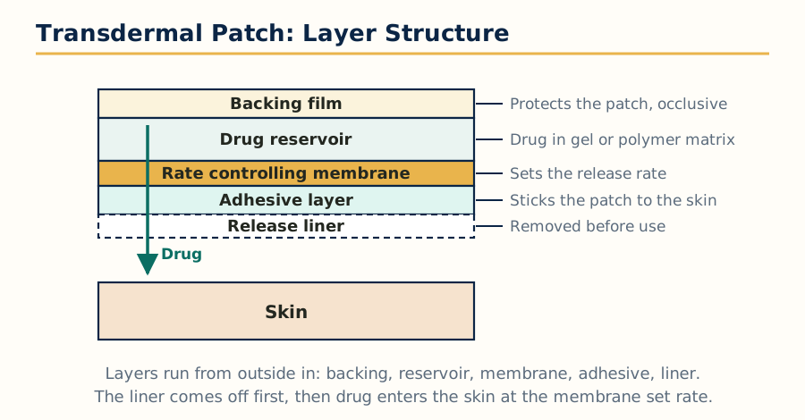 Figure: Layers of a membrane controlled transdermal patch, outside in, with the release liner removed before use.
The four standard architectures:
Transdermal patch architectures
1. Reservoir system
   Backing | Drug reservoir (gel/liquid) | Rate membrane | Adhesive | Liner
2. Matrix system
   Backing | Drug dispersed in polymer matrix | Adhesive | Liner
3. Drug in adhesive system
   Backing | Drug dissolved in the adhesive layer itself | Liner
4. Microreservoir system
   Backing | Drug in microscopic aqueous reservoirs within a
   lipophilic polymer matrix | Adhesive | Liner
Rule: a reservoir patch must never be cut (dose dumping risk);
most matrix and drug in adhesive patches state their own rule on the label.
Reservoir systems give the most constant rate but carry dose dumping risk if the membrane is damaged. Matrix and drug in adhesive systems are thinner, simpler and safer on failure, with rate controlled jointly by the matrix and the skin itself.
5.5 Formulation and evaluation of transdermal systems
Formulation variables: drug loading and its physical state (dissolved versus suspended decides whether activity stays constant), polymer or adhesive choice (acrylic, silicone and polyisobutylene adhesives dominate), enhancer type and level, backing and liner compatibility, and the patch area, which is the dose dial: rate per square centimetre times area equals daily input, so strengths of one product are often just different patch sizes.
Evaluation set:
- Physical and adhesive tests: thickness and weight uniformity, drug content, moisture content and uptake, flatness, peel adhesion, tack and shear strength, and cold flow (adhesive creeping beyond the patch edge on storage).
- In vitro permeation: Franz diffusion cell studies across excised animal or human skin or a synthetic membrane, reporting flux, lag time and permeability coefficient; in vitro release testing through a membrane for quality control of the patch itself.
- Stability and safety: stability under ICH conditions with adhesion rechecked, skin irritation and sensitisation testing, and residual drug assay of used patches, because a fentanyl patch after use still holds a dangerous amount of drug and its disposal is a safety topic in itself.
- In vivo confirmation: pharmacokinetic studies showing the plateau profile, and adhesion performance scoring in volunteers under real wearing conditions (bathing, exercise, heat), since heat over a patch can raise absorption dangerously, a counselling point printed on most labels.
Unit 6: Protein and Peptide Delivery
6.1 Why proteins are a special delivery problem
Proteins and peptides are large, fragile and active in tiny amounts. Insulin (51 amino acids), growth hormone, erythropoietin, interferons and monoclonal antibodies are all proteins or peptides, and nearly all of them are still given by injection. This unit explains exactly which barriers force that, and what formulation science is doing about it.
6.2 Barriers for protein delivery
Physicochemical and stability barriers:
- Size and polarity: molecular weights from about 1,000 (small peptides) to 150,000 (antibodies) daltons. They are too large and too hydrophilic to cross lipid membranes by passive diffusion in useful amounts.
- Chemical instability: hydrolysis of peptide bonds, deamidation of asparagine and glutamine, oxidation of methionine and cysteine, and racemisation proceed in solution, faster at extremes of pH and temperature.
- Physical instability: proteins unfold (denaturation) at interfaces, on shaking and on heating, and unfolded molecules aggregate into inactive, sometimes immunogenic clumps. Adsorption onto glass, plastic and tubing silently removes drug from solution.
- Enzymatic barriers: pepsin and pancreatic proteases (trypsin, chymotrypsin, carboxypeptidases) in the gut, peptidases in every mucosal surface, blood and tissue (including the brush border aminopeptidases) dismantle peptides within minutes. Insulin given orally is digested like food protein.
- Absorption and distribution barriers: poor membrane permeation means oral bioavailability of most proteins is under 1 to 2 percent; after injection, small peptides are cleared rapidly by the kidney (glomerular filtration under about 30 to 60 kDa effective range), and the liver and reticuloendothelial system remove carriers and aggregates.
- Immunogenicity: aggregates, impurities and non human sequences can provoke antibodies that neutralise the drug or, worst case, cross react with the body's own protein; formulation and purity are therefore safety issues, not only potency issues.
6.3 Formulation of delivery systems for proteins and macromolecules
Stabilisation inside the formulation comes first: buffer at the pH of maximum stability, stabilisers such as sugars and polyols (trehalose, sucrose, mannitol) that protect during drying and storage, surfactants in small amount (polysorbates) that stop adsorption and interface damage, antioxidants and chelators where oxidation is the risk, and presentation as a lyophilised (freeze dried) powder when solution stability is too short, reconstituted before use.
Delivery strategies by route:
- Parenteral depots and carriers (the working present): biodegradable PLGA microspheres and in situ forming gels turn daily or weekly injections into monthly ones (leuprolide depot for prostate cancer and endometriosis is the landmark example); liposomes and nanoparticles protect the protein and alter distribution; PEGylation, attaching polyethylene glycol chains, shields the protein from enzymes and kidney filtration and extends half life (pegfilgrastim, peginterferons); implants and osmotic mini pumps give steady input for potent peptides.
- Pulmonary delivery: the lung offers a huge thin surface; inhaled insulin reached the market (Exubera, later withdrawn for commercial and device reasons, then Afrezza) and proves both feasibility and the difficulty of patient acceptance.
- Nasal delivery: a small but real route for peptides that act centrally or systemically in small doses (desmopressin, calcitonin), limited by mucociliary clearance, enzymes and dose size.
- Oral delivery (the research frontier): enzyme inhibitors co formulated, permeation enhancers that open tight junctions reversibly, mucoadhesive and nanoparticle carriers that protect the cargo to the intestinal wall, and device based approaches (microneedle capsules that inject the gut wall from inside). One oral peptide success exists in a related small molecule like class: oral semaglutide uses an absorption enhancer (SNAC) to carry a peptide analogue across the stomach wall, the teaching proof that enhancer assisted oral peptide delivery can work commercially.
- Transdermal and buccal attempts: iontophoresis and microneedles bypass the skin barrier physically (see Unit 5); buccal films place the peptide against a friendlier mucosa (see Unit 3), but enzyme activity and low flux keep doses tiny.
- Gene and cell based production approaches (recombinant expression in the body, encapsulated cell implants that secrete the protein) sit at the edge of this subject; one line of awareness is enough here.
6.4 Evaluation of protein delivery systems
- Identity, purity and potency: electrophoresis (SDS PAGE), chromatography (size exclusion for aggregates, reverse phase for related impurities), mass spectrometry for identity, and a biological potency assay, because chemical content alone does not prove an active folded protein.
- Aggregation and particle checks: size exclusion chromatography, light scattering and subvisible particle counting; aggregates are tracked as a critical quality attribute throughout.
- Delivery performance: in vitro release from depots and carriers (with the caution that release media must not denature the protein being measured), encapsulation efficiency and loading, residual solvent in microspheres, syringeability of suspensions, and dose uniformity of inhalation and nasal devices.
- Stability programme: real time and accelerated studies watching potency, aggregation, moisture of lyophilised cakes and reconstitution time; cold chain behaviour (2 to 8 degrees Celsius typically, freezing is damaging for many proteins) is part of the product design, not an afterthought.
- In vivo evaluation: pharmacokinetics with immunoassays or LC MS that distinguish intact active protein, duration of action for depots, and immunogenicity monitoring (anti drug antibodies) in animal and clinical studies.
| Barrier | Core problem | Main formulation responses |
|---|---|---|
| Gut enzymes | Peptide bonds hydrolysed in minutes | Injection routes, enzyme inhibitors, protective carriers |
| Size and polarity | No passive membrane crossing | Carriers, enhancers, physical methods, depot injection |
| Physical instability | Unfolding and aggregation | Stabilisers, lyophilisation, cold chain, gentle processing |
| Rapid clearance | Kidney filtration and proteolysis in blood | PEGylation, depot systems, albumin and Fc fusion designs |
| Immunogenicity | Antibodies against drug or aggregates | High purity, low aggregation, humanised sequences |
Unit 7: Vaccine Delivery Systems
7.1 Vaccines: the basics this unit builds on
A vaccine is a preparation of antigen (killed or live attenuated organism, a subunit or toxoid, a polysaccharide conjugate, or genetic instructions such as mRNA in a lipid carrier) given to produce active immunity before natural infection. The delivery problem is different from every other unit: the dose is meant to meet the immune system, in microgram amounts, usually once or a few times, and the response, not a plasma level, is the product.
Types in brief: live attenuated (measles, BCG, oral polio), inactivated whole organism (hepatitis A, inactivated polio), subunit and recombinant (hepatitis B surface antigen, HPV virus like particles), toxoids (tetanus, diphtheria), polysaccharide and conjugate vaccines (pneumococcal, Hib, meningococcal), viral vector vaccines, and nucleic acid vaccines (mRNA in lipid nanoparticles, the COVID 19 generation of products). Adjuvants such as aluminium salts, oil in water emulsions and newer immune stimulants are part of the delivery system in the wide sense: they shape how antigen is presented.
7.2 Uptake of antigens
For a vaccine to work, antigen must be taken up by antigen presenting cells (dendritic cells above all, also macrophages and B cells). The sequence to learn:
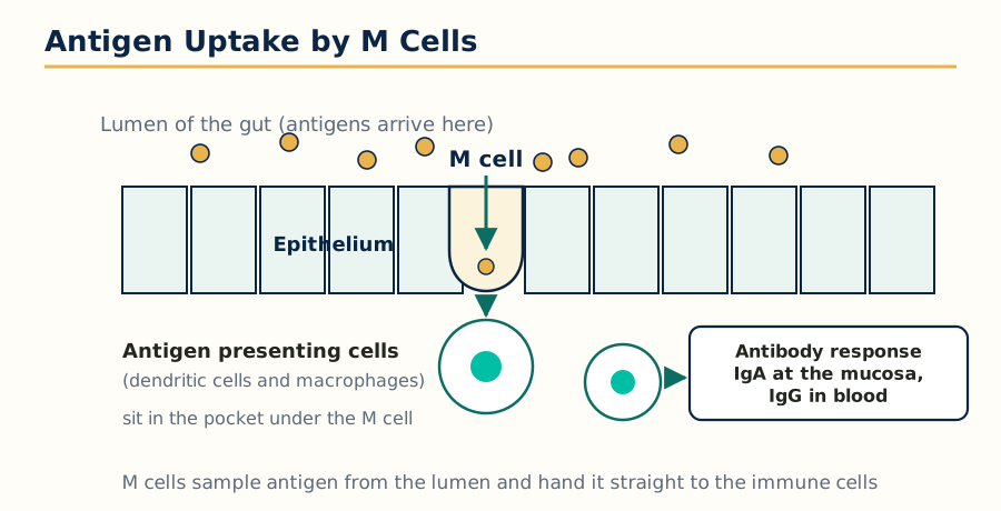 Figure: M cells sample antigen from the mucosal surface and hand it to antigen presenting cells, starting the antibody response.
Uptake of antigen and the start of the immune response
Antigen deposited (muscle, skin or mucosa)
  -> dendritic cells capture antigen by phagocytosis, endocytosis
     or (for particulate vaccines) size dependent uptake
  -> dendritic cells mature and migrate to the draining lymph node
  -> antigen is processed and presented on MHC molecules
     (MHC class II to helper T cells, class I cross presentation to killer T cells)
  -> T cells activate, help B cells, antibodies and memory cells are produced
Delivery levers: particle size (about 20 to 200 nm drains freely to lymph nodes,
larger particles are carried by cells), antigen persistence at the site,
adjuvant signals that mature the dendritic cell, and route of delivery.
Two formulation consequences follow. Particulate antigens are taken up far better than soluble ones, so adsorbing antigen on alum, forming virus like particles, liposomes and polymer particles is a delivery decision, not decoration. And the route decides the flavour of immunity: injection gives strong systemic IgG, mucosal delivery can add secretory IgA at the surface where many pathogens enter.
7.3 Single shot vaccines
A single shot vaccine aims to deliver, in one administration, the effect of a prime dose plus its boosters. The design logic: the first fraction of antigen is available immediately (the prime), and later fractions are locked inside biodegradable microspheres or particles engineered to break down at staggered times (weeks to months later), releasing pulse doses that act as boosters without another clinic visit.
- Technology used: PLGA and related biodegradable polymer microspheres of different polymer composition, molecular weight and size, each population timed to erode and release its antigen pulse at a set interval; antigen stability inside the degrading, acidifying microsphere is the central technical difficulty and is managed with stabilisers and buffering excipients.
- Why it matters: missed booster doses are a leading cause of incomplete immunisation in real programmes; a single visit product also reduces cold chain trips, needles and record keeping.
- Honest status: single shot tetanus and other candidates have been demonstrated in animals and early studies, but antigen damage during encapsulation and storage, and dose control of each pulse, have kept routine marketed products limited; the concept remains an active goal of vaccine delivery research.
7.4 Mucosal delivery of vaccines
Most pathogens enter through mucosal surfaces (gut, respiratory tract, genital tract), so a vaccine applied at the mucosa can raise secretory IgA at the entry point plus systemic immunity, something injected vaccines do poorly.
- Working examples: oral polio vaccine, oral rotavirus vaccine, oral cholera and typhoid vaccines, and the intranasal live influenza vaccine (a fine spray, no needle).
- Barriers: dilution and clearance by mucus and cilia, enzymes and acid (for oral delivery the stomach is the first wall), tolerance, because the mucosal immune system is trained to ignore harmless food and commensal antigens, and the need for mucosal adjuvants that are safe on delicate surfaces.
- Formulation answers: mucoadhesive and particulate carriers that linger and are sampled by the M cells overlying Peyer's patches in the gut (the natural antigen sampling route), enteric protection for oral products, and dry powder or spray devices that deposit a controlled dose on the nasal mucosa.
7.5 Transdermal delivery of vaccines
Skin is immunologically rich: the epidermis carries Langerhans cells and the dermis carries dendritic cells, so antigen placed in the skin meets antigen presenting cells immediately. This is why intradermal vaccination works at lower doses than intramuscular (dose sparing, used for rabies and some influenza and BCG practice, BCG being given intradermally by design).
- Methods: microneedle patches are the flagship technology: arrays of tiny solid, coated, hollow or dissolving needles that painlessly cross the stratum corneum and deposit antigen in the epidermis and upper dermis. Dissolving microneedle patches (sugar or polymer needles that melt into the skin) can be self applied, are stable without a deep cold chain in some designs, generate no sharp waste, and have shown strong responses in influenza and other trials.
- Other skin routes: needle free jet injectors, powder and particle bombardment devices, and topical application after barrier disruption (tape stripping, abrasion) in research settings.
- Programme value: painless, potentially self administered, dose sparing and sharps free delivery attacks the practical barriers of vaccination campaigns: fear of needles, trained staff, cold chain volume and waste disposal. That combination is why transdermal vaccine delivery closes this subject: it is delivery science applied to the largest preventive intervention in medicine.
Frequently Asked Questions
Q: What is the difference between sustained release and controlled release? Sustained release prolongs the release of drug over time and reduces dosing frequency, but the rate is not necessarily constant. Controlled release is stricter: the system releases the drug at a predetermined, ideally constant zero order rate, largely independent of the body environment, so plasma levels stay nearly flat.
Q: Which drugs are ideal candidates for SR and CR formulations? Drugs with a half life of about 2 to 8 hours, moderate solubility, adequate absorption from the whole gut, a dose that fits one unit (generally up to about 500 mg), a reasonable therapeutic index, and stability in gastrointestinal fluids. Very short or very long half life drugs, large dose drugs and drugs absorbed from one narrow gut region are poor candidates.
Q: How does an osmotic pump give zero order release, and how do the elementary and push pull designs differ? Water enters the tablet through a semipermeable cellulose acetate coat at a constant rate, drawn by the osmotic core. The pressure pushes drug solution out of a laser drilled orifice at the same constant rate while solid core material remains. Because water inflow is constant and independent of gut pH and motility, drug outflow is constant too: zero order. The elementary pump has a single core of drug with osmogen and works best for moderately soluble drugs. The push pull pump has two layers, a drug layer and a swelling push layer, so even poorly soluble drugs can be pushed out as a suspension at a constant rate; most modern once daily osmotic products use the push pull design.
Q: How do floating gastro-retentive systems work, and how are they evaluated? Floating systems keep their density below that of gastric fluid, either by trapped gas generated inside the form (effervescent type) or by a swollen gel that traps air. They are evaluated by floating lag time, total floating duration, swelling behaviour and in vitro release in simulated gastric fluid, with in vivo imaging such as gamma scintigraphy used to prove real retention.
Q: What is mucoadhesion and which polymers are most used for it? Mucoadhesion is the attachment of a dosage form to a mucosal surface, occurring by wetting and contact followed by interpenetration of polymer and mucin chains and formation of hydrogen bonds and other weak forces. Carbopol (polycarbophil) is the benchmark anionic polymer, chitosan is the main cationic one, and HPMC, sodium carboxymethyl cellulose and alginate are common blending partners.
Q: Why is buccal delivery said to avoid first pass metabolism? Blood from the buccal mucosa drains into the internal jugular vein and reaches the systemic circulation directly, without passing through the liver first. The drug also escapes stomach acid and gut enzymes. This is why the buccal route suits potent drugs that are heavily metabolised on first pass or degraded in the gut.
Q: Why does so little of an eye drop reach inside the eye? The tear film holds only a few microlitres while a drop is 25 to 50 microlitres, so most of the dose spills or drains through the nasolacrimal duct within minutes, and the corneal layers then block much of what remains. Typically less than about 5 percent of the instilled dose enters the eye.
Q: What makes a drug suitable for transdermal delivery? It should be potent (milligram daily doses, ideally under 10 to 20 mg per day), small (molecular weight under about 500), moderately lipophilic (log P about 1 to 4), low melting, stable in the patch, and non irritating to skin. Only a small set of drugs meets all of these conditions, which is why the transdermal catalogue is short but valuable.
Q: Why are proteins and peptides almost always injected instead of given orally? In the gut they are hydrolysed by proteases within minutes, they are too large and polar to cross the intestinal membrane in useful amounts, and what little is absorbed faces further peptidase attack. Oral bioavailability is usually under 1 to 2 percent, so injection, increasingly in depot and carrier systems that protect and prolong the protein, remains the standard route.
Q: What is a single shot vaccine and how is it designed? It is a vaccine designed to replace a prime dose plus scheduled boosters with one administration. Part of the antigen is immediately available, and the rest is sealed in biodegradable polymer microsphere populations that erode at staggered times, releasing booster pulses weeks to months later. Antigen stability inside the degrading microspheres is the main technical challenge.
Complete MPH102T notes, all 7 units in one PDF, free to keep.
Other M.Pharm subjects: MPH101T · MPC101T · MPL101T · MPH102T · MPH103T · MPH104T · M.Pharm