
MPC104T — Chemistry of Natural Products
4 hrs/week, 4 credits, 60 hours, 100 marks. Notes follow the M.Pharm syllabus, unit by unit.
MPC104T Chemistry of Natural Products notes for M.Pharm Semester 1 Pharmaceutical Chemistry: natural products as drug leads, alkaloids, flavonoids, steroids, terpenoids, vitamins, rDNA in drug discovery and spectral structure elucidation, with free unit wise PDF downloads on PharmacoLearn.
Units in this subject
- Unit 1 — Natural Products as Leads for New Pharmaceuticals
- Unit 2 — Alkaloids, Flavonoids and Steroids
- Unit 3 — Terpenoids and Vitamins
- Unit 4 — Recombinant DNA Technology, Drug Discovery and Indigenous Crude Drugs
- Unit 5 — Structural Characterization of Natural Compounds
Download MPC104T PDF Notes
Free PDFs for this subject, one per unit:
Chemistry of Natural Products, subject code MPC104T, is a core Semester 1 subject of M.Pharm Pharmaceutical Chemistry. It carries 4 credits and 60 hours of theory, taught at 4 hours per week, for 100 marks.
This subject studies the molecules that living organisms make, and above all the ones that became medicines. Unit 1 makes the central argument with case histories: morphine, paclitaxel, lovastatin, the curare alkaloids and the great antibiotic families all began as natural products and were turned into drugs by chemical modification. Units 2 and 3 then study the great structural families one by one: alkaloids, flavonoids and steroids, then the terpenoids and the vitamins, each with its isolation methods and its classical structure proofs. Unit 4 changes scale, from molecules to the technology that now produces and discovers them: recombinant DNA, hybridomas, oligonucleotide and gene therapy, and the active constituents of the crude drugs of the indigenous system used for diabetes, liver dysfunction and tumors. Unit 5 closes the loop with the modern answer to the question the classical chemists answered by degradation: how IR, NMR and mass spectra together characterize a natural compound, worked through penicillin, morphine, camphor, vitamin D, quercetin and the digitalis glycosides. A student who completes this subject should be able to classify a natural product from its skeleton, outline how it would be isolated and proved, and explain how a natural lead was modified into the drug that patients actually receive.
Unit 1: Natural Products as Leads for New Pharmaceuticals
1.1 Why natural products make leads
A natural product is a chemical compound produced by a living organism: a plant, a microbe, a fungus, a marine animal or a venomous creature. Most are secondary metabolites, molecules the organism makes not for its basic growth chemistry but for its life in the world: to poison a predator, kill a competing microbe, attract a pollinator or signal to its own kind. That ecological purpose is exactly why they interest pharmacologists. A molecule that evolved to bind a protein in another organism arrives pre-tested by evolution for biological activity, and its target is often a protein family that human physiology shares.
Four practical advantages follow:
- Novel skeletons: natural products occupy regions of chemical space that synthetic libraries rarely reach: bridged rings, dense stereochemistry, unusual heteroatom patterns. A new skeleton can mean a new mechanism.
- Built in bioactivity: the hit rate of natural extracts in bioassays is far higher than that of random synthetic compounds, because the molecules were selected to do something.
- Lead, not drug: the natural molecule itself is usually too toxic, too scarce, too unstable or too complex to be the medicine. Its role is to show where activity lives; chemistry then simplifies, strengthens or detoxifies it. This lead to analog sequence is the pattern of almost every section of this unit.
- Supply can be engineered: once a lead is valuable, supply is solved by cultivation, fermentation, semi-synthesis from an abundant relative, total synthesis, or biotechnology (Unit 4).
The development path is standard enough to draw:
FROM ORGANISM TO DRUG: THE NATURAL PRODUCT PATH
  Source organism         (plant, microbe, fungus, venom)
        |
  Extraction and          (bioassay guided fractionation:
  fractionation            activity tracked at every split)
        |
  Isolation and           (chromatography, crystallisation)
  structure proof
        |
  LEAD COMPOUND           (active, but flawed)
        |
  Molecular modification  (semi-synthesis, analog series, SAR)
        |
  Candidate drug          (preclinical, then clinical trials)
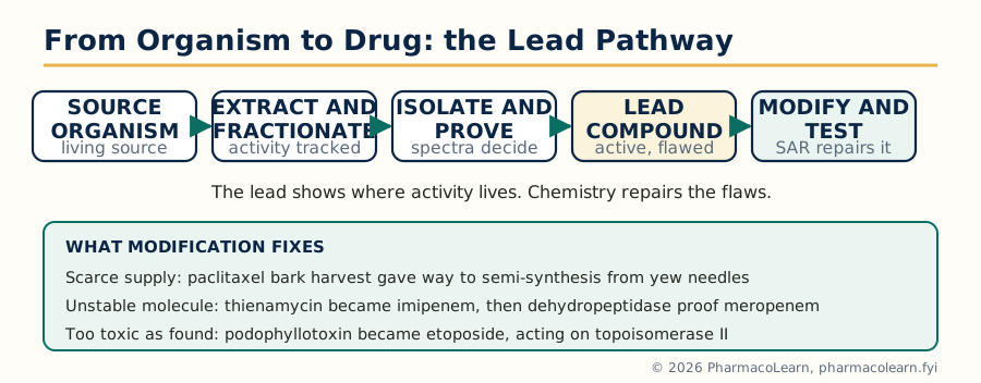 Figure: The natural product path from source organism to candidate drug, with the lead compound as the central hand over from biology to chemistry.
The honest limitation is the mirror image of the advantage: complex natural molecules are hard to make, hard to modify at one position only, and often bind several targets. The history of this unit is the history of chemists paying that price only where the activity was worth it.
1.2 CNS drugs: the morphine alkaloids
Source and chemistry. Opium is the dried latex of the unripe seed capsule of the opium poppy, Papaver somniferum. It contains a family of benzylisoquinoline derived alkaloids, of which morphine (about 10 percent of opium) is the principal analgesic, with codeine, thebaine, papaverine and noscapine alongside. Morphine, C17H19NO3, is a phenanthrene alkaloid: three fused six membered rings and a dihydrofuran oxygen bridge, a phenolic hydroxyl, a secondary alcoholic hydroxyl, and a basic tertiary nitrogen held in a piperidine ring (the full structure proof is in Unit 2, section 2.6).
Action. Morphine is an agonist at opioid receptors, principally the mu receptor, in the brain, spinal cord and gut. Receptor activation reduces pain signalling and produces analgesia, sedation and euphoria, and also the adverse pattern that limits it: respiratory depression, constipation, nausea, tolerance and dependence.
The lead at work. Morphine's modification history is the textbook of medicinal chemistry:
| Compound | Change from morphine | Result |
|---|---|---|
| Codeine | Phenolic OH methylated (3-O-methyl) | Weaker oral analgesic and antitussive; a prodrug, converted by CYP2D6 to morphine |
| Diamorphine (heroin) | Both hydroxyls acetylated | Crosses into the brain faster, then hydrolyses to morphine; potency and abuse liability both rise |
| Oxycodone, hydrocodone | Ring changes from the thebaine series | Oral analgesics of the same family |
| Nalorphine | N-methyl replaced by N-allyl | Mixed agonist and antagonist; showed the path to pure antagonists |
| Naloxone, naltrexone | N-allyl or N-cyclopropylmethyl, 14-hydroxy series | Pure opioid antagonists: naloxone reverses overdose, naltrexone is used in dependence |
| Etorphine | Bulky lipophilic side chain (from thebaine) | Extraordinary potency, used only in veterinary large animal work |
The SAR lesson is permanent: small changes at the nitrogen switch the same skeleton between agonist and antagonist, because the receptor reads the substituent as part of the activation signal.
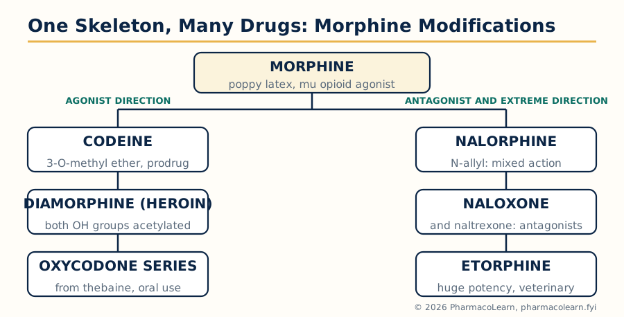 Figure: Morphine as a lead: small edits at the oxygen and nitrogen positions produce agonists, antagonists and extremes of potency from one skeleton.
1.3 Anticancer drugs: taxanes and podophyllotoxin derivatives
Paclitaxel and docetaxel. Paclitaxel (Taxol) was isolated from the bark of the Pacific yew, Taxus brevifolia, in a US screening programme; stripping bark kills the tree, and the original supply crisis was solved chemically: the abundant needle constituent 10-deacetylbaccatin III, from the renewable yew Taxus baccata, is converted by semi-synthesis into paclitaxel, and the same route with a different side chain gives docetaxel. Both are diterpenoids of the taxane family (Unit 3). Their mechanism is the opposite of the vinca alkaloids: where vincristine prevents microtubule assembly, paclitaxel stabilises the microtubule polymer and prevents the disassembly a dividing cell needs, so mitosis arrests and the cell dies. Clinically the taxanes are mainstays in ovarian, breast and lung cancers; the price of the mechanism is neutropenia and peripheral neurotoxicity, and paclitaxel's poor solubility historically required a castor oil derived vehicle.
Etoposide and teniposide. Podophyllotoxin is a lignan from the roots and rhizomes of Podophyllum species (the American mayapple, Podophyllum peltatum, and the Himalayan Podophyllum hexandrum). The natural compound itself binds tubulin and is too toxic to the gut for systemic use. Semi-synthetic glycosidic modification gave etoposide and teniposide, and the modification also changed the target: these derivatives act mainly by stabilising the complex between topoisomerase II and DNA, so the enzyme leaves double strand breaks behind. Etoposide is used in testicular, lung and blood cancers. Two exam points deserve emphasis: a natural lead's derivative can work by a different mechanism than the lead, and glycosylation here was a property fix (solubility and handling) that turned into a pharmacology change.
1.4 Cardiovascular drugs: lovastatin, teprotide and dicoumarol
Lovastatin. Lovastatin is a fungal polyketide metabolite, isolated from Aspergillus terreus (a close relative, mevastatin, came first from Penicillium species). It is given as a lactone, which hydrolyses in the body to the open beta-hydroxy acid; that acid is a competitive inhibitor of HMG-CoA reductase, the rate limiting enzyme of cholesterol synthesis, because it mimics the enzyme's natural intermediate. The result is reduced hepatic cholesterol, increased LDL receptor expression, and lower blood LDL. The whole statin class descends from this fungal lead: the later members (simvastatin by simple modification, then fully synthetic atorvastatin and rosuvastatin) kept the enzyme binding pharmacophore the natural product revealed.
Teprotide and the ACE inhibitors. Teprotide is a nonapeptide from the venom of the Brazilian pit viper Bothrops jararaca. Venom peptides that potentiated bradykinin were found to inhibit the angiotensin converting enzyme (ACE), and the peptide itself lowered blood pressure but could not be given orally. Studying how its C-terminal proline and carboxylate bound the enzyme's zinc guided the design of captopril, the first oral ACE inhibitor: a small sulfhydryl compound built to present the same binding features. Teprotide never became a drug; it is one of the purest examples in this course of a natural product whose value was the information it carried.
Dicoumarol and the coumarin anticoagulants. Cattle fed spoiled sweet clover (Melilotus species) developed a haemorrhagic disease; the cause was traced to dicoumarol, a bis(4-hydroxycoumarin) formed when fungi act on the plant's coumarin. Dicoumarol antagonises vitamin K, blocking the gamma-carboxylation that activates clotting factors II, VII, IX and X. It was used clinically, and its structure led to the simpler synthetic warfarin, for decades the standard oral anticoagulant, whose action is now known to centre on inhibition of the vitamin K epoxide reductase complex (VKORC1).
1.5 Neuromuscular blocking drugs: the curare alkaloids
Curare is the arrow poison of South American hunting peoples, prepared from plants such as Chondrodendron tomentosum and Strychnos toxifera. Its principal alkaloid, tubocurarine, is a bulky benzylisoquinoline alkaloid carrying two nitrogen atoms, one quaternary. Tubocurarine is a competitive antagonist at the nicotinic acetylcholine receptor of the neuromuscular junction: it occupies the receptor without activating it, so the muscle cannot be signalled to contract. Surgeons turned the poison into a tool: controlled, reversible paralysis for anaesthesia.
Tubocurarine's faults (ganglion blockade, histamine release, long and variable action) drove the analog programme: atracurium was designed to self-destruct by Hofmann elimination at body pH and temperature, giving predictable offset in organ failure; vecuronium and rocuronium are steroidal neuromuscular blockers built on an androstane skeleton carrying the same two nitrogen pharmacophore, a bridge to Unit 2's steroid chemistry. The lesson: the pharmacophore of a natural poison can be transplanted onto a wholly different, better behaved skeleton.
1.6 Antimalarial drugs and analogues
Quinine and the synthetic quinolines. Quinine, from the bark of the Cinchona tree, was the first effective antimalarial and is a quinoline methanol alkaloid whose structure (Unit 2 territory) set the template for the synthetic 4-aminoquinolines, above all chloroquine: the quinoline nucleus concentrates in the parasite's food vacuole and blocks the detoxification of haem into haemozoin, so free haem kills the parasite. Widespread chloroquine resistance ended its reign in most regions, but the natural product had defined both target space and scaffold.
Artemisinin and its derivatives. Artemisinin (qinghaosu) was isolated from Artemisia annua, a herb of the Chinese materia medica, by the group of Tu Youyou, who found that a low temperature ether extraction preserved the activity that hot preparations destroyed. Chemically it is a sesquiterpene lactone containing an endoperoxide bridge, the essential feature: inside the parasite, iron from digested haemoglobin cleaves the peroxide and generates carbon centred radicals that damage parasite proteins and membranes. Artemisinin itself is poorly soluble in both water and oil and has a short half life, so the drugs used are its derivatives: dihydroartemisinin (reduction of its lactone carbonyl), artemether (its methyl ether) and artesunate (a water soluble hemisuccinate ester for injection). Artemisinin based combination therapies (ACTs), pairing a fast artemisinin derivative with a longer acting partner drug, are the World Health Organization recommended first line treatment for Plasmodium falciparum malaria; emerging partial resistance in several regions is the standing warning of this section.
1.7 Antibiotics I: the macrolides
Erythromycin, from the soil actinomycete Saccharopolyspora erythraea, is built on a 14 membered macrocyclic lactone ring (erythronolide) carrying two sugars, one of them an amino sugar. It binds the 50S bacterial ribosomal subunit at the 23S rRNA of the peptide exit tunnel and blocks translocation of the growing peptide chain. Erythromycin is acid labile: stomach acid degrades it to inactive products, and its bitterness and gut motility effects limit it.
The semi-synthetic answers are a model exam answer:
| Drug | Modification | What it fixed |
|---|---|---|
| Clarithromycin | 6-O-methylation of erythromycin | Blocks the acid degradation pathway; better stability, exposure and tissue levels |
| Roxithromycin | Oxime ether extension at C-9 | Improved acid stability and pharmacokinetics |
| Azithromycin | Ring expanded to 15 members, nitrogen inserted (an azalide) | Long tissue half life, once daily short courses, strong intracellular and tissue penetration |
The ribosome reads all four almost identically; the patient experiences four quite different drugs. That is molecular modification doing pharmacokinetic, not pharmacodynamic, work.
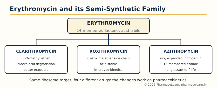 Figure: The macrolide family: three semi-synthetic repairs of erythromycin, all aimed at stability and exposure rather than a new target.
1.8 Antibiotics II: beta-lactams, cephalosporins and carbapenems
All beta-lactam antibiotics share the strained four membered beta-lactam ring, whose carbonyl acylates the active site serine of bacterial penicillin binding proteins (PBPs), the enzymes that cross link peptidoglycan; the wall weakens and the bacterium lyses. The penicillins themselves are the subject of the spectral study in Unit 5; this section covers the two families the syllabus names beside them.
Cephalosporins descend from cephalosporin C, from the fungus Acremonium chrysogenum (formerly Cephalosporium acremonium). Its nucleus, 7-aminocephalosporanic acid (7-ACA), differs from the penicillin nucleus by a six membered dihydrothiazine ring fused to the beta-lactam, and it offers two positions for modification: the 7-acyl side chain (spectrum and beta-lactamase stability) and the 3-substituent (pharmacokinetics and some spectrum). Successive clinical generations traded on these positions: early members stronger against Gram positive cocci, later generations reaching Gram negative bacilli including, for ceftazidime and cefepime, Pseudomonas, and ceftaroline regaining activity against MRSA. The natural nucleus was a platform, not a finished drug.
Carbapenems descend from thienamycin, from Streptomyces cattleya: the same beta-lactam warhead on a five membered ring in which carbon replaces the sulfur of penicillin, with a trans hydroxyethyl side chain that confers remarkable stability to most beta-lactamases. Thienamycin itself was too unstable in concentrated solution and is degraded in the kidney by dehydropeptidase I. The fixes define the family: imipenem (a stabilised N-formimidoyl derivative) is given with cilastatin, an inhibitor of that renal enzyme; meropenem carries a 1-beta-methyl group that makes it intrinsically stable to dehydropeptidase, needing no partner. Carbapenems hold the broadest spectrum of the beta-lactams and are reserved accordingly, which is why carbapenem resistant organisms are a first order clinical threat.
Unit 2: Alkaloids, Flavonoids and Steroids
2.1 Alkaloids: introduction and classification
Alkaloids are naturally occurring nitrogen containing compounds, mostly of plant origin, that are basic and usually physiologically potent. The classic definition carries standard exceptions worth quoting in an exam: colchicine is barely basic, some alkaloids come from animals, fungi and microbes (ergot, Unit 1's venom peptides aside), and the nitrogen of piperine sits in an amide. Most alkaloids are biosynthesised from amino acids, and that fact organises the whole family.
| Class (by precursor) | Ring skeleton | Examples from this course |
|---|---|---|
| Ornithine and lysine derived | Tropane, pyrrolizidine, quinolizidine | Hyoscyamine and atropine, cocaine (tropane) |
| Phenylalanine and tyrosine derived | Benzylisoquinoline, phenanthrene, aporphine | Morphine, codeine, tubocurarine, papaverine, emetine, berberine |
| Tryptophan derived | Indole, beta-carboline, quinoline (via strictosidine) | Reserpine, ergot alkaloids, strychnine, quinine, vinca alkaloids |
| Histidine derived | Imidazole | Pilocarpine |
| Purine and pseudoalkaloids | Xanthine, steroid and terpene skeletons with nitrogen added late | Caffeine, solasodine, aconitine |
An older chemical classification simply names the heterocyclic ring present (pyrrole, pyridine, isoquinoline, indole, imidazole, purine). Both schemes survive because both answer real questions: the ring scheme helps analysis and synthesis, the precursor scheme explains why related structures occur together in a plant. Alkaloids occur in the plant largely as salts of plant acids (oxalic, tannic, citric) or bound to tannins, concentrated in particular organs: bark, root, seed or latex depending on species.
2.2 Isolation and purification of alkaloids
Alkaloid isolation is applied acid-base chemistry. The free base is soluble in organic solvents and poorly soluble in water; the salt reverses both. The working sequence:
ALKALOID SEPARATION BY ACID-BASE SWITCHING
  Powdered plant material
        |
  Extract with dilute acid        (alkaloids dissolve as salts)
  or with alcohol, then acidify
        |
  Filter, concentrate
        |
  Basify with ammonia or          (free bases liberated)
  sodium carbonate
        |
  Extract into chloroform,        (alkaloid bases move organic;
  dichloromethane or ether         tannins and salts stay aqueous)
        |
  Wash, dry, evaporate
        |
  Separate the mixture            (fractional crystallisation of salts,
        |                           column and preparative chromatography,
        |                           counter current distribution)
  Pure alkaloid                   (constant melting point, single spot
                                   on TLC, spectral identity)
Purification details examiners favour: salts differ in solubility (picrates, oxalates, tartrates crystallise well), so a salt is often made to be recrystallised and then broken back to the base; vacuum distillation serves the few liquid alkaloids such as nicotine and coniine; and modern practice follows every fraction by TLC and increasingly by LC-MS, which is bioassay guided fractionation (Unit 1) with better eyes. Identity is fixed by melting point, mixed melting point with an authentic sample, and the spectra of Unit 5.
2.3 Molecular modification and biological activity of alkaloids
Because alkaloid skeletons are potent and complex, modification usually means semi-synthesis: keep the skeleton, edit its edges. The morphine series (Unit 1) showed methylation, acetylation and N-substitution producing a whole pharmacopoeia from one skeleton. General principles:
- Quaternary salts (as in tubocurarine) stay out of the brain and act peripherally; tertiary bases cross membranes and reach the CNS. Basicity and charge are distribution switches.
- N-demethylation often gives more polar, differently active metabolites (nortropane and normorphine series); N-alkyl size tunes agonist to antagonist character at opioid receptors.
- Ester hydrolysis and exchange changes duration: cocaine's benzoyl ester is its activity, and its hydrolysis products are inactive, which is also why local anaesthetic design copied its pharmacophore synthetically (procaine, lignocaine) discarding the tropane ring entirely.
- Stereochemistry is decisive: the alkaloid families are heavily stereogenic, and one epimer may be drug and the other near inert (section 2.5 and 2.9).
- Activity targets cluster: neurotransmitter receptors (nicotine, morphine), transporters (cocaine, reserpine), enzymes (physostigmine at acetylcholinesterase) and structural proteins (colchicine and the vinca alkaloids at tubulin). A potent alkaloid almost always has a protein it was evolved to hit.
2.4 General methods of structural determination
The classical proof of an alkaloid's structure, the method this unit's five case studies used, ran in a fixed order:
- Purity and formula: elemental analysis and molecular weight fix the molecular formula; today HRMS does both in one measurement.
- Functional group inventory: tests and spectra for hydroxyl, phenol, methoxyl (Zeisel), N-methyl (Herzig-Meyer), methylenedioxy, olefinic bonds, ester and lactone groups; the degree of unsaturation from the formula tells how many rings and multiple bonds must be accounted for.
- Skeleton by degradation: controlled reactions that break the molecule at informative points: Hofmann exhaustive methylation opens nitrogen rings and counts ring attachments to nitrogen; Emde and von Braun degradations cut C-N bonds differently; zinc dust distillation, sulfur or selenium dehydrogenation strips hydrogens to reveal an aromatic parent hydrocarbon (a phenanthrene from the morphine series, a carbazole or beta-carboline from indole alkaloids); oxidation with permanganate or chromic acid cuts side chains to identifiable acids; hydrolysis separates esters, glycosides and amides into known halves.
- Assembly: the fragments are fitted into one structure consistent with every degradation and with the biosynthetic logic of the family.
- Proof by synthesis: total synthesis of the proposed structure, identical in all properties (including spectra and optical rotation) with the natural compound, was the classical gold standard; Gates's morphine synthesis confirmed the morphine structure in this way.
- Stereochemistry: relative configuration from chemical interconversions and coupling behaviour, absolute configuration by correlation with compounds of known configuration and, in the modern era, by X-ray crystallography of salts.
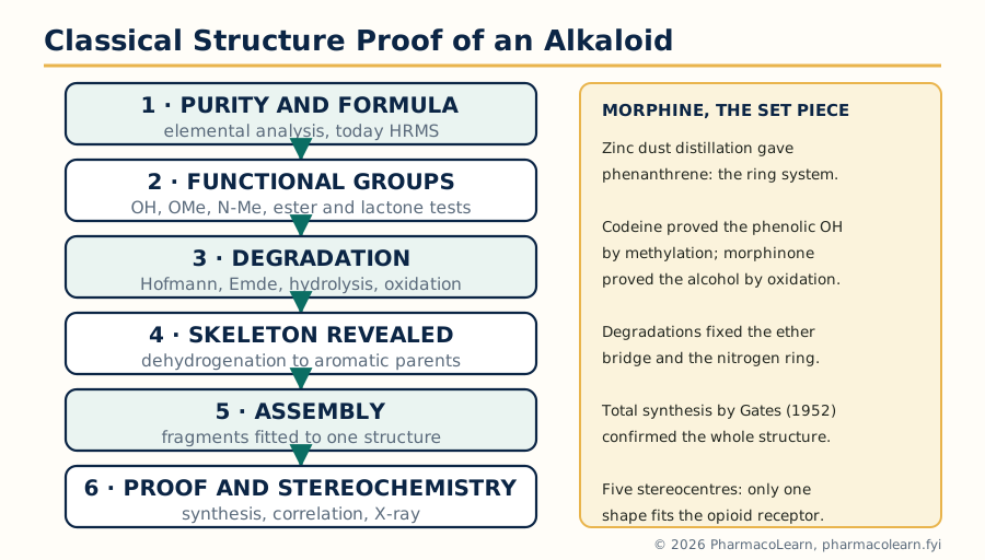 Figure: The six steps of a classical alkaloid structure proof, with morphine as the worked historical example.
Modern spectroscopy (Unit 5) now obtains in days what degradation took years to win, but examiners still teach the classical logic because it is the clearest training in structural reasoning: every reaction is an experiment whose result the proposed structure must explain.
2.5 Ephedrine: structure and stereochemistry
Ephedrine, the principal alkaloid of Ephedra species (ma huang in Chinese medicine), is a phenylpropanolamine, C10H15NO: a benzene ring bearing a three carbon chain with a hydroxyl on the benzylic carbon (C-1) and a methylamino group on C-2. It is a protoalkaloid: the nitrogen is not in a ring. Structurally it is the plant world's close copy of adrenaline, missing only the catechol hydroxyls, which is why it acts as a mixed sympathomimetic, releasing noradrenaline and stimulating adrenergic receptors directly, and why removing the ring hydroxyls makes it orally active and longer acting than adrenaline itself.
Elucidation logic. Oxidation gave benzoic acid, fixing a monosubstituted benzene with an oxidisable side chain. The formula and functional tests accounted for one secondary alcohol and one secondary amine. Degradation and synthesis from benzaldehyde based routes confirmed the chain as Ph-CHOH-CH(NHMe)-CH3.
Stereochemistry is the examinable core. Two stereocentres (C-1 and C-2) give four stereoisomers: the natural product is (1R,2S)-ephedrine, levorotatory; its C-1 epimer, (1S,2S), is pseudoephedrine, a different drug (a milder nasal decongestant) despite the identical constitution. The pair teach the permanent lesson that diastereomers are different substances with different pharmacology, and that a natural product structure is not fully stated until configuration is stated.
2.6 Morphine: structural elucidation and stereochemistry
Morphine's proof (Gulland and Robinson, 1923 to 25, confirmed by Gates's total synthesis, 1952) remains the classical set piece.
The evidence trail:
- Formula and groups: C17H19NO3; one phenolic hydroxyl (soluble in alkali, gives colour reactions, methylates to codeine), one secondary alcoholic hydroxyl (oxidises to the ketone morphinone), one inert ether oxygen, one tertiary N-methyl base. Codeine is morphine's 3-methyl ether, and heroin its diacetyl derivative (Unit 1), relations proved by hydrolysis back to morphine.
- The phenanthrene skeleton: exhaustive methylation and degradation gave trimethylamine and nitrogen free fragments; zinc dust distillation of the alkaloid gave phenanthrene itself, proving the three fused ring carbocyclic system.
- Locating the oxygen bridge and the nitrogen ring: a long campaign of degradations (the morphine to apomorphine rearrangement under acid, Hofmann degradations of codeine derivatives to compounds such as morphenol, a phenanthrene bearing the ether oxygen) fixed the dihydrofuran bridge across the ring junction and the ethanamine bridge that makes the piperidine ring.
- The double bond: uptake of one molar equivalent of hydrogen and the behaviour on oxidation placed one olefinic bond in the third ring, allylic to the alcohol.
Stereochemistry. Morphine has five stereogenic centres (C-5, C-6, C-9, C-13, C-14) and a rigid, T-shaped three dimensional structure; the absolute configuration was established by correlation and confirmed by synthesis, and synthetic efforts that produced the mirror skeleton produced inactive material: the opioid receptor accepts one shape only. For the spectral re-examination of morphine see Unit 5, section 5.4.
2.7 The ergot alkaloids
Ergot is the dark sclerotium of the fungus Claviceps purpurea growing on rye and other grasses; its alkaloids explained both the historical poisonings of ergotism and a rich pharmacology. All share lysergic acid, a tetracyclic ergoline structure built from tryptophan, with an indole ring fused into a four ring system bearing a carboxyl group whose substituent divides the family:
- Peptide ergot alkaloids: ergotamine, ergocristine and relatives, in which lysergic acid is joined to a tripeptide; complex, poorly soluble, vasoconstrictor.
- Simple amide: ergometrine (ergonovine), lysergic acid joined to alaninol; water soluble, the oxytocic used against postpartum haemorrhage.
- Clavines: smaller relatives such as agroclavine, intermediates on the biosynthetic path.
Elucidation proceeded by hydrolysis: every member yields lysergic acid (or its C-8 epimer, isolysergic acid, an easy isomerisation that complicated early work and is itself an exam favourite), plus ammonia and, for the peptide series, identifiable amino acids and pyruvic acid from the unusual tripeptide. Pharmacology follows the family chemistry: partial agonism at serotonin, adrenergic and dopamine receptors in different proportions, hence uses from migraine (ergotamine) to prolactin suppression by the semi-synthetic bromocriptine, and hence also the toxicity that pushed practice towards the synthetic triptans. The semi-synthetic lysergic acid diethylamide (LSD) is the famous cautionary member of the series.
2.8 Emetine
Emetine is the principal alkaloid of ipecacuanha, the root of Carapichea ipecacuanha (long named Cephaelis ipecacuanha), accompanied by cephaeline, its phenolic relative. Emetine is a bis-isoquinoline alkaloid: two 6,7-dimethoxy tetrahydroisoquinoline units linked through a substituted ethyl bridge, with four methoxyl groups, one secondary and one tertiary nitrogen, and four stereogenic centres whose configuration was settled by degradation to known fragments and final synthesis.
Elucidation headlines: methoxyl estimation fixed four OMe groups; oxidation and Hofmann type degradations split the molecule into isoquinoline fragments, from which the two halves and their junction were reconstructed. Pharmacologically emetine inhibits protein synthesis by blocking ribosomal translocation in both parasite and host: historically the drug of choice for invasive amoebiasis and, in syrup of ipecac, an emetic; both uses have retreated before safer agents (metronidazole for amoebiasis), making emetine a clean case of a natural drug retired by its own therapeutic index rather than by loss of activity.
2.9 Reserpine
Reserpine, from the root of Rauwolfia serpentina (sarpagandha, used in Ayurvedic practice for agitation and hypertension), is a complex indole alkaloid of the yohimbane family, built from tryptamine and the monoterpene secologanin. Its structure carries an indole fused pentacyclic system with a methoxycarbonyl group and a 3,4,5-trimethoxybenzoyl ester on the E ring: hydrolysis therefore yields reserpic acid, methanol and trimethoxybenzoic acid, the first degradation result from which the assembly was worked out. The molecule has six stereogenic centres, and the configuration at C-3, set by the biosynthetic coupling, is critical: its epimer, isoreserpine, is inactive, another stereochemistry decides activity case for the file beside ephedrine.
Reserpine irreversibly blocks the vesicular monoamine transporters (VMAT) that load noradrenaline, dopamine and serotonin into storage vesicles; the transmitters are then destroyed by monoamine oxidase, sympathetic tone falls, and blood pressure falls with it. It was among the first effective antihypertensives, and its mood lowering side effect, depression in a significant minority of patients, both limited its use and historically seeded the monoamine hypothesis of depression. It survives today mainly as a pharmacological tool and a teaching structure.
2.10 Flavonoids: introduction, isolation and purification
Flavonoids are the yellow and coloured polyphenols of the plant world: a C6-C3-C6 skeleton in which two benzene rings (A and B) are joined by a three carbon unit that usually closes into an oxygen heterocycle (the C ring). They are biosynthesised from the shikimate pathway (the B ring and the three carbons, via a cinnamate) condensed with three acetate units (the A ring). The oxidation level and substitution of the C ring define the classes:
| Class | C ring character | Examples |
|---|---|---|
| Flavones | 2,3 double bond, 4-keto, no 3-OH | Apigenin, luteolin |
| Flavonols | Flavone plus 3-OH | Quercetin, kaempferol, myricetin |
| Flavanones | No 2,3 double bond, 4-keto | Naringenin, hesperetin |
| Flavanols (catechins) | No 4-keto, 3-OH | Catechin, epicatechin |
| Anthocyanidins | Flavylium cation, coloured | Cyanidin, delphinidin |
| Isoflavones | B ring at C-3 instead of C-2 | Genistein, daidzein |
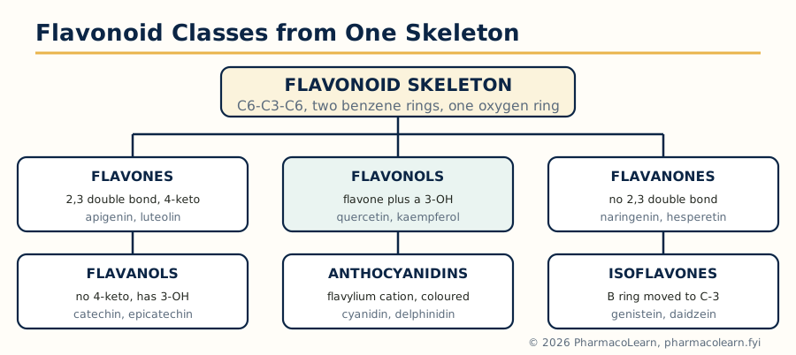 Figure: The flavonoid classes differ only in the oxidation pattern of the central oxygen ring built on the C6-C3-C6 skeleton.
In the plant they occur as glycosides (O-glycosides at the 3 or 7 hydroxyl usually; the aglycone is the sugar free part). Isolation: extract with aqueous alcohol or methanol, concentrate, clarify with lead acetate solution (which precipitates interfering tannins and acids, carefully, since it can carry flavonoids too), and separate by column, paper or preparative thin layer chromatography; glycosides are crystallised from alcohol, and hydrolysis with dilute acid releases the aglycone plus identifiable sugars. Flavonoids give diagnostic colour reactions used throughout their classical chemistry: the Shinoda test (magnesium and hydrochloric acid, pink to red for most classes), colour with ferric chloride, yellow solutions in alkali that intensify on standing, and fluorescence changes on chromatograms sprayed with aluminium chloride.
2.11 Structural determination of flavonoids and the structural elucidation of quercetin
The classical flavonoid proof combines spectroscopy with controlled destruction:
- UV spectroscopy with shift reagents was the great classical fingerprint: Band II (240 to 285 nm, A ring) and Band I (300 to 400 nm, B ring and cinnamoyl system) move in diagnostic directions with sodium methoxide (free 4 prime hydroxyl), sodium acetate (free 7 hydroxyl), aluminium chloride, stable to acid if a 5-hydroxyl or ortho dihydroxyl system is present. Methylation analysis (methylate, hydrolyse, identify which hydroxyls were free) locates glycosylation.
- Alkaline degradation: fusion or strong alkali splits a flavonoid at the C ring into an A ring fragment (usually a phloroglucinol or resorcinol derivative) and a B ring fragment (a benzoic or cinnamic acid), identifying the hydroxylation pattern of each ring separately.
- Hydrolysis and sugar analysis identify glycosides; NMR and MS (Unit 5) complete the modern proof.
Quercetin worked fully: formula C15H10O7; five acetylatable hydroxyls (pentaacetate); methylation then degradation gave phloroglucinol from the A ring and protocatechuic acid (3,4-dihydroxybenzoic acid) from the B ring, fixing the B ring diol pattern; alkaline fusion products and the flavonol colour and UV behaviour (free 3-hydroxyl shown by its aluminium chloride complex and by ready oxidation) place hydroxyls at 3, 5, 7, 3 prime and 4 prime. Quercetin is therefore 3,5,7,3 prime,4 prime-pentahydroxyflavone, and it occurs in plants chiefly as glycosides: rutin (3-O-rutinoside, from rue and buckwheat), quercitrin (3-O-rhamnoside) and isoquercitrin (3-O-glucoside). Onion, apple, tea and many leafy vegetables supply it in the diet; its antioxidant activity in vitro is strong, its oral bioavailability limited, and examiners expect that measured verdict rather than supplement advertising.
2.12 Steroids: sterols, sapogenins and cardiac glycosides
The steroid nucleus is the cyclopentanoperhydrophenanthrene (gonane) system: three six membered rings (A, B, C) fused to a five membered ring (D), carrying angular methyl groups at C-10 and C-13 in most natural members. Everything in the steroid industry is a variation on this frame.
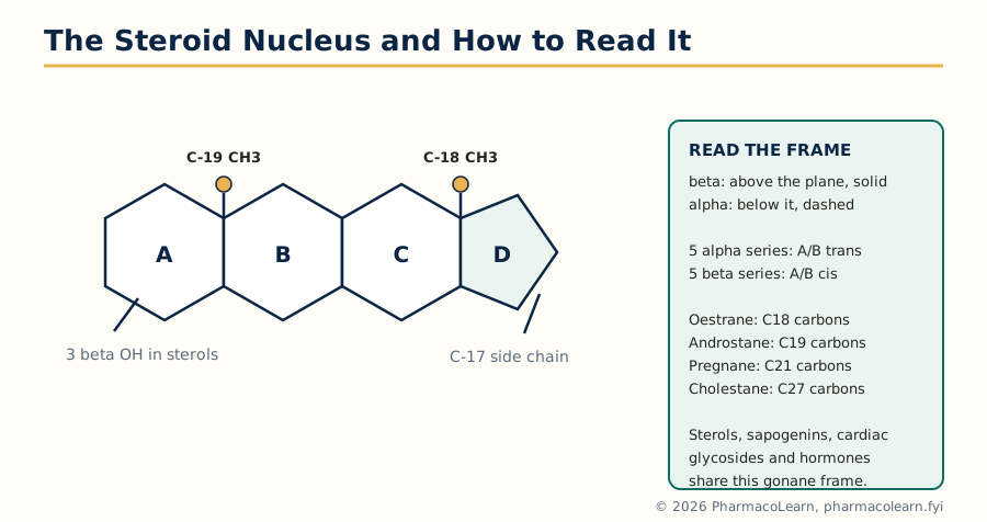 Figure: The gonane steroid nucleus: rings A to D, the angular methyl groups, and the alpha and beta convention used throughout steroid nomenclature.
Sterols are steroid alcohols with a 3 beta hydroxyl and a side chain at C-17: cholesterol in animals, beta-sitosterol and stigmasterol in plants, ergosterol in fungi (the vitamin D2 precursor, section 2.14). Cholesterol is the biosynthetic parent of every steroid hormone and of the bile acids.
Sapogenins are the aglycones of the saponins, glycosides that froth in water and haemolyse red cells. Steroidal sapogenins such as diosgenin (from Dioscorea species) carry a spiroketal side chain (the spirostane system). Diosgenin's industrial importance is hard to overstate: the Marker degradation converted it into progesterone and onward to cortisone and the contraceptive steroids, making yam derived diosgenin the feedstock that founded the steroid industry before total synthesis and fermentation routes matured.
Cardiac glycosides join a steroidal aglycone (genin) of the cardenolide type to sugars at the 3-hydroxyl. The genin carries an unsaturated five membered lactone (butenolide) ring at C-17 and a 14 beta hydroxyl; Digitalis purpurea gives digitoxin, Digitalis lanata gives digoxin (12 beta hydroxy digitoxin) and their relatives. They inhibit the sodium potassium ATPase, raising intracellular calcium in cardiac muscle: positive inotropy for heart failure, at a notoriously narrow therapeutic index the spectral study revisits in Unit 5.
2.13 Steroid nomenclature and stereochemistry
Steroid stereochemistry is described relative to the plane of the ring system. The angular methyls at C-10 and C-13 are taken as beta (above the plane, drawn solid), and any substituent is alpha (below, dashed) or beta by comparison. Parent hydrocarbon names encode the framework and the A/B ring junction:
| Parent | Feature |
|---|---|
| Oestrane | 18 carbons, no C-10 methyl, aromatic A ring in the hormone series |
| Androstane | 19 carbons, methyls at C-10 and C-13 |
| Pregnane | 21 carbons, androstane plus a two carbon side chain at C-17 |
| Cholestane | 27 carbons, full cholesterol side chain |
Within this frame: 5 alpha steroids have the A/B junction trans (H at C-5 alpha, rings extended and near planar in effect); 5 beta steroids have it cis (epimeric at C-5, the A ring bent under), which distinguishes the bile acid and cardiac glycoside series (5 beta) from the androstane hormone series (5 alpha). Hydroxyls are cited as 3 beta, 11 beta, 17 beta and so on; ketones as oxo positions (androstan-3-one); double bonds by locants with the Greek delta in older literature. A structure written as pregn-4-ene-3,20-dione is, by these rules alone, already nearly drawn: that is progesterone.
2.14 Sex hormones, contraceptive agents, adrenocorticoids and vitamin D
Testosterone is androst-4-en-17 beta-ol-3-one, the principal androgen, made in the testes from cholesterol through pregnenolone and dehydroepiandrosterone; its 5 alpha reduced metabolite dihydrotestosterone is the more potent androgen in prostate and skin. Oral testosterone is destroyed by first pass oxidation of the 17 beta hydroxyl, so the clinic uses 17 esters by injection or 17 alpha alkylated analogs (hepatotoxic, hence disfavoured).
Oestradiol (estradiol) is oestra-1,3,5(10)-triene-3,17 beta-diol: the A ring is aromatic, the C-10 methyl is absent (lost during aromatisation of an androgen precursor), and the phenolic 3-hydroxyl is its binding handle. Progesterone, pregn-4-ene-3,20-dione, is the natural progestogen of the corpus luteum and placenta, preparing and maintaining the endometrium; it is orally weak, which is why contraception was built from analogs.
Contraceptive agents. The combined oral contraceptive pairs an estrogen, ethinylestradiol (17 alpha ethinyl estradiol, the ethinyl group blocking 17 oxidation and so conferring oral activity), with a progestin: 19-nor steroids such as norethisterone and levonorgestrel (from the testosterone frame with the C-10 methyl removed and a 17 alpha ethinyl added) or later pregnane and spironolactone derived progestins such as drospirenone. The mechanism is endocrine feedback: suppression of the midcycle LH surge, so ovulation does not occur, with cervical mucus and endometrial effects in support. The whole family tree (sterol to diosgenin to progesterone to norethisterone) is the steroid industry's founding story, told in section 2.12.
Cortisone is the adrenal glucocorticoid frame: pregn-4-ene-17,21-diol-3,11,20-trione, its active form in tissue being the 11 beta alcohol cortisol (hydrocortisone), to which cortisone is reduced and back. The 11 oxygen function, so hard won in the early syntheses (the famous routes from bile acids and diosgenin existed largely to install it), is the glucocorticoid signature; modifications such as the 1,2 double bond (prednisone, prednisolone) and 9 alpha fluorine (dexamethasone, with a 16 methyl) raised anti-inflammatory potency stepwise.
Vitamin D belongs in this unit because it is a secosteroid: in skin, ultraviolet light opens the B ring of 7-dehydrocholesterol to give previtamin D3, which thermally isomerises to cholecalciferol (vitamin D3); ergosterol from fungi and yeast gives ergocalciferol (D2) the same way. Two hydroxylations, liver to calcidiol (25-OH) and kidney to calcitriol (1,25-(OH)2), convert it into a hormone regulating calcium and phosphate. Its chemistry is revisited spectroscopically in Unit 5 and nutritionally in Unit 3.
Unit 3: Terpenoids and Vitamins
3.1 Terpenoids: classification and isolation
The terpenoids (isoprenoids) are the largest family of natural products, built from five carbon isoprene units (C5H8) joined, with very few exceptions, head to tail. Carbon count therefore classifies the entire family at sight:
| Class | Isoprene units | Carbons | Examples from this unit |
|---|---|---|---|
| Hemiterpenoids | 1 | C5 | Isoprene itself, prenol |
| Monoterpenoids | 2 | C10 | Citral, menthol, camphor |
| Sesquiterpenoids | 3 | C15 | Artemisinin (Unit 1), farnesol |
| Diterpenoids | 4 | C20 | Retinol, phytol, taxol (paclitaxel) |
| Sesterterpenoids | 5 | C25 | A small marine and insect family |
| Triterpenoids | 6 | C30 | Squalene, ginsenosides, sterol precursors |
| Tetraterpenoids | 8 | C40 | Carotenoids, beta carotene |
| Polyterpenoids | Many | Long chains | Rubber, dolichols |
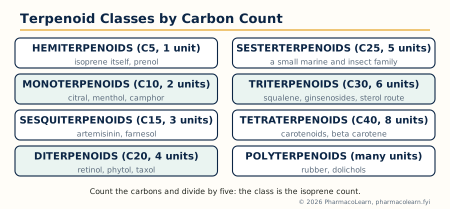 Figure: Terpenoid classification is carbon counting: each class is a whole number of C5 isoprene units.
Biosynthetically the C5 units are isopentenyl pyrophosphate (IPP) and its isomer dimethylallyl pyrophosphate (DMAPP), made by the mevalonate pathway in animals, fungi and plant cytosol, and by the methylerythritol phosphate (MEP) pathway in most bacteria and plant plastids; chain elongation and cyclisation by terpene synthases generate the family, and squalene (section 3.5) is the junction to the steroids.
Isolation. The volatile mono and sesquiterpenoids of essential oils are obtained by steam distillation or expression of the plant material, then separated by fractional distillation under reduced pressure and by chromatography; sensitive members demand low temperatures and protection from air, because terpenes oxidise and polymerise readily (old turpentine is the household example). The higher, non volatile terpenoids are solvent extracted and purified by column chromatography and crystallisation in the usual way of section 2.2.
3.2 The isoprene rule and general methods of structural elucidation
Wallach's isoprene rule observes that terpene skeletons can be dissected into isoprene units; Ruzicka's biogenetic isoprene rule goes further and proposes that all terpenoids arise from a few acyclic precursors (geraniol for C10, farnesol for C15, geranylgeraniol for C20, squalene for C30) by cyclisation, rearrangement and oxidation, so a proposed structure must be a plausible child of its acyclic parent. The rule is both a proof tool and a biosynthesis map: menthol and camphor must both be constructible from a geraniol chain folded two different ways, and they are.
TWO FOLDINGS OF ONE CHAIN: GERANIOL TO MENTHOL AND CAMPHOR
 Acyclic start: geraniol (C10, head to tail isoprene units)
        |
  Fold and ring close between C-1 and C-6
        |--- menthol line: keep the single ring, reduce and
        |    set three stereocentres (p-menthane skeleton)
        |
        |--- camphor line: second closure builds the bridged
             bornane skeleton, oxidise one carbon to the ketone
General elucidation methods follow the Unit 2 logic with terpene specific tools: formula and unsaturation account; catalytic hydrogenation counts double bonds; ozonolysis and oxidative cleavage cut at double bonds and identify the carbonyl fragments, mapping the chain; dehydrogenation (sulfur, selenium, palladium) converts cyclic terpenes to known aromatic compounds (p-cymene from many monoterpenes), exposing the carbon skeleton; functional group chemistry and spectra complete the proof, and synthesis or X-ray analysis confirms. Where the molecule is an oil that will not crystallise, solid derivatives (semicarbazones of carbonyls, esters of alcohols) were the classical handles, a trick that recurs in the camphor and citral proofs below.
3.3 Monoterpenoids: citral, menthol and camphor
Citral is the lemon scented aldehyde of lemongrass and lemon peel oils, in fact a pair of geometric isomers: geranial (citral a, E) and neral (citral b, Z), an acyclic monoterpene, 3,7-dimethylocta-2,6-dienal. The proof pattern: formula C10H16O, aldehyde reactions andready semicarbazone formation; oxidation and ozonolysis fragments (acetone from the isopropylidene end, levulinic acid derivatives from the chain) placed the two double bonds and the methyl branches exactly as the isoprene dissection predicts. Citral is also an industrial intermediate: its cyclisation chemistry underlies ionone and vitamin A synthesis.
Menthol is the cooling alcohol of peppermint oil (Mentha piperita and related), a saturated cyclic monoterpene alcohol of the p-menthane skeleton (a cyclohexane ring bearing methyl and isopropyl groups para to each other, hydroxyl at C-3 old numbering). Three stereogenic centres give eight stereoisomers, of which natural (minus)-menthol, (1R,2S,5R), is the potent cooling agent acting at the TRPM8 cold receptor; the others are weaker or differently flavoured, ephedrine's lesson (section 2.5) again. Oxidation of menthol gives menthone, the corresponding ketone, and dehydrogenation gives p-cymene, tying the skeleton proof together.
Camphor is the bridged bicyclic ketone of Cinnamomum camphora: 1,7,7-trimethylbicyclo[2.2.1]heptan-2-one, the bornane skeleton, a cyclisation of the geraniol chain that builds a bridge and so escapes flat ring chemistry. Its classical proof (Bredt, 1893) rested on oxidation to camphoric acid, a dicarboxylic acid whose own degradations mapped the ring system carbon by carbon; camphor became one of the earliest structures proved against serious alternatives and later a standard chiral starting material and resolving agent. Its IR carbonyl, unusually high for a ketone because of the strained ring, returns in Unit 5.
3.4 Diterpenoids: retinol, phytol and taxol
Retinol is vitamin A alcohol, treated chemically here and physiologically in section 3.7. A C20 molecule built of four isoprene units ending in a beta-ionone ring, with a conjugated pentaene side chain terminating in a primary alcohol; the all trans form is the natural standard. The conjugated system is its identity (intense UV absorption, the Carr-Price blue colour with antimony trichloride) and its weakness: light, air and heat isomerise and oxidise it, which is why vitamin A preparations are protected and why its aldehyde (retinal) and acid (retinoic acid) partners interconvert in vision and in gene regulation respectively.
Phytol is the acyclic diterpene alcohol, 3,7,11,15-tetramethylhexadec-2-en-1-ol, obtained by hydrolysis of chlorophyll, whose phytyl ester side chain it is. Only one double bond and one alcohol: a deliberately plain molecule whose elucidation (Fischer's school) by stepwise oxidation proved the regular head to tail assembly of four isoprene units and anchored the isoprene rule for the diterpene class. In industry phytol is a starting material for vitamins E and K synthesis.
Taxol (paclitaxel) returns from Unit 1 as the diterpenoid case study: a taxane skeleton of an eight membered ring fused into a strained tricyclic system with an oxetane ring, heavily oxygenated, bearing the N-benzoyl phenylisoserine side chain that its microtubule binding requires. Its elucidation (structure published 1971 by Wani and Wall) used exactly the classical armamentarium: degradation to baccatin fragments, spectroscopy and derivatisation, on milligram quantities of a molecule with eleven stereogenic centres. The supply sequel, semi-synthesis from 10-deacetylbaccatin III, is in section 1.3.
3.5 Triterpenoids: squalene and the ginsenosides
Squalene is the acyclic C30 triterpene hydrocarbon, six isoprene units joined with a centre of symmetry where two farnesyl units meet tail to tail. Isolated first from shark liver oil (Squalus), it occurs in olive oil, human sebum and every eukaryote that makes sterols, because it is the sterol precursor: squalene epoxidase and oxidosqualene cyclase fold and cyclise squalene 2,3-epoxide into lanosterol (in animals and fungi) or cycloartenol (in plants), and nineteen further steps trim lanosterol to cholesterol. Structure proof rested on its formula C30H50, six double bonds by hydrogenation, and oxidative fragments consistent with the symmetrical chain; its cyclisation products, predicted by the biogenetic isoprene rule with a chair boat chair folding, were found exactly as predicted, one of the rule's great confirmations.
Ginsenosides are the saponins of ginseng root (Panax ginseng and Panax quinquefolius): glycosides of triterpene aglycones, chiefly of the dammarane type (protopanaxadiol and protopanaxatriol series), named by their chromatography (Rb1, Rb2, Rc, Rd from the diol series; Re, Rg1 from the triol series). Acid or enzymatic hydrolysis gives the aglycone plus glucose, rhamnose, arabinose and xylose in member specific patterns. They are the chemical basis on which ginseng preparations are standardised, whatever view is taken of the adaptogen claims, and they show the triterpene family wearing the saponin dress of section 2.12: the same foam, haemolysis and sugar chemistry on a dammarane rather than spirostane frame.
3.6 Carotenoids: beta carotene
The carotenoids are C40 tetraterpenoids: eight isoprene units joined to a long conjugated polyene chain, the conjugation (eleven conjugated double bonds in beta carotene) producing the yellow, orange and red colours of carrots, tomatoes (lycopene) and autumn leaves. Beta carotene, C40H56, carries a beta-ionone ring at each end of the chain. Its structure was settled by degradation and by synthesis (Karrer, Nobel laureate for this and the vitamin work of section 3.7), and its physiology is its chemistry: oxidative cleavage at the central double bond by the intestinal enzyme beta carotene 15,15 prime dioxygenase yields two molecules of retinal, making beta carotene the principal provitamin A. The same conjugated system quenches singlet oxygen and radicals, the basis of carotenoid antioxidant discussion, and its intense absorption makes it a food colour (E160a) as well as a nutrient.
3.7 Vitamins: chemistry and physiological significance
The vitamins are organic micronutrients the body cannot make (or cannot make enough of) whose absence produces specific deficiency disease. Eight are in this syllabus; each entry below gives the chemistry first and the physiology as its consequence.
| Vitamin | Chemical identity | Physiological significance and deficiency |
|---|---|---|
| A (retinol) | Diterpenoid alcohol, conjugated pentaene, beta-ionone ring | Retinal is the visual pigment chromophore; retinoic acid regulates gene expression and epithelial differentiation. Deficiency: night blindness, xerophthalmia, a leading preventable cause of childhood blindness |
| B1 (thiamine) | Pyrimidine and thiazolium rings joined by a methylene bridge; the thiazolium carbon is the reactive centre | As thiamine pyrophosphate, coenzyme for oxidative decarboxylation (pyruvate and alpha-ketoglutarate dehydrogenases, transketolase). Deficiency: beriberi and Wernicke Korsakoff syndrome |
| B2 (riboflavin) | Isoalloxazine ring system with a ribityl side chain | Built into FMN and FAD, the redox coenzymes of dehydrogenases and oxidases; the isoalloxazine ring carries the electrons. Deficiency: ariboflavinosis, sore tongue, photophobia, cracked lips |
| B12 (cobalamin) | A cobalt containing corrin ring, the most complex vitamin; cyanocobalamin is the stable pharmaceutical form | Coenzyme for methionine synthase and methylmalonyl CoA mutase; absorbed only with intrinsic factor. Deficiency: megaloblastic anaemia and, distinctively, demyelinating neurological damage; pernicious anaemia is the absorption failure form |
| C (ascorbic acid) | A sugar like enediol lactone, a strong reducing agent; oxidises reversibly to dehydroascorbic acid | Cofactor for the hydroxylases of collagen synthesis (prolyl and lysyl hydroxylases) and for catecholamine and carnitine synthesis; antioxidant. Deficiency: scurvy, failed wound healing and bleeding gums |
| E (tocopherols) | Chromanol ring with a phytyl side chain; alpha tocopherol the most active of the four tocopherols | The principal lipid soluble chain breaking antioxidant of membranes, protecting polyunsaturated fatty acids from peroxidation. Deficiency is rare, seen in fat malabsorption: neuropathy and haemolysis |
| Folic acid | Pteridine ring plus para-aminobenzoic acid plus glutamate (pteroylglutamic acid) | As tetrahydrofolate, the carrier of one carbon units for purine and thymidylate synthesis, hence DNA synthesis. Deficiency: megaloblastic anaemia and fetal neural tube defects, the basis of periconceptional supplementation |
| Niacin | Vitamin B3: nicotinic acid and nicotinamide, pyridine derivatives | Built into NAD and NADP, the hydride carrying coenzymes of hundreds of dehydrogenases. Deficiency: pellagra, dermatitis, diarrhoea and dementia |
Two structural threads tie the table to the rest of the unit: retinol is this unit's own diterpenoid (section 3.4) viewed as a nutrient, and vitamin E's side chain is phytol's (section 3.4), the chlorophyll tail recycled into an antioxidant. Vitamin D is deliberately not in this table: the syllabus places it with the steroids in Unit 2 (section 2.14) and in the spectral exercises of Unit 5.
Unit 4: Recombinant DNA Technology, Drug Discovery and Indigenous Crude Drugs
4.1 Recombinant DNA technology: the toolkit
Recombinant DNA technology joins DNA from different sources into one molecule and maintains it in a host. The toolkit elements, in the order a practical exam expects:
- Restriction endonucleases cut DNA at specific short sequences, many leaving staggered (sticky) ends that anneal to matching ends from any other DNA cut with the same enzyme.
- DNA ligase seals the joins. Vectors carry the recombinant molecule into cells: plasmids for bacteria, bacteriophage and cosmids for larger inserts, yeast and viral vectors for other hosts.
- Gene sources: genomic DNA, or cDNA copied from messenger RNA by reverse transcriptase, which for expression in bacteria is essential because bacteria cannot remove introns.
- Selection and screening: antibiotic resistance markers on the vector identify cells that took it up; probes, antibodies or activity assays identify the clone carrying the wanted gene.
- Amplification and sequencing: the polymerase chain reaction (PCR) amplifies a chosen sequence in vitro with a thermostable polymerase; Sanger and later high throughput sequencing read the result, so the construct is verified, not assumed.
4.2 rDNA in drug discovery and production
The first recombinant pharmaceutical, human insulin (1982), set the pattern: the human gene (as separate A and B chain genes in the first process, later as proinsulin) expressed in Escherichia coli, the product purified and folded to a protein identical to the human hormone, ending dependence on pig and cattle pancreas with their allergy and supply problems. The procession since includes human growth hormone (ending cadaver derived supply and its prion risk), erythropoietin, interferons, hepatitis B surface antigen vaccine (a recombinant subunit vaccine made in yeast), tissue plasminogen activator and the coagulation factors.
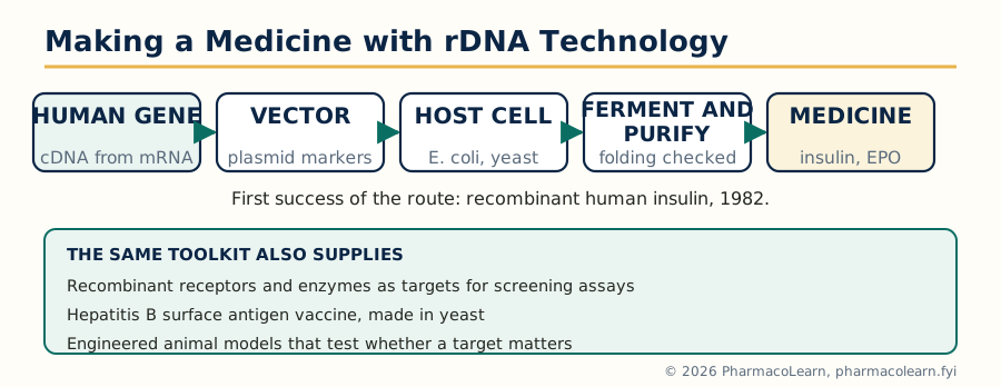 Figure: The recombinant production route from human gene to medicine, with recombinant human insulin as its first success.
For drug discovery the technology works at three further levels: it supplies pure human targets, receptors and enzymes, for screening assays (the kinases and GPCRs of the MPC103T syllabus are screened as recombinant proteins); it supplies disease proteins and antigens for structural studies that feed structure based design; and animal models engineered with human genes or gene deletions test whether a target matters in a whole organism before chemistry money is spent. The subject's earlier units isolated what organisms happened to make; this unit is about instructing organisms to make what is needed.
4.3 Hybridoma technology and monoclonal antibodies
A B lymphocyte makes one antibody but will not grow indefinitely in culture; a myeloma cell grows indefinitely but makes no useful antibody. Köhler and Milstein (1975) fused the two: immunise a mouse, fuse its spleen B cells with myeloma cells using polyethylene glycol, and select in HAT medium, in which the myeloma partner (deficient in the enzyme HGPRT) dies and unfused B cells die naturally, leaving only hybridomas. Screening and cloning of the survivors yields a monoclonal antibody: one specificity, one sequence, unlimited supply.
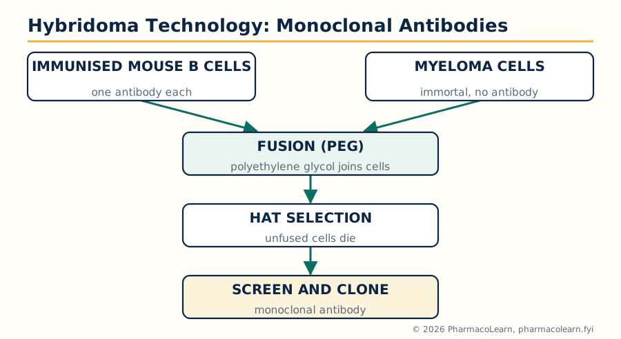 Figure: Hybridoma production: fusion, HAT selection and cloning convert a mouse immune response into an unlimited supply of one antibody.
Pharmaceutical consequences: monoclonal antibodies became a drug class in their own right (the trastuzumab, rituximab and adalimumab generation, progressively humanised and then fully human to escape the immunogenicity of mouse protein), and became the discovery tools of the trade: assay antibodies, target validation reagents, antibody drug conjugates that deliver cytotoxic payloads to antigen bearing cells, and the purification handles of downstream processing.
4.4 Oligonucleotide therapy
Oligonucleotide drugs are short synthetic DNA or RNA like strands, chemically stabilised, that act on nucleic acids rather than proteins:
- Antisense oligonucleotides bind a target messenger RNA by base pairing and either recruit RNase H to destroy it or block its processing. They must be protected against nucleases, hence backbone modification (phosphorothioate linkages) and sugar modifications (2 prime-O-methoxyethyl and relatives). Fomivirsen, against cytomegalovirus retinitis, was the first approved antisense drug (1998); nusinersen, which corrects the splicing of the SMN2 gene in spinal muscular atrophy, shows the splice switching branch of the family.
- Small interfering RNAs (siRNAs) load into the RISC complex and direct it to cleave a complementary mRNA; patisiran, delivered in a lipid nanoparticle to the liver, was the first approved siRNA (2018).
- Aptamers are folded oligonucleotides selected to bind a protein target in three dimensions, pegaptanib (anti-VEGF) being the approved example.
The class-wide obstacles are the delivery and stability problems of large charged molecules, which is why the liver, the eye and the spinal canal, all reachable compartments, host most successes so far.
4.5 Gene therapy: introduction, clinical applications and recent advances
Gene therapy treats disease by delivering a functional gene, or gene editing machinery, to the patient's cells. Ex vivo therapy removes cells, corrects them in the laboratory and returns them (the controllable route); in vivo therapy delivers the vector to the patient directly. Delivery vehicles divide into viral vectors, retrovirus and lentivirus (integrating, durable, with insertional mutagenesis risk that early trials tragically confirmed), adenovirus (efficient, transient, immunogenic) and adeno-associated virus (AAV, largely non integrating, the current in vivo favourite, limited cargo size), and non viral systems, chiefly lipid nanoparticles.
Clinical applications with regulatory approvals include: ex vivo correction of ADA deficient severe combined immunodeficiency; AAV delivered RPE65 gene for inherited retinal dystrophy (voretigene neparvovec), the first in vivo gene therapy for an inherited disease; AAV delivered SMN1 for spinal muscular atrophy (onasemnogene abeparvovec); lentiviral beta globin for transfusion dependent beta thalassaemia; and the CAR T cell therapies, in which a patient's T cells are engineered ex vivo with a chimeric antigen receptor against CD19, gene therapy wearing an immunotherapy uniform.
Recent advances are dominated by gene editing: CRISPR Cas9 editing of a patient's own blood stem cells ex vivo, reactivating fetal haemoglobin, won the first CRISPR approvals in 2023 for sickle cell disease and beta thalassaemia (exagamglogene autotemcel). Base editing and prime editing, which rewrite single letters without double strand breaks, in vivo editing trials, and steadily better capsids and lipid nanoparticles for tissue targeting mark the current frontier. The syllabus phrase recent advances should always be answered with the editing story plus the standing problems: durability, immune responses to vectors, manufacturing cost and equitable access.
4.6 Principles of RNA and DNA estimation
Nucleic acid estimation is a Unit 1 practical skill of this course (the MPC105P practical list includes RNA and DNA estimation), and its principles are chemical:
- Ultraviolet absorption: nucleic acids absorb maximally near 260 nm because of their bases. One absorbance unit at 260 nm corresponds to about 50 microgram per ml for double stranded DNA, 40 for RNA and 33 for single stranded DNA. The A260/A280 ratio (about 1.8 for pure DNA, 2.0 for pure RNA) reports protein contamination; it measures purity and quantity together in one reading, and it cannot tell DNA from RNA in a mixture.
- Colour reactions for specific sugars: the diphenylamine (Dische) reaction gives a blue colour with the deoxyribose of DNA under hot acid conditions, and the orcinol (Bial) reaction gives a green colour with the ribose of RNA; these classical methods estimate one nucleic acid in the presence of the other.
- Fluorescent dyes: modern quantitation uses dyes whose fluorescence rises sharply on binding double stranded DNA (ethidium bromide historically, safer intercalating and groove binding dyes now), giving sensitivity far beyond UV absorption for dilute samples.
4.7 Indigenous crude drugs I: the diabetic therapy group
The indigenous system (Ayurveda and allied practice) carries a large antidiabetic materia medica; the syllabus names five. In each case the active constituents, not vague virtue, are the chemistry:
| Crude drug | Principal active constituents | Chemical note |
|---|---|---|
| Gymnema sylvestre (gurmar) | Gymnemic acids, a family of oleanane triterpene saponins | The leaf famously abolishes sweet taste temporarily (gymnemic acids block sweet receptors); saponin chemistry of section 2.12 on an oleanane frame; intestinal glucose uptake effects reported |
| Salacia reticulata | Salacinol and kotalanol, sulfonium ion glycosidase inhibitors | Potent alpha glucosidase inhibitors of unusual sulfonium sulfate structure; the acarbose mechanism from a root bark |
| Pterocarpus marsupium (vijaysar) | (Minus)-epicatechin (a flavanol, section 2.10 chemistry), pterostilbene, marsupsin | Heartwood constituents; epicatechin is the constituent most discussed in beta cell studies |
| Swertia chirata (chirayata) | Amarogentin and swertiamarin, secoiridoid glycosides; xanthones such as mangiferin | Amarogentin is among the most bitter substances known; the drug is the classical intensely bitter tonic of the group |
| Trigonella foenum-graecum (fenugreek, methi) | The unusual amino acid 4-hydroxyisoleucine; galactomannan soluble fibre; steroidal saponins (diosgenin) | 4-hydroxyisoleucine is reported to support glucose dependent insulin release; the fibre slows carbohydrate absorption; the seed also links back to diosgenin, section 2.12 |
The measured professional position, which examiners credit: these are chemically defined constituents with plausible and in several cases demonstrated mechanisms in vitro and in animal models, and variable, usually modest human trial evidence; they are adjuncts studied alongside, not replacements for, standard diabetic therapy.
4.8 Indigenous crude drugs II: liver dysfunction and antitumor
Phyllanthus niruri (bhumyamalaki), the syllabus drug for liver dysfunction, is a small herb of the Euphorbia family carrying lignans (phyllanthin and hypophyllanthin), flavonoids including quercetin itself (section 2.11), and ellagitannins. It is a classical Ayurvedic drug for jaundice and liver complaints, and the related species Phyllanthus amarus drew wide attention from trials in chronic hepatitis B carriage; the chemical teaching points are the lignan skeleton (two phenylpropane units joined beta to beta, the same dimer family as podophyllotoxin, section 1.3) and the discipline of species identity, since the Phyllanthus species are easily confused and chemically distinct.
Curcuma longa Linn. (turmeric, haldi), the syllabus antitumor drug, owes its colour and activity to the curcuminoids of the rhizome, chiefly curcumin, a diarylheptanoid: two ferulic acid like aromatic rings joined by a seven carbon chain that exists largely as its conjugated enol, with demethoxy and bisdemethoxycurcumin beside it. Curcumin modulates several signalling pathways in cell studies (NF-kB among them) and shows antitumor activity in vitro and in animal models; its notorious limitation is pharmacokinetic: very poor aqueous solubility and rapid conjugation, so blood levels after oral dosing are low, and the literature must be read with that in mind. Formulation science (piperine combinations, liposomes, nanoparticles) is the attempt to close that gap, and the examinable verdict is research promise with unproven clinical benefit, on a molecule whose chemistry is thoroughly characterised.
Unit 5: Structural Characterization of Natural Compounds
5.1 The spectral workflow: what each technique answers
Structural characterization asks four questions, and the syllabus's four techniques answer one each, which is why they are taught as a set:
| Technique | Physical basis | Question it answers first |
|---|---|---|
| Mass spectrometry (MS) | Ionisation and separation by mass to charge ratio | What is the molecular weight, and (high resolution) the molecular formula? What fragments does it break into? |
| IR spectroscopy | Absorption by vibrating bonds | Which functional groups are present: hydroxyl, carbonyl and of what kind, aromatic ring, alkene, ether? |
| 1H NMR | Proton environments, integration, splitting and coupling | How many hydrogens of each kind, and what is next to what? Where are the methyls, the exchangeable protons, the olefinic and aromatic protons? |
| 13C NMR | Carbon environments (with DEPT editing) | What is the carbon skeleton: how many carbons of each type, CH3, CH2, CH and quaternary; carbonyls by their low field shifts |
The professional workflow runs: purity first (a spectrum of a mixture characterises nothing), then HRMS for formula and unsaturation, IR for the functional group inventory, NMR for the skeleton assembled proton by proton and carbon by carbon, with 2D experiments (COSY for proton neighbours, HSQC and HMBC for carbon proton connections) as the modern assembly tools, and MS fragmentation as the cross check that the assembled structure breaks the way the real molecule breaks:
STRUCTURAL CHARACTERIZATION WORKFLOW
  Pure compound        (chromatography, one spot, sharp behaviour)
        |
  HRMS: molecular      (formula, double bond equivalents)
  weight and formula
        |
  IR: functional       (OH or NH, carbonyl type, aromatic,
  group inventory       alkene, ether, lactone)
        |
  1H NMR: proton map   (count, environment, neighbours, J values)
        |
  13C NMR and DEPT:    (every carbon classified and placed)
  carbon skeleton
        |
  Assemble draft       (consistent with formula and all spectra)
        |
  Confirm: MS fragments, 2D NMR, chemical tests,
  comparison with authentic data, synthesis or X-ray
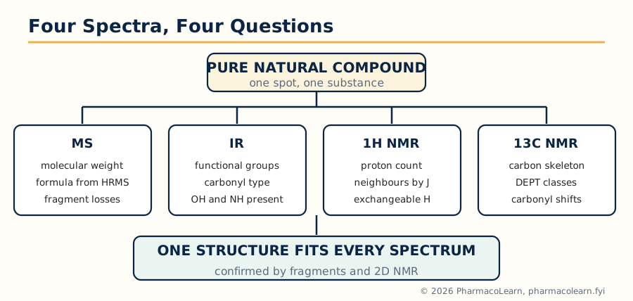 Figure: The four syllabus techniques answer four different questions; only a structure consistent with all four spectra is accepted.
5.2 Functional group frequencies and shifts to have by heart
| Observation | Interpretation |
|---|---|
| IR 3200 to 3600 cm-1, broad | Hydrogen bonded O-H or N-H stretch |
| IR around 1740 to 1780 cm-1 | Ester or strained lactam/lactone carbonyl; the higher the ring strain, the higher the frequency |
| IR around 1715 cm-1 | Ordinary ketone; around 1680 cm-1 if conjugated |
| IR 1600, 1580, 1500 cm-1 bands | Aromatic ring vibrations |
| 1H NMR 0.6 to 1.0 ppm singlets | Angular methyls on a steroid skeleton |
| 1H NMR 3.2 to 4.0 ppm | Protons on oxygen bearing carbon (OCH, OCH3 a sharp singlet near 3.3 to 3.9) |
| 1H NMR 4.5 to 6.5 ppm | Olefinic protons; 6.5 to 8.5 ppm aromatic protons; exchangeable OH and NH broad, disappearing on D2O shake |
| 13C NMR 170 to 220 ppm | Carbonyl carbons: ester and lactone near 170 to 180, ketone near 200 to 215 |
| MS: loss of 18, 28, 44 | Loss of water, carbon monoxide (or ethylene), carbon dioxide: first reading of a fragmentation |
Deuterium exchange deserves its own sentence: shaking the NMR sample with D2O makes OH, NH and COOH proton signals vanish, the cheapest experiment in spectroscopy and the standard way to count exchangeable protons in a natural product full of hydroxyls.
5.3 Penicillin
The problem historically: penicillin's structure was contested through the 1940s, with the fused beta-lactam thiazolidine structure competing against an oxazolone alternative; chemical degradation, IR and Dorothy Hodgkin's X-ray crystallography settled it for the strained four membered lactam fused cis to the thiazolidine ring, bearing a carboxyl group and the variable acyl side chain (benzyl in penicillin G).
Spectroscopic signature today:
- IR: the beta-lactam carbonyl absorbs unusually high, around 1760 to 1780 cm-1, because the four membered ring prevents normal amide resonance: the carbonyl behaves almost like a ketone's. Beside it sit the side chain amide carbonyl (around 1660 to 1680 cm-1) and the carboxylic acid or carboxylate bands. No other common drug class shows a carbonyl that high, so the band is practically diagnostic.
- 1H NMR: the two bridgehead protons of the fused system (H-5 and H-6) appear as characteristic doublets around 5.4 to 5.6 ppm with a small cis coupling (about 4 Hz) that itself reports the cis ring fusion; the gem dimethyl group of the thiazolidine ring gives two singlets near 1.5 ppm; the side chain benzyl CH2 and aromatic protons identify penicillin G's R group.
- 13C NMR: three carbonyl carbons in a row tell the story: lactam near 173 to 175 ppm, side chain amide near 170 ppm, carboxyl near 175 ppm, with the quaternary thiazolidine carbon near 65 ppm.
- MS: molecular ion (penicillin G, 334) with fragmentation dominated by cleavage around the side chain amide and the thiazolidine ring; the high resolution measurement gives C16H18N2O4S, the single sulfur itself confirmed by the characteristic M+2 isotope peak of about 4.4 percent.
5.4 Morphine
IR: a broad phenolic and alcoholic O-H envelope around 3200 to 3500 cm-1, aromatic bands near 1600 and 1500 cm-1, aromatic C-H out of plane bending consistent with a 1,2,4-trisubstituted ring, strong C-O stretching between 1000 and 1250 cm-1 for the ether bridge and the two hydroxyls, and significantly no carbonyl band, which at once excludes the oxidised relative morphinone.
1H NMR: two aromatic protons as an AB pair of doublets (J about 8 Hz) near 6.5 to 6.7 ppm, the isolated olefinic protons of the ring C double bond near 5.3 to 5.7 ppm, the N-methyl a sharp singlet near 2.4 ppm, and a crowded aliphatic region whose coupling network maps the rigid bridged skeleton; the phenolic and alcoholic protons exchange with D2O. Coupling constants around the allylic alcohol fix the configuration at C-6 relative to the ring system, spectroscopy doing the stereochemistry that once needed chemical correlation (section 2.6).
13C NMR and MS: seventeen carbons, six aromatic (two oxygen bearing near 143 to 146 ppm), two olefinic, the ether bridge carbon (C-5) characteristically downfield near 90 ppm, and the N-methyl near 43 ppm. Electron impact MS gives M+ at 285 (C17H19NO3 by high resolution), with the morphinan skeleton fragmenting reluctantly: a strong molecular ion is itself characteristic of these rigid polycyclic bases, in contrast to floppy molecules that shatter.
5.5 Camphor
Camphor is the cleanest teaching example of the set because it is small, rigid and symmetrical in nothing but its formula.
IR: the ketone carbonyl absorbs around 1740 cm-1, high for a saturated ketone (normally near 1715), because the carbonyl sits in the strained bicyclic framework; no O-H band and no C=C absorption, consistent with a saturated ketone.
1H NMR: three methyl singlets (the bridgehead methyl and the gem dimethyl pair, the latter two slightly different in environment) between about 0.8 and 1.0 ppm account for nine of the sixteen hydrogens without any splitting: three methyls on quaternary carbons, a fingerprint of the bornane skeleton; the remaining signals are the methylene and bridgehead protons of the bicyclic frame, the proton alpha to the carbonyl appearing downfield near 2.3 ppm.
13C NMR and MS: ten carbons with the ketone carbonyl far downfield near 218 ppm and the oxygen bridgehead quaternary carbon near 57 ppm. MS gives M+ at 152 (C10H16O); alpha cleavage beside the carbonyl and loss of carbon monoxide (152 to 124) and hydrocarbon fragments such as m/z 95 and 81 trace the ring opening of the bornane frame. The classical degradative proof (section 3.3) and this spectral reading arrive at the same bicyclo[2.2.1] structure by independent roads.
5.6 Vitamin D
Vitamin D's spectra are dominated by one feature: the conjugated triene created when the B ring of the sterol opens (section 2.14).
UV and IR: the triene gives intense absorption with a maximum near 265 nm, historically the basis of vitamin D assay, and its disappearance on irradiation or oxidation monitors degradation. IR shows the hydroxyl stretch, and characteristic triene C=C and out of plane C-H bands in the 1600 to 1650 and 880 to 900 cm-1 regions (the exocyclic methylene of the triene system).
1H NMR: the olefinic region is the signature: the exocyclic methylene protons as two broad signals near 4.8 and 5.0 ppm, and the triene protons between 5.9 and 6.3 ppm with coupling patterns that distinguish the vitamin from its inactive isomers (the 5,6-trans isomer and tachysterol differ exactly here, which is why NMR polices vitamin D preparations). The angular methyl singlet near 0.55 ppm and the side chain methyl doublets place the molecule in the sterol family the triene came from.
13C NMR and MS: twenty seven carbons for D3 (twenty eight for D2, whose side chain carries an extra methyl and double bond); the triene carbons spread between 112 and 145 ppm. Molecular weights 384 (D3, C27H44O) and 396 (D2, C28H44O) by MS, with ready loss of water (M minus 18) from the allylic alcohol. The spectral lesson: vitamin D is identified less by any single peak than by the combination of a sterol carbon count with a triene proton and UV pattern.
5.7 Quercetin
Quercetin (section 2.11) is where the classical and modern methods are most profitably compared, because its classical UV shift reagent method remains standard practice for flavonoids.
UV with shift reagents: in methanol, quercetin shows Band II near 255 nm and Band I near 370 nm, the long wavelength band reflecting the flavonol 3-hydroxyl conjugation. Sodium methoxide pushes Band I strongly to longer wavelength (a free 4 prime hydroxyl); sodium acetate shifts Band II (a free 7 hydroxyl); aluminium chloride shifts Band I and the shift largely survives added acid, reporting the 5-hydroxy 4-keto chelation system and the ortho dihydroxyl B ring. In twenty minutes this battery locates free hydroxyls that methylation analysis once took weeks to place.
IR and NMR: IR shows the hydrogen bonded carbonyl of the flavonol near 1660 cm-1 (lowered by conjugation and chelation to the 5-hydroxyl), aromatic bands and a broad phenolic envelope. In 1H NMR (DMSO-d6) the chelated 5-OH appears far downfield near 12.5 ppm, a signal so characteristic it is practically a name tag; the A ring protons appear as two meta coupled doublets near 6.2 and 6.4 ppm; the B ring shows the 3 prime, 4 prime dihydroxy pattern (a doublet near 6.9, a doublet near 7.5 and a double doublet near 7.6 ppm). 13C NMR gives fifteen carbons with the carbonyl (C-4) near 176 ppm and the oxygenated C-2 near 147 ppm. MS gives M+ at 302 (C15H10O7), with retro Diels-Alder cleavage of the C ring producing the diagnostic A ring fragment at m/z 153 that locates the A ring dihydroxylation.
5.8 Digitalis glycosides
The digitalis glycosides (digoxin, digitoxin; section 2.12) are the hardest of the six because they are three part molecules: a steroidal genin, an unsaturated lactone and a chain of unusual sugars (digitoxose, a 2,6-dideoxy sugar).
IR: the butenolide lactone carbonyl absorbs near 1740 to 1780 cm-1 with its C=C band near 1620 cm-1; hydroxyls give the broad high region band; and the absence of aromatic bands separates these steroids from the oestrane family at a glance.
1H NMR: recognition signals: the lactone vinyl proton near 5.9 ppm; the 21 methylene of the lactone as a pair of doublets of doublets near 4.6 to 4.9 ppm; two angular methyl singlets (C-18 near 0.85, C-19 near 0.95 ppm); anomeric protons of the sugar chain between 4.8 and 5.2 ppm; and a signature that belongs to the digitoxose sugars alone, their 6-methyl doublets near 1.2 ppm, one per sugar unit, so counting them counts the sugars.
13C NMR and MS: the lactone carbonyl appears near 174 ppm and the olefinic lactone carbons near 117 and 174 ppm; the anomeric carbons sit between 95 and 100 ppm. Digoxin (C41H64O14, mass 780) and digitoxin (C41H64O13, mass 764) differ by one oxygen, the 12 beta hydroxyl of digoxin, which MS states in the molecular weight alone. Soft ionisation MS (ESI, giving [M+Na]+ and [M+NH4]+ rather than a naked molecular ion) shows stepwise losses of 130 mass units, one digitoxose at a time, reading the sugar sequence off the spectrum; electron impact, by contrast, destroys these molecules, which is itself a teaching point: the ionisation method must be chosen for the fragility of the natural product, and glycosides demand soft ionisation.
| Compound | One line spectral identity |
|---|---|
| Penicillin | IR carbonyl near 1770 cm-1 (strained lactam) plus sulfur isotope pattern in MS |
| Morphine | No carbonyl; phenolic OH; strong M+ at 285; ether carbon near 90 ppm in 13C |
| Camphor | Ketone at 1740 cm-1 and near 218 ppm; three methyl singlets; M+ 152 |
| Vitamin D | Triene UV at 265 nm and olefinic 1H pattern; sterol methyls below 1 ppm |
| Quercetin | UV shift reagent battery; chelated 5-OH at 12.5 ppm; RDA fragment at m/z 153 |
| Digitalis glycosides | Lactone vinyl proton near 5.9 ppm; anomeric signals; MS sugar losses of 130 |
Frequently Asked Questions
Q: What is a natural product lead, and why is the natural compound itself often not the drug? A lead is a natural compound whose activity shows where a drug can be built. The compound itself is usually flawed for medicine: paclitaxel was scarce and insoluble, thienamycin was unstable, podophyllotoxin was too toxic. Chemical modification, semi-synthesis and analog design repair those faults while keeping the activity the natural product revealed.
Q: How did teprotide lead to captopril if teprotide was never a drug? Teprotide, a peptide from pit viper venom, proved that inhibiting the angiotensin converting enzyme lowers blood pressure and showed how a peptide carboxylate and proline engage the enzyme's zinc site. Captopril was designed as a small, orally active molecule presenting the same binding features, so the natural product contributed the mechanism and the binding map rather than the structure.
Q: Why are alkaloids extracted by switching between acid and base? Because the two forms have opposite solubility. As salts in dilute acid, alkaloids dissolve in water and leave fat and resin behind; basified, the free bases move into organic solvents and leave salts and tannins behind. Switching twice purifies the bases from most of the plant matrix before chromatography or crystallisation begins.
Q: Morphine and ephedrine both come from plants. Why is only morphine called a true alkaloid in the strict sense? Morphine's nitrogen sits inside a ring system biosynthesised into a complex polycyclic alkaloid. Ephedrine's nitrogen is in an open side chain as a methylamino group; it belongs to the protoalkaloids, plant amines of amino acid origin whose nitrogen is not part of a heterocyclic ring. The pharmacology overlaps, the structural class does not.
Q: What does the isoprene rule actually claim, and how did squalene confirm it? It claims terpenoid skeletons are assembled from C5 isoprene units through a few acyclic precursors, so structures must be derivable from the right precursor by cyclisation and rearrangement. Squalene, the C30 acyclic precursor, cyclises in the laboratory of the cell to lanosterol and then cholesterol exactly along the folding the rule predicted, confirming the scheme for the whole triterpene and steroid family.
Q: Quercetin has five hydroxyl groups. How do spectroscopists show where they are? Three tools combine: the UV shift reagent battery locates free hydroxyls at 7, 4 prime and the 5 position by the direction of band shifts; 1H NMR shows the chelated 5-OH far downfield near 12.5 ppm and the aromatic proton patterns of both rings; and mass spectrometry's retro Diels-Alder fragment at m/z 153 reports the A ring hydroxylation. Together they fix 3,5,7,3 prime,4 prime.
Q: Why does vitamin D appear in both the steroid unit and the vitamins unit? Because it is both. Biosynthetically and structurally it is a secosteroid, a sterol whose B ring has opened, which places it with the steroids in Unit 2 of the syllabus; nutritionally and physiologically it is a vitamin whose hydroxylated metabolite is a calcium regulating hormone, so it is examined alongside the vitamins as well.
Q: What is the difference between a recombinant protein drug and a monoclonal antibody from hybridoma technology? A recombinant protein drug such as insulin is made by inserting the human gene into a production organism and purifying the protein it expresses. A monoclonal antibody comes from a hybridoma, a fused cell line that secretes one antibody of one specificity indefinitely; modern antibody drugs are then engineered and produced recombinantly as well, but their specificity was selected by the hybridoma route.
Q: Why does the beta-lactam carbonyl of penicillin absorb at such a high IR frequency? In an ordinary amide, resonance with nitrogen flattens and weakens the carbonyl, lowering its stretching frequency. The four membered beta-lactam ring twists the amide out of planarity and prevents that resonance, so the carbonyl behaves more like a ketone's and absorbs near 1760 to 1780 cm-1. The same strain is what makes the ring reactive towards penicillin binding proteins.
Q: The indigenous diabetic drugs contain real active molecules. Why are they still described as adjuncts? Because constituent chemistry is only the first test. Gymnemic acids, salacinol and 4-hydroxyisoleucine have credible mechanisms, but human trial evidence for the crude drugs is variable and usually modest, preparations are not standardised like medicines, and diabetes carries risks that demand proven therapy. The professional position is study and adjunctive use with disclosure, never replacement of prescribed treatment.
Complete MPC104T notes, all 5 units in one PDF, free to keep.
Other M.Pharm subjects: MPH101T · MPC101T · MPL101T · MPH102T · MPH103T · MPH104T · MPL102T · MPL103T · MPL104T · MPC102T · MPC103T · MPC104T · MPH201T · MPH202T · MPH203T · MPH204T · MPC201T · MPC202T · MPC203T · MPC204T · M.Pharm