
MPC103T — Advanced Medicinal Chemistry
4 hrs/week, 4 credits, 60 hours, 100 marks. Notes follow the M.Pharm syllabus, unit by unit.
MPC103T Advanced Medicinal Chemistry notes for M.Pharm Semester 1 Pharmaceutical Chemistry: drug discovery and targets, prodrug and analog design, bioisosterism, drug class SAR, enzyme inhibitor design and peptidomimetics, with free unit wise PDF downloads on PharmacoLearn.
Units in this subject
- Unit 1 — Drug Discovery
- Unit 2 — Prodrug Design and Analog Design
- Unit 3 — Medicinal Chemistry Aspects of Drug Classes and Stereochemistry and Drug Action
- Unit 4 — Rational Design of Enzyme Inhibitors
- Unit 5 — Peptidomimetics and Eicosanoid Chemistry
Download MPC103T PDF Notes
Free PDFs for this subject, one per unit:
Advanced Medicinal Chemistry, subject code MPC103T, is a core Semester 1 subject of M.Pharm Pharmaceutical Chemistry. It carries 4 credits and 60 hours of theory, taught at 4 hours per week, for 100 marks.
This subject is where organic chemistry meets biology. MPC102T taught how molecules are made; this subject teaches how molecules are designed to act on the body. Unit 1 sets out the modern drug discovery pipeline: how a disease target is identified and validated, where lead molecules come from, and how drugs interact with their main biological targets, the receptors, including the classical theories of drug receptor interaction and the difference between agonists and antagonists. Unit 2 covers the two great lead modification strategies: prodrug design, in which a temporary chemical group carries a drug past a pharmaceutical or pharmacokinetic barrier, and analog design, in which a lead is systematically varied by bioisosteric replacement, rigidification, ring changes and stereochemical design, together with the strategies used to combat drug resistance. Unit 3 applies the method of systematic study (classification, SAR, mechanism of action and synthesis) to eight major drug classes, from antihypertensives to antivirals, and closes with stereochemistry and drug action. Unit 4 is the rational design of enzyme inhibitors, built on enzyme kinetics. Unit 5 treats peptidomimetics, the art of making peptide like molecules that survive in the body, and the chemistry of the eicosanoids: prostaglandins, leukotrienes and thromboxanes. A student who completes this subject should be able to outline the stages of a drug discovery programme, propose a prodrug or analog strategy for a given problem, read the SAR of a drug class, and explain how a new generation inhibitor or peptidomimetic was designed.
Unit 1: Drug Discovery
1.1 Stages of drug discovery
Modern drug discovery is a funnel. Thousands of compounds enter at the top, and typically one reaches the market. The stages, in order, are:
- Target identification and validation: a biological molecule or pathway is chosen and evidence gathered that changing its activity will change the disease (Unit 1, section 1.3).
- Hit discovery: large numbers of compounds are screened against the target to find hits, compounds with measurable, reproducible activity.
- Hit to lead: hits are confirmed, checked for selectivity and obvious liabilities, and grouped into chemical series; the most promising series become leads.
- Lead optimisation: the lead is systematically modified (Unit 2) to improve potency, selectivity, solubility, metabolic stability and safety together. This is the home ground of medicinal chemistry.
- Preclinical development: the optimised candidate is tested in animals for pharmacokinetics and toxicology and a formulation is developed.
- Clinical development and approval: Phase I (safety in volunteers), Phase II (first efficacy in patients), Phase III (large confirmatory trials), then regulatory review, marketing, and Phase IV surveillance after launch.
The economics explain the funnel shape: most candidates fail, and they fail mostly for lack of efficacy or for toxicity, so every early test that removes a weak compound cheaply saves money later. A useful distinction is between the lead, a compound with proven activity and a workable structure but many faults, and the drug candidate, a compound in which those faults have been engineered out.
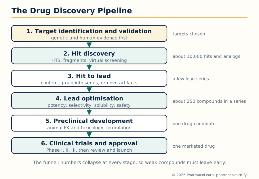 Figure: The drug discovery funnel. Numbers fall steeply at every stage, which is why weak compounds must be removed as early and as cheaply as possible.
1.2 Lead discovery: where leads come from
A lead can come from many sources, and most marketed drugs trace back to one of a few classic routes:
- Natural products and their derivatives: morphine, the penicillins, lovastatin and paclitaxel all began as natural leads (their chemistry is the subject of MPC104T).
- Screening of synthetic compound collections: high throughput screening (HTS) tests hundreds of thousands of stored compounds against the target in automated assays. Fragment based screening tests very small molecules (fragments) at high concentration with sensitive biophysical methods and grows the best fragments into leads.
- Existing drugs and their side effects: many leads began as an observed side effect of a known drug; the sulfonamide antibacterials revealed hypoglycaemic sulfonylureas, and chlorpromazine grew out of work on antihistamines.
- The endogenous ligand or substrate: designing from the body's own messenger, as histamine guided the design of both H1 and H2 antagonists, and as angiotensin guided the ACE inhibitors.
- Rational and structure based design: when the three dimensional structure of the target is known, compounds can be designed to fit its binding site directly; this is developed in Unit 4 for enzymes.
- Serendipity: penicillin, librium (chlordiazepoxide) and sildenafil (developed for angina, redirected by an observed effect) all arrived partly by chance, although chance only helps a prepared screening programme.
Whichever the source, a hit becomes a lead only after confirmation: the activity must repeat in a fresh sample of verified identity and purity, show a sensible dose response, and not be an assay artifact (some compounds interfere with assays themselves, for example by aggregating or by reacting with assay reagents).
1.3 Drug targets: identification, validation and diversity
A drug target is the biological molecule whose activity the drug changes to produce its therapeutic effect. Target identification draws on genomics and disease biology (genes linked to disease by mutation or expression studies), on pathway analysis, and on the targets of drugs that already work in the disease.
Target validation asks whether the target deserves a full discovery programme. The evidence, in increasing strength:
TARGET VALIDATION SEQUENCE
  Genetic evidence        : disease linked to the gene or pathway
        |
  Expression evidence     : target present in the diseased tissue
        |
  Tool compound or        : changing the target in cells changes
  genetic knockdown         the disease relevant behaviour
        |
  Animal model evidence   : changing the target helps in vivo
        |
  Human evidence          : human genetics or an existing drug
                            acting on the target works in patients
The human evidence line is the strongest: the cholesterol target HMG-CoA reductase was validated by the statins themselves, and targets supported by human genetic evidence succeed in the clinic at a clearly higher rate than targets without it.
Diversity of drug targets. Most marketed drugs act on a small set of target classes:
| Target class | Share of drug action (approx.) | Examples of drug and target |
|---|---|---|
| Receptors (mostly GPCRs) | Largest single class | Propranolol at beta adrenoceptors, morphine at opioid receptors |
| Enzymes | Second largest class | Statins at HMG-CoA reductase, methotrexate at dihydrofolate reductase |
| Ion channels | Smaller but important | Amlodipine at L-type calcium channels, phenytoin at sodium channels |
| Transporters (carriers) | Smaller | Fluoxetine at the serotonin transporter, omeprazole at the proton pump (an ATPase) |
| Nucleic acids and structural proteins | Smallest | Alkylating agents and intercalators on DNA, colchicine on tubulin |
The diversity point the syllabus makes is that the druggable genome is much larger than the few hundred targets of current drugs, and new target classes (protein protein interactions, RNA targets) keep opening as techniques improve.
1.4 Receptors: the major biological drug target
A receptor is a protein that recognises an endogenous chemical messenger (a neurotransmitter, hormone or local mediator) and converts that recognition into a cellular response. Receptors are classified into four superfamilies by structure and signalling mechanism:
| Superfamily | Structure | Signalling | Response speed | Examples |
|---|---|---|---|---|
| Ligand gated ion channels | Multi subunit membrane protein with a central pore | Ion flow across the membrane | Milliseconds | Nicotinic acetylcholine receptor, GABA-A receptor |
| G protein coupled receptors (GPCRs) | Single protein, seven transmembrane helices | G proteins, then second messengers (cAMP, IP3, DAG) or ion channels | Seconds | Adrenoceptors, histamine, dopamine, opioid receptors |
| Kinase linked receptors | Single transmembrane protein with intracellular enzyme (kinase) activity | Phosphorylation cascades | Minutes to hours | Insulin receptor, growth factor receptors |
| Nuclear receptors | Intracellular (cytoplasmic or nuclear) protein | Direct control of gene transcription | Hours | Steroid, thyroid hormone and vitamin D receptors |
Binding and activation. A drug binds to a receptor at a defined binding site, usually the site used by the endogenous agonist (the orthosteric site) or a separate regulatory site (an allosteric site). Binding alone is not enough: the receptor must also change shape and signal. Section 1.5 separates these two properties, affinity and efficacy.
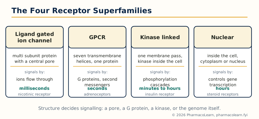 Figure: The four receptor superfamilies. Structure decides signalling: a pore, a G protein, a kinase, or the genome itself.
1.5 Drug receptor interactions: binding forces, affinity and efficacy
Drug receptor binding is held together by the same non-covalent forces that hold any molecular complex together, acting together over the surface of the binding site:
- Ionic (electrostatic) interactions: between oppositely charged groups, for example a protonated amine of the drug and a carboxylate of the receptor. Strong, and often the first anchor point.
- Hydrogen bonds: directional, moderate strength; donors and acceptors must be correctly positioned, which is one source of selectivity.
- Van der Waals forces: weak individually, but summed over a well fitting surface they contribute greatly; fit must be close, since the force falls off very steeply with distance.
- Hydrophobic interactions: non-polar surfaces of drug and pocket exclude water and associate; a major driver of binding for lipophilic drugs.
- Covalent bonds: rare in receptor binding, common in enzyme inhibition (Unit 4); usually irreversible.
Two independent properties describe what a bound drug does:
- Affinity is how tightly the drug binds, measured by the equilibrium dissociation constant Kd (or its negative log, pKd). Low Kd means high affinity.
- Efficacy (intrinsic activity) is the ability of the bound drug to activate the receptor and produce a response. It is a property of the drug receptor complex, not of binding strength.
A drug can therefore have high affinity and no efficacy: it occupies the site, activates nothing, and blocks the agonist. That combination is exactly an antagonist (section 1.7).
1.6 Theories of drug receptor interaction
The classical theories were attempts to connect binding with response mathematically:
| Theory | Core idea | What it explains | Limitation or extension |
|---|---|---|---|
| Occupancy theory (Clark) | Response is proportional to the fraction of receptors occupied | Graded dose response, the basis of Kd and Emax thinking | Cannot explain partial agonists, which occupy fully but respond partially |
| Rate theory (Paton) | Response depends on the rate of drug receptor encounters, not occupancy | Why some agonists act briefly; dissociation rate matters | Hard to test directly; largely of historical interest |
| Induced fit theory (Koshland) | Binding changes the shape of the receptor; the fit is induced, not preformed | Activation as a conformational event; applies to enzymes too | Extended by later conformational models |
| Two state (allosteric) model | Receptors exist in equilibrium between inactive (R) and active (R) states; agonists stabilise R, antagonists bind both equally, inverse agonists stabilise R | Partial agonism, constitutive activity and inverse agonism in one scheme | The working modern framework |
The Easson-Stedman hypothesis belongs in this section for stereochemistry: a chiral drug needs at least three points of attachment to a receptor to distinguish its enantiomers, because two points can be matched by either mirror image. Unit 3 returns to this with case studies.
1.7 Agonists, antagonists and dose response
Response is plotted against the logarithm of dose or concentration, giving the sigmoidal log dose response curve. Two numbers are read from it: EC50, the concentration giving half the maximal response (a measure of potency), and Emax, the maximal response the drug can produce (a measure of efficacy in that system).
- Full agonist: high efficacy, reaches the system maximum. Adrenaline at beta receptors is the physiological example.
- Partial agonist: lower efficacy; even at full occupancy its Emax stays below the system maximum, and it antagonises a full agonist when both are present. Pindolol at beta adrenoceptors and aripiprazole at dopamine D2 receptors are standard examples.
- Competitive antagonist: binds the agonist site reversibly without activating; the agonist curve shifts to the right in parallel, with Emax unchanged, and the block is surmountable by more agonist. Naloxone at opioid receptors is the classic example. The dose ratio it produces underlies the Schild analysis and the pA2 measure of antagonist affinity.
- Non-competitive antagonist: binds a separate site, or irreversibly, and reduces Emax; the curve is depressed, not just shifted, and more agonist cannot restore the maximum. Phenoxybenzamine at alpha adrenoceptors (covalent, irreversible) is the textbook case.
- Inverse agonist: reduces the basal (constitutive) activity of a receptor that signals without any agonist; predicted by the two state model and real for several GPCRs, for example rimonabant at cannabinoid CB1 receptors.
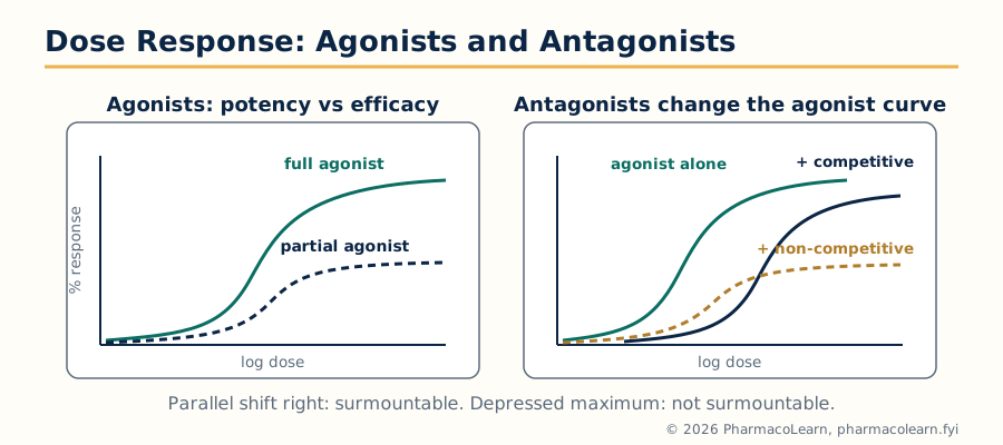 Figure: Reading a log dose response curve. A parallel shift right means competitive antagonism; a depressed maximum means the block cannot be surmounted.
1.8 Artificial enzymes
An artificial enzyme (enzyme mimic) is a synthetic molecule designed to do what an enzyme does: bind a substrate selectively and catalyse its reaction. The syllabus includes them because they are drug target chemistry viewed from the other side, and because several are used as tools and scavenger drugs:
- Cyclodextrins: ring shaped sugars with a hydrophobic cavity that binds guest molecules; used mainly as solubilising hosts, and in one case as a drug itself, sugammadex, which encapsulates the neuromuscular blocker rocuronium and reverses it.
- Catalytic antibodies (abzymes): antibodies raised against a transition state analog of a reaction bind the transition state and can accelerate the reaction; proof that binding energy alone can produce catalysis.
- Molecularly imprinted polymers: a polymer is formed around a template molecule; removing the template leaves a shaped cavity with selective binding, used in separations and sensing.
- Synthetic hosts and metallomimics: macrocycles and metal complexes designed to reproduce one step of an enzyme's chemistry, for example superoxide dismutase mimics that remove reactive oxygen species.
Artificial enzymes rarely match natural enzymes in rate, but they are robust, designable, and they teach the central lesson of this unit: selective binding, correctly arranged, is the foundation of all drug action.
Unit 2: Prodrug Design and Analog Design
2.1 The prodrug concept
A prodrug is a compound that is itself inactive, or much less active, and is converted in the body into the active drug. The idea is credited to Albert, who used the word in 1958, and to Harper, who framed drug latentiation: a drug is deliberately masked with a temporary group, the promoiety, chosen so that a chemical or enzymatic step in the body removes it at the right place and rate.
Prodrugs are designed against three classes of barrier:
THE THREE BARRIERS A PRODRUG CAN CROSS
  Pharmaceutical barriers : poor solubility, instability, bad taste
        |
  Pharmacokinetic barriers: poor absorption, first pass loss,
        |                   short action, wrong distribution
  Pharmacodynamic barriers: toxicity at non-target sites,
                            no selectivity for the target tissue
Classification by structure:
- Carrier linked prodrugs: the active drug is joined to a carrier group through a bond that the body breaks. Subtypes: bipartite (drug plus one carrier), tripartite (drug, linker and carrier, where the linker self-destructs after the first cleavage) and mutual prodrugs (two active drugs joined to each other, each the other's carrier).
- Bioprecursors: no carrier at all; the molecule itself is metabolically transformed into the active species, for example by oxidation, reduction or decarboxylation.
2.2 Carrier linked prodrugs and bioprecursors
Carrier linked examples. Enalapril is the ethyl ester of enalaprilat: the diacid enalaprilat inhibits the angiotensin converting enzyme well but is poorly absorbed, so one carboxyl group is masked as an ester, absorbed, and hydrolysed by liver esterases to the active diacid. Valaciclovir is the L-valyl ester of aciclovir: the amino acid ester is carried across the gut wall by a peptide transporter, then hydrolysed, giving several fold better oral delivery of aciclovir. Fosphenytoin is a phosphate ester prodrug of phenytoin that is freely water soluble for injection where phenytoin itself is not.
Bioprecursor examples. Levodopa crosses the blood brain barrier on an amino acid transporter and is decarboxylated to dopamine in the brain; dopamine itself cannot be given because it does not cross. Codeine is O-demethylated by CYP2D6 to morphine, which provides most of its analgesia, which is why CYP2D6 poor metabolisers get little effect and ultra rapid metabolisers are at risk of opioid toxicity. Cyclophosphamide is oxidised in the liver to an intermediate that breaks down to the cytotoxic phosphoramide mustard. Sulindac is reduced to its active sulfide.
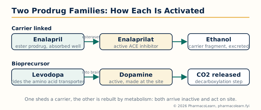 Figure: The two prodrug families. A carrier linked prodrug sheds a promoiety; a bioprecursor is itself rebuilt by metabolism into the active molecule.
2.3 Prodrugs of functional groups
The promoiety is chosen to fit the functional group being masked:
| Functional group on the drug | Common prodrug forms | Cleavage |
|---|---|---|
| Carboxylic acid | Alkyl and aryl esters, (acyloxy)alkyl esters | Esterases |
| Alcohol or phenol | Esters, carbonates, carbamates, phosphate esters | Esterases, phosphatases |
| Amine | Amides, carbamates, imines (Schiff bases), N-Mannich bases | Amidase or hydrolysis |
| Carbonyl (ketone, aldehyde) | Oximes, imines, cyclic derivatives | Hydrolysis or oxidation |
| Phosphate or phosphonate on the drug | Aryl and acyloxyalkyl masking groups | Esterases, giving the charged parent for cell entry problems |
Two practical rules follow. First, the masking bond must be stable enough to survive formulation and the stomach, yet labile enough to break in plasma, gut wall, liver or target tissue. Second, the promoiety itself must be safe: pivalate esters, for example, release pivalic acid, which is excreted as a carnitine conjugate and can lower body carnitine on prolonged use.
2.4 Prodrugs for patient acceptability
Some drugs fail simply because patients will not take them:
- Taste: chloramphenicol is intensely bitter; its palmitate ester is tasteless because it does not dissolve in saliva, and is hydrolysed in the gut to release the drug. This is the classical taste masked prodrug.
- Pain and irritation on injection: long chain esters and water soluble phosphate prodrugs reduce the irritation of insoluble or irritating drugs at the injection site.
- Gastric irritation: ester prodrugs of some acidic anti-inflammatory drugs were designed to pass the stomach intact and release the acid later, although in practice systemic prostaglandin inhibition still contributes to gastric damage, a useful warning that a prodrug cannot fix a mechanism based side effect.
- Odour and formulation acceptability: masking volatile or unstable groups improves handling and shelf presentation as well as compliance.
2.5 Prodrugs for solubility, absorption and distribution
Solubility. Adding an ionisable promoiety converts an insoluble drug into a salt forming, water soluble prodrug for parenteral use: chloramphenicol sodium succinate and fosphenytoin sodium are the standard examples. The prodrug dissolves, is injected, and hydrolysis releases a drug whose low solubility no longer matters because it is already in solution at low concentration in blood.
Absorption. Masking a polar group raises membrane permeability: enalapril (oral) versus enalaprilat (poorly absorbed) is the cleanest pair. Transporter targeting goes further: valaciclovir uses an amino acid ester to ride the intestinal peptide transporter PEPT1.
Distribution. Dipivefrin, the dipivalate ester of adrenaline, crosses the cornea far better than adrenaline itself and releases adrenaline inside the eye, giving the same effect at a fraction of the dose, with less systemic exposure. Levodopa is a distribution prodrug aimed at the blood brain barrier.
2.6 Site specific delivery and sustained action
Site specific delivery. Sulfasalazine carries 5-aminosalicylic acid (5-ASA) through the small intestine joined by an azo bond to sulfapyridine; colonic bacteria reduce the azo bond and release 5-ASA exactly where inflammatory bowel disease needs it, a prodrug switched on by the local microflora. Olsalazine joins two 5-ASA molecules the same way, removing the sulfapyridine carrier. The ADEPT concept (antibody directed enzyme prodrug therapy) generalises the idea: an enzyme is targeted to a tumour by an antibody, and a harmless prodrug given afterwards is converted to a cytotoxic drug only at the tumour.
Sustained action. Esterifying a drug with a long chain fatty acid makes an oily depot: fluphenazine decanoate and haloperidol decanoate release their antipsychotic slowly from an intramuscular oil depot over weeks, a prodrug answer to the compliance problem in chronic schizophrenia. Insulin analogs that precipitate or bind albumin after injection apply a related slow release logic at the protein level.
2.7 Rationale and practical considerations of prodrug design
PRODRUG DESIGN DECISION SEQUENCE
  Name the barrier precisely        (solubility? absorption? site?)
        |
  Choose the group to mask          (which functional group, which
        |                            promoiety fits it?)
  Check the cleavage step           (which enzyme or pH, where, how
        |                            fast relative to the journey?)
  Check the promoiety               (inactive, non toxic, excreted)
        |
  Test stability vs activation      (shelf stable, body labile)
The ideal prodrug is chemically stable in the bottle, converts quantitatively and predictably at the intended site, produces an inert promoiety, and does not create a new problem while solving the old one. Practical failures come from conversion that is too fast (the prodrug behaves like the drug, barrier unsolved), too slow (drug levels never reached), or dependent on an enzyme that varies between patients, as codeine depends on CYP2D6.
2.8 Combating drug resistance
Drug resistance is the inherited ability of a microbe or a cancer cell to survive a drug concentration that previously killed it. Its main mechanisms:
| Mechanism | Bacterial example | Cancer example |
|---|---|---|
| Target modified so the drug binds poorly | Altered penicillin binding proteins in MRSA | Mutated BCR-ABL gatekeeper (T315I) resisting imatinib |
| Drug destroyed or modified | Beta-lactamases hydrolyse penicillins | Enzyme induction and detoxification in tumour cells |
| Drug pumped out (efflux) | Tetracycline efflux pumps | P-glycoprotein (MDR1) expelling many anticancer drugs |
| Drug kept out (reduced uptake) | Porin loss in Gram negative bacteria | Reduced folate carrier loss resisting methotrexate |
| Bypass of the blocked step | Alternative folate routes | Alternative signalling pathways bypass a blocked kinase |
Genetic principles. Resistance genes arise by mutation and, decisively in bacteria, spread by horizontal gene transfer: plasmids carrying resistance genes pass between bacteria by conjugation, and transformation and transduction move genes as well. Selection does the rest: every course of antibiotic kills the susceptible and leaves the resistant. In cancer, resistance arises from the genetic instability of the tumour, which generates resistant subclones that treatment then selects.
Strategies in antibiotics: combine a drug with an inhibitor of the resistance enzyme (amoxicillin with clavulanic acid, a suicide inhibitor of beta-lactamases, Unit 4); use combinations that require two independent resistance events at once (tuberculosis therapy); design analogs that the resistance enzyme handles poorly (later generation cephalosporins); and reduce selection pressure by correct dosing and duration.
Strategies in anticancer therapy: combination chemotherapy with non cross resistant drugs (the logic of regimens such as CHOP); next generation inhibitors designed against the resistant mutant itself (osimertinib was designed for the T790M mutant EGFR that resists first generation inhibitors); and attacking the efflux or bypass mechanism, although P-glycoprotein inhibitors have so far disappointed in the clinic because the pump also protects normal tissues.
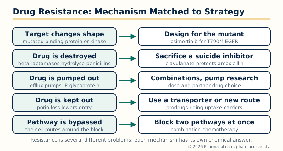 Figure: Resistance is a set of distinct mechanisms, and each has its own chemical answer. One drug rarely answers two mechanisms at once, which is why combinations work.
2.9 Analog design: modifying a lead molecule
Analog design (lead modification) is the systematic structural variation of a lead to improve the properties the lead lacks: potency, selectivity, duration, solubility, safety. The medicinal chemist works around a pharmacophore, the arrangement of features (hydrogen bond donors and acceptors, charged groups, hydrophobic regions, at defined distances) that the target recognises, and varies everything else one change at a time so that the effect of each change can be read. The strategies in the rest of this unit are the working tools: bioisosteric replacement (2.10), and the structural variations of 2.11.
2.10 Bioisosteric replacement strategies
A bioisostere is a group that can replace another group in a molecule with a similar biological result, because the target reads the two as alike in the property that matters (size, charge, hydrogen bonding, electron distribution), while other properties such as stability or solubility improve.
Classical bioisosteres come from the old isosterism rules, atoms and groups with the same number of valence electrons (Grimm's hydride displacement law) or similar size and shape (Erlenmeyer's ring equivalents):
| Classical replacement | Examples |
|---|---|
| -CH3, -NH2, -OH, -SH (one row of the hydride series) | Swapping along the series changes acidity and hydrogen bonding in small steps |
| -CH= exchanged for -N= in a ring | Pyridine for benzene in a ring system |
| Ring equivalents | Benzene, thiophene, furan and pyridine rings exchanged for one another |
| Halogen for hydrogen or CF3 | Fluorine for hydrogen (similar size, very different electronics) |
| -O-, -S-, -NH-, -CH2- in a chain | Linker exchanges that change bond angles and metabolism |
Non-classical bioisosteres do not look alike on paper but present the same key interaction: the tetrazole ring for a carboxylic acid is the famous case, since both are acidic and anionic at body pH, but the tetrazole is more lipophilic and resists glucuronidation; replacing the acid of an early angiotensin antagonist lead with tetrazole produced losartan. Others: the cyanoguanidine group for thiourea in cimetidine (section 3.5), sulfonamide and carboxamide exchanges, and replacing an ester with a 1,2,4-oxadiazole or other ring that keeps the geometry but refuses hydrolysis.
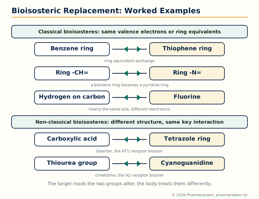 Figure: Bioisosteres keep the interaction the target reads and change the properties the body imposes: stability, solubility, duration.
2.11 Other analog design strategies
- Rigid analogs (conformational restriction): locking a flexible lead into the shape it adopts at the target removes the entropic cost of folding on binding and often raises potency and selectivity; it also tests the active conformation. Ring closure and added double bonds are the usual tools.
- Alteration of chain branching: adding or moving a methyl branch changes fit and blocks or exposes metabolic sites; branching near a functional group also slows hydrolysis by steric shielding.
- Changes in ring size: expanding or contracting a ring repositions every substituent on it; a six membered ring lead may become a better five or seven membered analog.
- Ring position isomers: moving a substituent around a ring (ortho, meta, para) scans the geometry of the pocket at almost no synthetic cost and is one of the first scans in any program.
- Design of stereoisomers and geometric isomers: making and testing each enantiomer separately (Unit 3), and locking cis or trans geometry, as in the stilbene framework where the trans isomer diethylstilbestrol carries the oestrogenic activity.
- Fragments of a lead molecule (molecular dissection): cutting a complex lead down to the smallest fragment that keeps activity. Morphine dissected stepwise gave the simpler pethidine: the anilinopiperidine fragment retained analgesia and was far easier to make and vary.
- Variation in interatomic distance (homologation): lengthening or shortening the chain between two key groups finds the distance the target prefers. The antipsychotic phenothiazines need three carbons between ring nitrogen and side chain nitrogen; two carbons gives a different (antihistaminic) profile, a classical distance lesson.
Unit 3: Medicinal Chemistry Aspects of Drug Classes and Stereochemistry
3.1 How a drug class is studied systematically
Every class in this unit is studied the same way, and examination answers should follow the same order:
- Classification: the chemical or pharmacological families inside the class.
- Mechanism of action: the target, and how engaging it produces the clinical effect.
- SAR: the structural features that activity requires, tolerates or forbids, read from analog series.
- Synthesis: one representative, preferably new generation, drug built step by step, because a synthesis is SAR made practical: it shows which ring and which group is installed when, and why.
A new generation drug in each class below means the later member whose design fixed a named fault of the older drugs: poorer selectivity, shorter action, worse side effects or resistance.
3.2 Anti-hypertensive drugs
Groups: ACE inhibitors, angiotensin receptor blockers (ARBs), calcium channel blockers, beta blockers, diuretics, direct renin inhibitors and others.
ACE inhibitors. Mechanism: block the angiotensin converting enzyme, so less angiotensin II (vasoconstrictor) is made and less bradykinin is destroyed. SAR: the inhibitor must present a group that binds the active site zinc ion (a carboxylate, or the sulfhydryl of the first drug captopril), a carbonyl that hydrogen bonds in the site, and a carboxylate that mimics the substrate C-terminus. Captopril's free sulfhydryl gave rash and taste disturbance; enalapril replaced it with a second carboxylate and is given as its monoethyl ester prodrug (Unit 2). Lisinopril is the lysine analog of enalaprilat and needs no activation.
ARBs. Losartan blocks the angiotensin II AT1 receptor. Its design is a Unit 2 case study: an imidazole based lead carried a carboxylic acid; replacing the acid with a tetrazole (a non-classical bioisostere) and mounting it on a biphenyl frame gave losartan. A hydroxymethyl group on the imidazole is oxidised in the body to a carboxylic acid metabolite (EXP3174) that is more potent and longer acting than losartan itself.
Calcium channel blockers. Amlodipine is a 1,4-dihydropyridine that blocks L-type calcium channels in vascular smooth muscle. Dihydropyridine SAR: the ring nitrogen must stay unsubstituted, ester groups at the 3 and 5 positions tune potency, and different esters on the two sides (as in amlodipine) give a long acting, once daily drug.
Synthesis of amlodipine (outline): a Hantzsch dihydropyridine synthesis (the named reaction is in MPC102T) in which 2-chlorobenzaldehyde, two beta-keto ester components (one carrying the protected aminoethoxy side chain) and ammonia close the dihydropyridine ring in one multicomponent step; deprotection then frees the side chain amine, which is what makes amlodipine basic, long lived and well tolerated.
3.3 Psychoactive drugs
Antipsychotics. Typical agents block dopamine D2 receptors. Chlorpromazine SAR is classical: a phenothiazine ring with an electron withdrawing substituent at position 2 (chlorine), and a three carbon chain to a terminal dimethylamino group (section 2.11). Atypical agents such as risperidone combine D2 blockade with strong serotonin 5-HT2A blockade, giving fewer extrapyramidal effects; risperidone joins a benzisoxazole ring to a piperidine bearing a pyrimidinone system and is converted in the body to the equally active paliperidone.
Antidepressants. The selective serotonin reuptake inhibitors (SSRIs) block the serotonin transporter. Fluoxetine is a phenoxypropylamine: SAR tolerates the trifluoromethylphenoxy group and a secondary methylamine; it is a racemate whose enantiomers and the long lived metabolite norfluoxetine all contribute. Tricyclics (imipramine) block both serotonin and noradrenaline uptake but also hit many other receptors, which is why the selective agents displaced them.
Synthesis of fluoxetine (outline): a Mannich type route builds the 3-(methylamino)-1-phenylpropan-1-ol skeleton (acetophenone, formaldehyde and methylamine give the Mannich base, which is reduced to the amino alcohol); the alcohol is then etherified with 4-chlorobenzotrifluoride under basic conditions to install the trifluoromethylphenoxy group.
3.4 Anticonvulsant drugs
Mechanisms: most anticonvulsants stabilise neuronal membranes by blocking voltage gated sodium channels in their inactivated state (phenytoin, carbamazepine, lamotrigine), enhancing GABA inhibition (benzodiazepines, vigabatrin), or both (valproate). Newer agents use new targets: levetiracetam binds the synaptic vesicle protein SV2A.
Phenytoin is 5,5-diphenylhydantoin; the hydantoin ring with two phenyl groups is its pharmacophore, and it is made by the Bucherer-Bergs reaction of benzil chemistry routes or from benzophenone with urea and cyanide chemistry in the classical preparation. Carbamazepine is a tricyclic relative of the antidepressants with a carbamoyl group on the ring nitrogen. Lamotrigine, a new generation agent, is a diaminotriazine that blocks sodium channels and reduces glutamate release, with a broad spectrum including absence seizures where phenytoin fails.
3.5 H1 and H2 receptor antagonists
H1 antagonists (antihistamines). First generation agents (chlorpheniramine, promethazine) cross into the brain and sedate. Second generation agents were designed not to: cetirizine is the carboxylic acid metabolite of hydroxyzine, a zwitterion at body pH that crosses the blood brain barrier poorly, so it blocks peripheral H1 receptors with little sedation, and it is sold as the single active enantiomer levocetirizine as well (Unit 3.10).
H2 antagonists suppress gastric acid by blocking histamine H2 receptors on parietal cells, and their discovery is the classic of rational design. Burimamide, built by modifying histamine itself, worked but was weak orally. Adding a methyl group and the thioether chain gave metiamide, potent but causing granulocytopenia from its thiourea group. Replacing thiourea with its bioisostere cyanoguanidine gave cimetidine, safe and effective: the design chain histamine to burimamide to metiamide to cimetidine is analog design taught by example. Ranitidine then replaced the imidazole ring with a furan bearing a dimethylaminomethyl group and used a nitroethenediamine in place of cyanoguanidine, giving a more potent agent; famotidine is more potent still.
3.6 COX-1 and COX-2 inhibitors
Mechanism: non-steroidal anti-inflammatory drugs inhibit cyclo-oxygenase (COX), the enzyme that converts arachidonic acid to prostaglandins (Unit 5). Aspirin is unique: it acetylates a serine (Ser530) in the COX channel irreversibly, so its antiplatelet effect lasts the life of the platelet.
SAR of the classical agents: the arylalkanoic acids (ibuprofen, naproxen, diclofenac) need the acidic group held at a defined distance from a flat aromatic surface that sits in the hydrophobic COX channel; ibuprofen's alpha methyl group is largely a spacer, and its inactive R enantiomer is converted to the active S form in the body (section 3.10).
COX-2 selective inhibitors. COX-1 protects the stomach lining while COX-2 is induced at inflammation sites, so selective COX-2 blockade promised anti-inflammatory action without gastric damage. Celecoxib achieves it with a pyrazole ring carrying a sulfonamide group that fits a side pocket present in COX-2 but blocked in COX-1. The class lesson includes the failure: rofecoxib was withdrawn because selective COX-2 blockade also suppresses protective prostacyclin in blood vessels, raising cardiovascular risk, the comparison that every student of this unit should be able to explain.
3.7 Adrenergic and cholinergic agents
Adrenergic agonists. The catecholamine frame (phenethylamine with catechol hydroxyls) is read by all adrenoceptors; selectivity is engineered by substitution. Salbutamol is the model: replacing one catechol hydroxyl by a hydroxymethyl group (a bioisosteric change) makes it resist COMT methylation, and a bulky tert-butyl group on nitrogen directs it to beta-2 receptors, giving a long acting bronchodilator. Beta blockers share the aryloxypropanolamine frame of propranolol; atenolol's para substituent pattern gives beta-1 selectivity and water solubility, hence renal excretion and once daily dosing.
Cholinergic agents. Acetylcholine itself is useless as a drug (hydrolysed in seconds, active everywhere). Bethanechol fixes both faults of the muscarinic agonist: a carbamate ester resists cholinesterase where acetate fails, and a beta methyl group gives muscarinic over nicotinic selectivity and blocks hydrolysis further. Anticholinesterases work from the other side: donepezil reversibly inhibits acetylcholinesterase in the brain and is the standard symptomatic treatment in Alzheimer's disease, while the carbamate physostigmine carbamylates the enzyme reversibly and organophosphates phosphorylate it essentially irreversibly.
3.8 Antineoplastic agents
Groups and mechanisms: alkylating agents damage DNA directly; antimetabolites impersonate the building blocks of DNA synthesis; intercalators and topoisomerase inhibitors block DNA handling; antimitotics attack the spindle; targeted small molecules block the specific signalling protein a cancer depends on.
Antimetabolites: methotrexate mimics folic acid and inhibits dihydrofolate reductase (Unit 4), starving dividing cells of thymidylate; 5-fluorouracil is converted to a metabolite that irreversibly inhibits thymidylate synthase and is also built into RNA. Alkylating agents: cyclophosphamide is a bioprecursor prodrug (Unit 2) oxidised in the liver to phosphoramide mustard, which cross links DNA.
New generation, targeted design: imatinib inhibits the BCR-ABL tyrosine kinase that drives chronic myeloid leukaemia, a drug designed against a defined molecular target rather than against dividing cells generally. Its resistance story (the T315I gatekeeper mutation, section 2.8) and the later inhibitors designed to defeat that mutation make it the complete case study of this subject: target, design, success, resistance, redesign.
3.9 Antiviral agents
Aciclovir is a guanosine analog missing part of the sugar ring (an acyclic side chain instead of ribose). Its selectivity is built in two steps: it is phosphorylated efficiently only by the herpes virus thymidine kinase, so the active form concentrates in infected cells, and the resulting triphosphate then both inhibits the viral DNA polymerase and terminates the growing DNA chain because the chain has no 3-prime hydroxyl to continue from.
Antiretrovirals: zidovudine (AZT), a thymidine analog with an azido group in place of the 3-prime hydroxyl, terminates HIV reverse transcription the same way; modern therapy combines nucleoside analogs with non-nucleoside reverse transcriptase inhibitors (which bind an allosteric pocket of the enzyme) and protease inhibitors (Unit 4 transition state design, saquinavir).
Oseltamivir inhibits influenza neuraminidase, the enzyme that releases new virus particles from the cell; it was designed as a carbocyclic mimic of the sialic acid transition state, and is given as an ethyl ester prodrug hydrolysed to the active carboxylate.
3.10 Stereochemistry and drug action
Molecules with a chiral centre exist as enantiomers, mirror images with identical physical properties in an ordinary environment but different behaviour in a chiral one, and the body is chiral throughout: its enzymes, receptors and transporters are built from L-amino acids. The Easson-Stedman three point model (section 1.6) explains why one enantiomer can fit its receptor at three points while its mirror image manages only two.
| Stage | Enantioselectivity example |
|---|---|
| Absorption | L-DOPA is carried by an amino acid transporter; D-DOPA is not, one reason levodopa is the drug |
| Distribution | Plasma protein binding differs between warfarin enantiomers, changing free concentrations |
| Metabolism | S-warfarin is the more potent anticoagulant and is cleared mainly by CYP2C9; R-warfarin uses other enzymes, so interactions differ by enantiomer |
| Elimination and safety | D-penicillamine is the therapeutic chelator; the L form is far more toxic. Naproxen is marketed as the single S enantiomer, the active COX inhibitor |
Case studies every student should know:
- Thalidomide: sold as a racemate; one enantiomer is sedative, the other teratogenic, but the two interconvert in the body, so giving a single enantiomer would not have prevented the tragedy. The honest lesson is about testing in pregnancy, not just separation.
- Ibuprofen: only the S enantiomer inhibits COX, but the body inverts much of the R dose into S (a unidirectional metabolic inversion), so the racemate works and the single enantiomer (dexibuprofen) is an option, not a necessity.
- Chiral switches: omeprazole was remarketed as its S enantiomer esomeprazole, cetirizine as levocetirizine, and ofloxacin as levofloxacin (the antibacterial activity of ofloxacin lives almost entirely in the S form). A chiral switch is justified when the single enantiomer is genuinely cleaner, faster or safer, not merely newer.
- Geometric isomerism: diethylstilbestrol is active in the trans form, whose two hydroxyls sit at the distance the oestrogen receptor expects; the cis form is far weaker, a distance and geometry lesson that links back to section 2.11.
Unit 4: Rational Design of Enzyme Inhibitors
4.1 Enzyme kinetics in brief
Enzymes are the second great class of drug targets, and inhibitor design starts from their kinetics. For the simple scheme in which enzyme E binds substrate S, forms the ES complex and releases product P, the Michaelis-Menten equation gives the rate:
v = Vmax x [S] / (Km + [S])

  Km   : substrate concentration at half maximal rate; low Km
         means tight substrate binding
  Vmax : rate when the enzyme is saturated with substrate
  kcat : turnover number, products per enzyme per second at
         saturation (Vmax = kcat x total enzyme)
Reciprocal plotting (Lineweaver-Burk: 1/v against 1/[S]) turns the curve into a straight line whose intercepts give Km and Vmax, and, more importantly for this unit, turns each inhibition pattern into a recognisable picture (section 4.2). An inhibitor is quantified by its IC50 in an assay and, more fundamentally, by its inhibition constant Ki.
4.2 Principles of enzyme inhibition
| Type | Binds | Effect on Km | Effect on Vmax | Diagnostic picture (Lineweaver-Burk) |
|---|---|---|---|---|
| Competitive | Active site, free enzyme only | Increased (apparent) | Unchanged | Lines meet on the 1/v axis |
| Non-competitive | Separate site, enzyme or ES complex | Unchanged | Decreased | Lines meet on the 1/[S] axis |
| Uncompetitive | ES complex only | Decreased | Decreased | Lines are parallel |
| Mixed | Either, with different affinities | Changed either way | Decreased | Lines meet off both axes |
| Irreversible | Covalent bond at or near the active site | Time dependent loss of active enzyme | Falls with time | Activity does not return on dilution or dialysis |
Two distinctions cut across the table. Reversible versus irreversible is about the bond: non-covalent complexes dissociate, covalent ones do not. Suicide substrates (mechanism based inactivators) are a special irreversible class: the enzyme's own catalytic machinery converts the harmless inhibitor into the reactive species that destroys it, as clavulanic acid does to beta-lactamases (section 4.6).
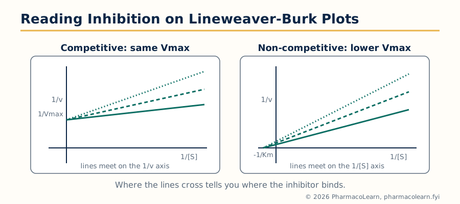 Figure: Inhibition patterns are read off reciprocal plots. Where the lines cross tells you where the inhibitor binds.
4.3 Enzyme inhibitors in medicine
| Inhibitor drug | Enzyme inhibited | Mode | Clinical use |
|---|---|---|---|
| Enalaprilat, lisinopril | Angiotensin converting enzyme | Competitive, zinc binding | Hypertension, heart failure |
| Atorvastatin and other statins | HMG-CoA reductase | Competitive with HMG-CoA | Cholesterol lowering |
| Benzylpenicillin (penicillin G) | Bacterial DD-transpeptidase (penicillin binding protein) | Irreversible acylation of the active serine | Bacterial infection |
| Aspirin | Cyclo-oxygenase | Irreversible acetylation | Pain, inflammation, antiplatelet |
| Allopurinol | Xanthine oxidase | Converted to oxypurinol, a tight binding product | Gout |
| Methotrexate | Dihydrofolate reductase | Tight binding competitive | Cancer, rheumatoid arthritis |
| Clavulanic acid | Beta-lactamases | Suicide substrate | Protects amoxicillin |
| Saquinavir and later protease inhibitors | HIV protease | Transition state mimic | HIV infection |
The statin story shows lead discovery inside this unit: compactin (mevastatin), isolated from a fungus, already contained the HMG-like portion that blocks the reductase; synthetic statins kept that warhead geometry and improved everything around it.
4.4 Enzyme inhibitors in basic research
Inhibitors are also laboratory instruments. A selective inhibitor lets a researcher switch one enzyme off in a living system and read its function from what stops, which is target validation done chemically (section 1.3). Affinity labels and activity based probes, irreversible inhibitors carrying a reporter group, tag the active enzymes in a mixture and are used to map enzyme families and to fish out the unknown target of a drug found by screening. Kinetic patterns (section 4.2) run in reverse are how an unknown inhibitor's binding site is located before any structure is available. Many drug targets were first taken seriously because a research inhibitor showed a clean, interpretable effect in cells.
4.5 Rational design of non-covalent inhibitors
The design cycle, once the target enzyme is chosen and validated:
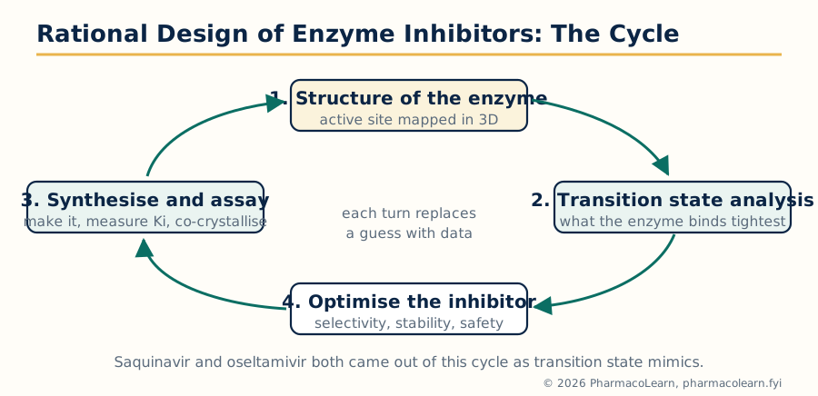 Figure: The design cycle. Every turn replaces a guess with a structure and an assay result.
The strongest non-covalent strategy is the transition state analog: enzymes bind the transition state of their reaction far more tightly than the substrate, so a stable molecule shaped and charged like the transition state is bound with exceptional affinity. Saquinavir carries a hydroxyethylene unit that mimics the tetrahedral intermediate of peptide bond cleavage by the HIV protease; oseltamivir (section 3.9) mimics the flattened sialic acid transition state of neuraminidase. Other non-covalent strategies design into allosteric pockets beside the active site (the non-nucleoside pocket of HIV reverse transcriptase) or exploit a single differing residue between two related enzymes to buy selectivity, as the COX-2 side pocket was exploited in section 3.6.
4.6 Rational design of covalent inhibitors
Covalent inhibition was avoided for decades from fear of indiscriminate reactivity, then returned deliberately once the design rules were understood:
- Two step logic: the inhibitor first binds reversibly and selectively in the pocket (a good non-covalent inhibitor), and only then does a mildly reactive warhead group react with a nearby amino acid side chain. Selectivity comes from the binding step; the covalent step adds duration.
- Warheads and targets: the acrylamide group of afatinib and osimertinib reacts with a cysteine at the rim of the EGFR ATP site; ibrutinib's acrylamide reacts with Cys481 of Bruton's tyrosine kinase. Aspirin's acetyl group and penicillin's beta-lactam ring are the classical covalent warheads, and the nitrile of some modern inhibitors (for example in cathepsin and DPP-4 chemistry) forms reversible covalent bonds, a halfway house.
- Advantages: potency at low dose, pharmacological action that outlives the drug's plasma presence (effect ends when the body resynthesises the enzyme, as with aspirin and platelets), and activity against some resistant mutants because the bond does not depend on binding affinity alone.
- Risks, stated honestly: a warhead that is too reactive modifies off target proteins and can trigger immune toxicity; a cysteine that mutates removes the anchor (the C481S mutation is a known ibrutinib resistance route). Modern covalent design therefore keeps warheads weakly reactive and lets binding do the work.
- Suicide substrates as the limiting case: clavulanic acid is processed by beta-lactamase into a species that permanently disables it, so the enzyme destroys itself with its own mechanism; gamma-aminobutyric acid transaminase is inactivated the same way by vigabatrin.
Unit 5: Peptidomimetics and Eicosanoid Chemistry
5.1 Why peptides make difficult drugs
Peptides are attractive starting points because biology uses them as precise signals (hormones, neurotransmitters), so a peptide lead often arrives with potency and selectivity built in. As drugs they are poor: digestive and plasma proteases hydrolyse their amide bonds within minutes; their size and hydrogen bonding surface keep them out of membranes, so oral absorption is negligible; kidneys clear them quickly; and many are expensive to make. A peptidomimetic is a molecule designed to reproduce the peptide's active shape and side chain display on a scaffold the body cannot digest so easily. The design strategies of this unit answer the three faults in order: stop the enzymes, improve the journey, keep the shape.
5.2 Peptidomimetics: concept and therapeutic value
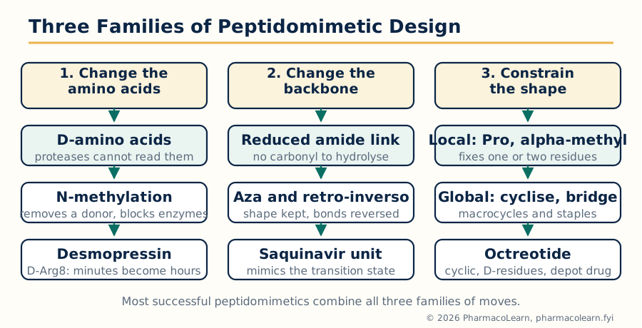 Figure: Three families of moves turn a fragile peptide into a drug-like molecule. Most successful peptidomimetics combine all three.
The therapeutic value is concrete: peptidomimetics have delivered renin and HIV protease inhibitor chemistry, thrombin inhibitors, and the whole class of constrained peptide drugs. Desmopressin, a deaminated arginine vasopressin analog with a D-amino acid, lasts hours where vasopressin lasts minutes. Octreotide, a cyclic octapeptide analog of somatostatin with D-amino acids and a reduced terminal alcohol, turned a two minute hormone signal into a monthly depot drug. Neither is a small molecule; both are peptides rebuilt by the strategies below until they behaved like drugs.
5.3 Design by manipulation of the amino acids
PEPTIDE OPTIMISATION LOOP
  Replace one residue at a time (alanine scan finds which
  side chains the target actually reads)
        |
  Protect the cleavage sites those assays reveal
        |
  Re-test activity, stability and solubility together
        |
  Repeat: every gain in stability must not cost the shape
- D-amino acids: proteases are built for L-amino acids; a D-residue at or near the cleavage site is not recognised, and it also bends the chain differently, which can help or must be compensated. Desmopressin's D-Arg8 is the model case.
- N-methylation: methylating a backbone nitrogen removes a hydrogen bond donor, blocks protease binding and can improve membrane passage; cyclosporine is extensively N-methylated by nature.
- Unnatural amino acids: residues outside the coded twenty add side chains no protease expects and new interactions with the target; alpha-methyl amino acids also restrict local conformation (section 5.5).
- Side chain surgery: shortening, lengthening or capping side chains, and replacing a charged terminus, tunes potency and stability independently of the backbone.
5.4 Design by modification of the peptide backbone
The amide bond is both the protease target and a strong hydrogen bonding unit, so backbone isosteres trade one property against the other:
| Backbone change | What it does | Cost to watch |
|---|---|---|
| Reduced amide, -CH2-NH- in place of -CO-NH- | Not hydrolysed by proteases; keeps chain length | Loses the carbonyl hydrogen bond acceptor; the amine is basic and may be protonated |
| Ketomethylene, -CO-CH2- | Keeps the carbonyl, removes the nitrogen | Loses the NH donor |
| Azapeptide (nitrogen replaces the alpha carbon) | Resists proteolysis, biases conformation | Changes side chain presentation geometry |
| Depsipeptide (ester replaces one amide) | Alters solubility and conformation; found in natural products | The ester is itself hydrolysable, but by different enzymes |
| Retro-inverso (reversed sequence of D-amino acids) | Side chains keep roughly the original display while every amide is reversed | End groups need correction; not a perfect mirror of binding |
| Alkene or alkane isostere for the amide | Fully non hydrolysable link | Removes both hydrogen bonding partners of that bond |
The transition state analogs of Unit 4 (the hydroxyethylene unit in saquinavir) are this table in action: one backbone unit of a peptide substrate was replaced by a non cleavable mimic of the reaction's high point, and a drug resulted.
5.5 Conformational constraints, local and global
A flexible peptide pays an entropy penalty when it folds into its bound shape, and presents many shapes to proteases. Constraining it pre-pays that cost:
- Local constraints act on one or two residues: proline and its analogs fix the preceding bond angle; alpha-methyl and other alpha,alpha-disubstituted amino acids narrow the allowed shapes; a D-amino acid in a turn position stabilises beta turns.
- Global constraints tie distant parts of the molecule together: macrocyclisation head to tail or side chain to side chain (cyclosporine is a natural macrocycle, and its ring is a large part of why it is orally available at all); disulfide bridges (oxytocin and insulin use them, although disulfides can be reduced in the body and are sometimes replaced by carbon or thioether bridges); lactam bridges between lysine and acidic side chains; and hydrocarbon staples that brace an alpha helix in peptide inhibitors of protein protein interactions.
- The balance to strike: constraint helps only when the locked shape is the active shape; locking the wrong conformation is worse than flexibility, which is why constraint follows structure determination of the bound peptide, not guesswork.
5.6 Chemistry of prostaglandins
The eicosanoids are local hormones made from arachidonic acid, a 20 carbon polyunsaturated fatty acid released from membrane phospholipids by phospholipase A2. Two enzyme families then compete for it:
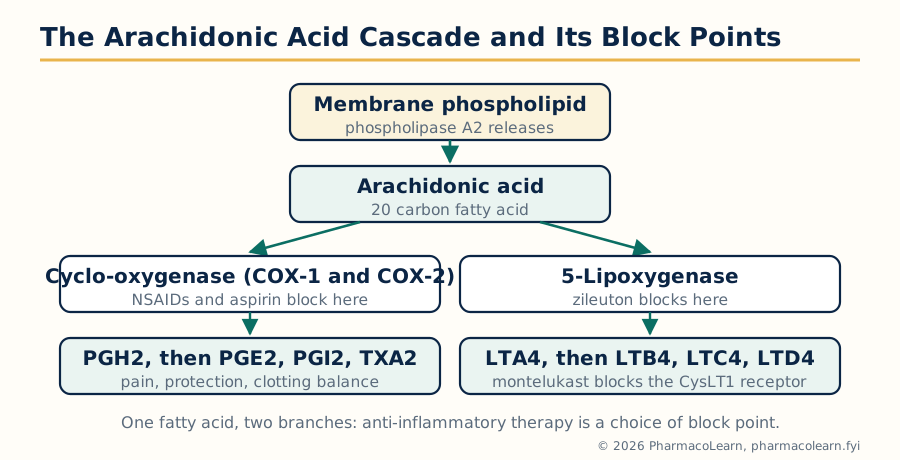 Figure: One fatty acid, two great branches. Most anti-inflammatory pharmacology is a choice of where to block this diagram.
Prostaglandin structure and chemistry. Prostaglandins are 20 carbon fatty acids carrying a five membered ring: the letter (E, F, D, I and others) names the ring's oxygen pattern, the subscript counts the double bonds. The cyclo-oxygenase pathway first builds the endoperoxide PGH2, the shared parent, which tissue specific enzymes convert to PGE2 (pain sensitisation, fever, gastric protection), PGF2alpha (uterine and luteal actions, and intraocular pressure control), PGD2, prostacyclin PGI2 and thromboxane A2 (next sections).
Prostaglandin drugs. Natural prostaglandins are destroyed in minutes by 15-hydroxyprostaglandin dehydrogenase; analog design (Unit 2 methods) fixed that: misoprostol is a PGE1 analog with a methyl ester and a moved hydroxyl group that resists the dehydrogenase, used for gastric protection and in obstetrics; latanoprost is a PGF2alpha analog given as an isopropyl ester prodrug eye drop for glaucoma. Carboprost (a 15-methyl PGF2alpha) resists oxidation at the 15 position, the literal weak point, because the added methyl group blocks the enzyme that starts prostaglandin destruction.
5.7 Leukotrienes
The 5-lipoxygenase branch converts arachidonic acid to the unstable epoxide LTA4, the parent of the leukotrienes. LTB4 is a powerful neutrophil attractant. The cysteinyl leukotrienes LTC4, LTD4 and LTE4 carry amino acid derived substituents (the peptide part is what cysteinyl means here), are released from mast cells and eosinophils, and are the substances once called slow reacting substance of anaphylaxis (SRS-A): potent bronchoconstrictors and drivers of mucus secretion and vascular leak in asthma.
Drugs of this branch: montelukast and zafirlukast block the CysLT1 receptor and are taken orally for asthma prophylaxis; zileuton inhibits 5-lipoxygenase itself, cutting off production of all leukotrienes at the branch point, at the price of liver monitoring. The comparison is a clean design contrast: block the factory, or block the receptor that reads its main product.
5.8 Thromboxanes and the prostacyclin balance
Platelets convert PGH2 to thromboxane A2 (TXA2), a potent platelet aggregator and vasoconstrictor with a half life of about 30 seconds before it hydrolyses to the inactive TXB2. Blood vessel endothelium converts the same PGH2 to prostacyclin (PGI2), which opposes TXA2 point for point: it inhibits platelet aggregation and dilates vessels. Haemostasis reads the balance between them.
Low dose aspirin exploits the asymmetry of the two systems: it irreversibly acetylates platelet COX (section 3.6), the anucleate platelet cannot make new enzyme, so TXA2 production stays off for the platelet's life, while nucleated endothelial cells resynthesising COX restore PGI2. The same balance explains the rofecoxib lesson of section 3.6: blocking COX-2 alone suppresses endothelial PGI2 while leaving platelet COX-1 and TXA2 intact, tipping the scale towards clotting.
Frequently Asked Questions
Q: What is the difference between a hit, a lead and a drug candidate? A hit is a compound with confirmed, reproducible activity against the target in a screening assay. A lead is a hit series judged worth optimising: it has real potency, a workable structure and no fatal liability. A drug candidate is the optimised end product of lead optimisation, good enough in potency, selectivity, pharmacokinetics and early safety to enter preclinical and then clinical development.
Q: Why is target validation done before screening begins? Because screening and optimisation are the expensive stages. Validation gathers genetic, expression, tool compound, animal and ideally human evidence that changing the target's activity changes the disease, so the programme spends its chemistry budget on a target that can actually deliver a medicine. Targets with human genetic support succeed in the clinic at a notably higher rate.
Q: What separates affinity from efficacy at a receptor? Affinity is how tightly a drug binds, measured by Kd. Efficacy is what the bound drug does, whether it activates the receptor. A full agonist has both, an antagonist has affinity with zero efficacy, and a partial agonist has affinity with submaximal efficacy, which is why a partial agonist can also antagonise a full agonist.
Q: How do carrier linked prodrugs and bioprecursors differ? Give one example of each. A carrier linked prodrug joins the drug to a removable promoiety: enalapril is the ethyl ester that esterases convert to enalaprilat. A bioprecursor has no carrier and is itself metabolically rebuilt into the active drug: levodopa is transported into the brain and decarboxylated to dopamine.
Q: Tetrazole in losartan is called a non-classical bioisostere of a carboxylic acid. Why does the exchange work? Both groups are acidic and exist as anions at body pH, with a similar spread of negative charge, so the AT1 receptor reads them alike. The tetrazole is more lipophilic and is not cleared by glucuronidation the way the acid was, so duration and exposure improve while binding is retained: the definition of a successful bioisosteric replacement.
Q: How does clavulanic acid combat beta-lactam resistance? Clavulanic acid is itself a weak antibacterial but an excellent suicide substrate for bacterial beta-lactamases: the enzyme processes it into a species that permanently disables the enzyme. Given with amoxicillin, it sacrifices itself to the resistance enzyme so the antibiotic survives to reach its penicillin binding protein targets.
Q: What does SAR mean, and how is it established? Structure activity relationship: the pattern connecting structural features of a series of analogs to their biological activity. It is established by varying one feature at a time around a lead, synthesising and testing the series, and reading which groups are essential (removing them kills activity), which are tolerated, and which improve potency or properties. Every class study in Unit 3 is presented as classification, mechanism, SAR and synthesis in that order.
Q: Why would a drug be sold as a single enantiomer instead of the racemate? Because enantiomers can differ at every pharmacokinetic and pharmacodynamic stage: levofloxacin carries almost all the antibacterial activity of ofloxacin, levocetirizine is the active half of cetirizine, and D-penicillamine avoids the toxicity of its mirror image. A single enantiomer is justified when it is genuinely more active, cleaner or safer, as in those cases, not merely when it is patentably newer.
Q: Competitive and non-competitive inhibition look different on a Lineweaver-Burk plot. How? A competitive inhibitor raises apparent Km and leaves Vmax unchanged, so the lines for different inhibitor concentrations meet on the 1/v axis. A non-competitive inhibitor lowers Vmax and leaves Km unchanged, so the lines meet on the 1/[S] axis. Uncompetitive inhibition lowers both and gives parallel lines.
Q: What three design moves turn a peptide into a peptidomimetic drug? Manipulate the amino acids (D-residues, N-methylation, unnatural side chains) to defeat proteases; modify the backbone (reduced amide, ketomethylene, aza and other isosteres) so the cleavable bonds disappear while the shape is kept; and constrain the conformation, locally with proline or alpha-methyl residues or globally by cyclisation and bridges, so the molecule is pre-organised into its active shape. Desmopressin and octreotide combine all three.
Complete MPC103T notes, all 5 units in one PDF, free to keep.
Other M.Pharm subjects: MPH101T · MPC101T · MPL101T · MPH102T · MPH103T · MPH104T · MPL102T · MPL103T · MPL104T · MPC102T · MPC103T · MPC104T · MPH201T · MPH202T · MPH203T · MPH204T · MPC201T · MPC202T · MPC203T · MPC204T · M.Pharm