
MPP102T — Pharmacotherapeutics-I
4 hrs/week, 4 credits, 60 hours, 100 marks. Notes follow the M.Pharm syllabus, unit by unit.
MPP102T Pharmacotherapeutics-I notes for M.Pharm Pharmacy Practice Semester 1: hypertension, heart failure, asthma, COPD, diabetes, thyroid, peptic ulcer, hepatitis, cirrhosis, anemia, thrombosis, arthritis, gout, skin diseases and glaucoma, with free unit wise PDF downloads on PharmacoLearn.
Units in this subject
- Unit 1 — Cardiovascular System
- Unit 2 — Respiratory System and Endocrine System
- Unit 3 — Gastrointestinal System, First Part
- Unit 4 — Gastrointestinal System, Second Part, and Hematological Diseases
- Unit 5 — Bone and Joint Disorders, Dermatological Diseases and Ophthalmology
Download MPP102T PDF Notes
Free PDFs for this subject, one per unit:
Pharmacotherapeutics-I, subject code MPP102T, is the second core theory subject of M.Pharm Pharmacy Practice, Semester 1. It carries 4 credits and 60 hours of theory, taught at 4 hours per week, for 100 marks, and its five units are of equal length at 12 hours each in the PCI scheme. Its scope, as the syllabus states it, is to enable students to understand the different treatment approaches in managing various disease conditions, and to impart skills in optimizing drug therapy by individualizing the treatment plan through evidence based medicine. Its objectives follow: to describe the rationale for drug therapy, to summarize the therapeutic approach for various disease conditions with reference to the latest evidence, to discuss clinical controversies and evidence based medicine, to prepare individualized therapeutic plans based on diagnosis, and to identify the patient specific parameters relevant in initiating and monitoring therapy. The subject head over the whole syllabus defines the method of every section: etiopathogenesis and pharmacotherapy of diseases of the following systems. The units follow the syllabus order: Unit 1 cardiovascular, Unit 2 respiratory and endocrine, Units 3 and 4 gastrointestinal and hematological, Unit 5 bone and joint, dermatological and ophthalmology. Every disease is therefore taught in the same order: what it is, what causes it, how it develops, how it presents, how it is recognised, and how its drug therapy works in principle. One discipline runs through the whole subject: these notes teach diseases and their drug therapy at standard textbook level, naming the standard drug classes and their roles and describing stepped management qualitatively. No doses or dose schedules are stated, and no patient specific advice is given. Drug therapy is always individualised on diagnosis, severity, comorbidity and response, which is the habit this subject exists to teach.
Unit 1: Cardiovascular System
1.1 The pharmacotherapeutics method: from etiopathogenesis to an individualised plan
Pharmacotherapeutics is the study of drug therapy as it is actually chosen for a disease in a patient, and its method is the discipline that separates this subject from a list of drugs. The method begins with etiopathogenesis: the causes of the disease and the mechanism by which those causes produce the disordered physiology, because rational drug therapy is therapy aimed at a mechanism. A drug class earns its place in a treatment plan when its action interrupts, replaces or compensates for a named step in that mechanism, and the first question about any therapy in this subject is therefore which step it acts on. The second stage is presentation and diagnosis: the symptoms, signs and investigations that establish which disease is present, how severe it is, and what else will condition therapy, including other diseases, other medicines, organ function, allergy and adherence. The third stage is the therapeutic plan itself, built in the stepped pattern most chronic diseases in this subject follow: non drug measures and treatment of causes first and throughout, then drug therapy started with the class or classes the mechanism and the evidence put first, then review against stated goals, then addition, substitution or escalation where the response is inadequate, with long term therapy, prevention of complications and monitoring for adverse effects planned from the start rather than added later. Examinations reward answers that keep this order visible, and penalise answers that name drugs without the disease process they are meant to change.
The pharmacotherapeutics method used in every section
  Etiopathogenesis: cause and mechanism of the disease
        |
        v
  Presentation and diagnosis: severity, comorbidity,
  organ function, other medicines, allergy, adherence
        |
        v
  Plan: non drug measures and causes treated throughout,
  first drug classes matched to the mechanism
        |
        v
  Review against goals: response, adverse effects,
  clinical and laboratory indices over time
        |
        v
  Adjust: add, substitute or escalate in steps,
  prevent complications, plan long term monitoring
1.2 Hypertension
Hypertension is persistently raised arterial blood pressure, in the standard textbook definition a clinic blood pressure of 140 over 90 mmHg or more on repeated measurement. Secondary hypertension has a specific cause that may be treatable in its own right: renal parenchymal and renovascular disease, endocrine causes such as primary aldosteronism, Cushing syndrome and phaeochromocytoma, thyroid disorders, coarctation of the aorta, obstructive sleep apnoea, and medicines including oral contraceptives, non steroidal anti inflammatory drugs, decongestants and corticosteroids. Young or abrupt onset, severe or resistant hypertension should always prompt the search for a secondary cause. In established hypertension resistance is raised, arterioles remodel and stiffen, and sympathetic and renin angiotensin activity sustain vasoconstriction and sodium retention. The harm is that of sustained pressure and accelerated atherosclerosis: left ventricular hypertrophy and heart failure, coronary disease, stroke, chronic kidney disease, retinopathy and peripheral arterial disease.
Hypertension is usually asymptomatic and found on measurement, so technique and repeated readings, supplemented by home or ambulatory monitoring where indicated, are the foundation of diagnosis. Diagnosis confirms sustained elevation, excludes white coat and masked patterns, grades severity, seeks secondary causes where clues exist, and assesses total cardiovascular risk and target organ damage: renal function, electrolytes, glucose, lipids, urine testing and an electrocardiogram are the standard core, because treatment intensity follows total risk, not the pressure figure alone. Pharmacotherapy rests on lifestyle measures for every patient, weight reduction, salt restriction, a diet rich in fruit, vegetables and low fat dairy produce, regular exercise, alcohol moderation and stopping smoking, and on four first line drug classes. Thiazide and thiazide like diuretics reduce sodium and volume and, with continued use, peripheral resistance. Calcium channel blockers relax arteriolar smooth muscle, dihydropyridines acting mainly on vessels and rate limiting agents also on the heart. ACE inhibitors and angiotensin receptor blockers remove angiotensin mediated vasoconstriction and aldosterone mediated retention and protect heart and kidney beyond pressure lowering. Beta blockers reduce cardiac output and renin release and are first line chiefly where a compelling cardiac indication coexists. Most patients need combination therapy, complementary classes combined, often in a single tablet to support adherence; resistant hypertension adds agents such as aldosterone antagonists in specialist hands. Monitoring follows pressure, renal function and electrolytes after renin angiotensin and diuretic changes, and class adverse effects: cough and angioedema with ACE inhibitors, ankle swelling with dihydropyridines, electrolyte disturbance with diuretics, bradycardia and bronchospasm risk with beta blockers.
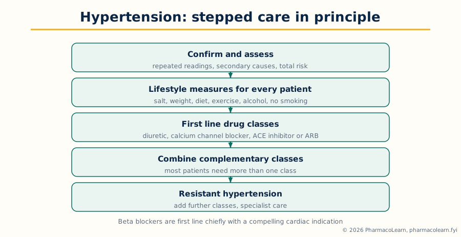 Figure: Hypertension stepped care. Lifestyle measures run throughout, drug classes are combined in steps, and resistant hypertension is a specialist problem.
1.3 Congestive cardiac failure
Congestive cardiac failure, in modern usage heart failure, is a syndrome in which the heart cannot pump blood at a rate sufficient for the body's needs, or can do so only at raised filling pressures, so congestion in the lungs and systemic veins is its defining physical consequence. Causes are the diseases that damage, overload or stiffen the ventricle: coronary disease and previous infarction are commonest, with hypertension, valvular disease, cardiomyopathies, myocarditis, arrhythmias, alcohol, cardiotoxic medicines and systemic conditions such as thyroid disease and anemia. It is classified by ejection fraction into reduced ejection fraction failure, where the ventricle contracts weakly and dilates, and preserved ejection fraction failure, where the ventricle is stiff and relaxes poorly, usually in older patients with hypertension, obesity and diabetes; the drug evidence base is strongest in the reduced form. The pathophysiology is a compensatory spiral that becomes the disease: falling output activates the sympathetic system and the renin angiotensin aldosterone system and increases antidiuretic hormone, retaining sodium and water and constricting vessels; chronically these overload the ventricle, drive hypertrophy, dilation and fibrosis, provoke arrhythmias and damage the kidneys. Natriuretic peptides, released from the stretched heart wall, oppose this system, and one modern class exists to strengthen them. Congestion explains the presentation: exertional and then resting breathlessness, orthopnoea and paroxysmal nocturnal dyspnoea, fatigue, ankle oedema, raised jugular venous pressure and, in acute decompensation, pulmonary oedema. Diagnosis combines the syndrome with examination, chest imaging, electrocardiogram, echocardiography defining structure, function and cause, natriuretic peptide measurement, and tests for cause and precipitants such as anemia, thyroid disease, infection and non adherence.
Pharmacotherapy in reduced ejection fraction failure is built on classes that block the maladaptive neurohormonal systems, because those classes improve survival, not only symptoms. Renin angiotensin inhibition, by an ACE inhibitor, an angiotensin receptor blocker, or the angiotensin receptor and neprilysin inhibitor combination pairing renin angiotensin blockade with natriuretic peptide strengthening, is foundational. The beta blockers proven in this condition slow the heart, protect against arrhythmia and reverse remodelling. Aldosterone antagonists block aldosterone's fibrotic and potassium losing actions. SGLT2 inhibitors, a class that began in diabetes care, reduce hospitalisation and death across the ejection fraction range and are now standard. Loop diuretics relieve congestion and are adjusted to fluid state, but are not themselves survival therapy. Further options in selected patients include hydralazine with a nitrate, digoxin, and device therapy where indicated. Management also treats cause and precipitants, and monitors weight, renal function and electrolytes, since the classes that protect the heart stress the kidney and potassium balance.
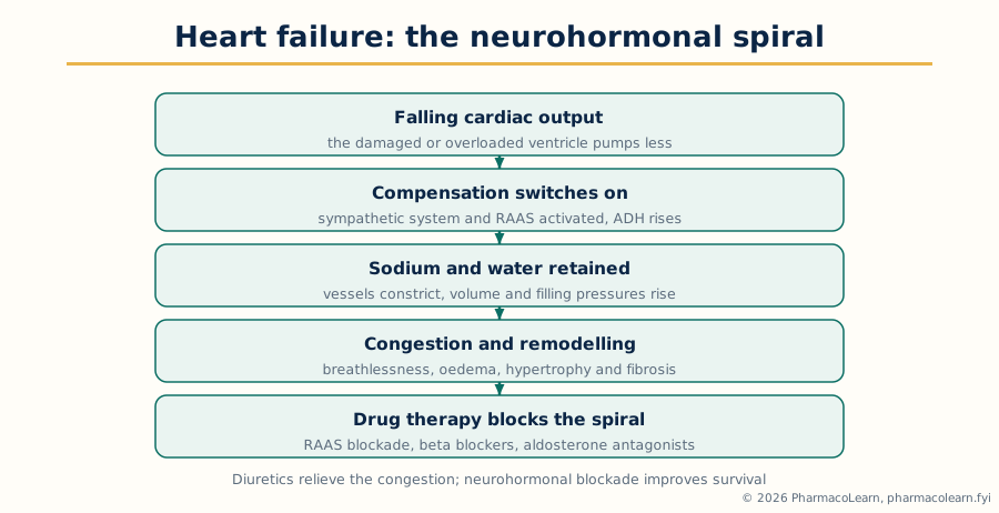 Figure: The heart failure spiral. The compensations that restore pressure in the short term produce congestion and remodelling in the long term, and the survival drug classes block them.
1.4 Acute coronary syndrome
Acute coronary syndrome is the spectrum of acute myocardial ischaemia caused by sudden disruption of a coronary atherosclerotic plaque: unstable angina, ischaemia without myocardial necrosis, and myocardial infarction, necrosis present, divided electrocardiographically into ST elevation infarction, with complete occlusion, and non ST elevation infarction, with partial or transient occlusion. Atherosclerosis develops over decades: endothelial injury and lipid accumulation form a plaque with a lipid core and fibrous cap, inflammation weakens the cap, and the precipitating event is plaque rupture or erosion exposing thrombogenic material. Platelets adhere and activate, coagulation generates thrombin and fibrin, and thrombus may occlude the artery completely, partially, or embolise downstream. Consequences follow the duration and territory of ischaemia: necrosis in a time dependent wavefront, electrical instability, pump failure and cardiogenic shock in large infarcts, and mechanical complications in the days that follow. Presentation is typically central chest pain or discomfort radiating to arm, neck or jaw, with sweating, nausea and breathlessness at rest or on minimal exertion; presentations are atypical more often in women, older patients and patients with diabetes, and some infarctions are silent. Diagnosis is urgent and structured: immediate electrocardiogram, which decides the ST elevation pathway, serial cardiac troponin, the biomarker of necrosis whose rise and fall defines infarction, risk assessment, and exclusion of mimics such as aortic dissection and pulmonary embolism where the picture suggests them.
Pharmacotherapy has two simultaneous aims: to limit necrosis by restoring flow, and to prevent thrombus extension and recurrence. Reperfusion is decisive in ST elevation infarction, by primary percutaneous coronary intervention where available in time or fibrinolytic therapy where it is not, and drug therapy supports it. Antiplatelet therapy pairs aspirin, blocking thromboxane mediated activation, with a P2Y12 receptor blocker, blocking ADP mediated activation; the dual combination protects the artery and any stent placed in it. Anticoagulation with heparin class agents in the acute phase prevents thrombus propagation. Nitrates relieve ischaemic pain and preload, beta blockers reduce myocardial oxygen demand where not contraindicated, high intensity statin therapy stabilises plaque and begins the long term lipid strategy of Section 1.6, and renin angiotensin inhibition follows in patients with ventricular impairment, hypertension or diabetes. Oxygen is given only where saturation is low, a standard examination point. Long term secondary prevention, continued antiplatelet therapy, statin and risk factor treatment including smoking cessation, is part of the same answer, because the syndrome declares a systemic arterial disease.
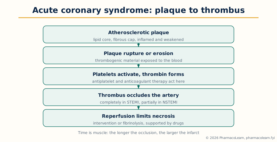 Figure: From plaque rupture to reperfusion. Antiplatelet and anticoagulant therapy act on the thrombus steps, reperfusion on the occlusion itself.
1.5 Arrhythmias
Arrhythmias are disorders of heart rate, rhythm or conduction, arising from abnormal impulse formation, abnormal conduction, or re entry circuits in which an impulse circles repeatedly through tissue with differing conduction and refractory properties. The standard classification divides them by rate and site: bradyarrhythmias, including sinus bradycardia and the degrees of heart block, and tachyarrhythmias, divided into supraventricular forms, including atrial fibrillation, atrial flutter and paroxysmal supraventricular tachycardia, and ventricular forms, ventricular tachycardia and fibrillation, which are life threatening. Causes span ischaemic and structural heart disease, hypertension, heart failure, thyroid disease, electrolyte disturbance, infection, alcohol and medicines; several antiarrhythmic and non cardiac drugs themselves prolong repolarisation and provoke arrhythmia, a drug induced mechanism the pharmacist must watch for. Diagnosis rests on capturing the rhythm, a 12 lead electrocardiogram during symptoms and longer monitoring where episodes are intermittent, with blood tests for precipitants and assessment of the underlying heart. Atrial fibrillation, the commonest sustained arrhythmia, deserves its own emphasis: loss of organised atrial contraction causes an irregularly irregular pulse and, more importantly, atrial stasis and a substantial embolic stroke risk, so its management always has two parts, rate control or rhythm restoration, and anticoagulation assessed against stroke and bleeding risk.
Pharmacotherapy follows the mechanism and the aim. For rate control in atrial fibrillation, beta blockers and rate limiting calcium channel blockers slow atrioventricular nodal conduction, digoxin being an additional agent in sedentary patients and in heart failure. Antiarrhythmic drugs are classified by dominant action in the Vaughan Williams scheme: Class I agents block sodium channels, Class II are beta blockers, Class III agents block potassium channels and prolong repolarisation and the refractory period, Class IV are the rate limiting calcium channel blockers, with digoxin, adenosine and magnesium outside the scheme, adenosine being the acute terminator of paroxysmal supraventricular tachycardia by brief atrioventricular block.
1.6 Hyperlipidemias
The hyperlipidemias are raised levels of the blood lipids carried in lipoproteins: raised low density lipoprotein cholesterol, the principal atherogenic fraction, raised triglycerides, and often low high density lipoprotein cholesterol. They are primary, the familial and polygenic disorders, of which familial hypercholesterolaemia, an inherited defect of low density lipoprotein clearance presenting young with tendon xanthomas and premature coronary disease, is the standard example, or secondary, where diabetes, hypothyroidism, nephrotic syndrome, cholestasis, obesity, alcohol and medicines including thiazides and corticosteroids raise lipids and must be sought and treated. The pathophysiology is the atherosclerosis of Section 1.4 read from the lipid side: low density lipoprotein enters the arterial wall, is retained and modified, is taken up by macrophages to form foam cells, and builds the plaque whose rupture causes acute events; triglyceride rich remnants contribute to the same process, and severe hypertriglyceridaemia adds a distinct danger, acute pancreatitis. Hyperlipidemia is silent, found by testing and presenting through its complications, and is assessed as part of total cardiovascular risk, with a lipid profile, exclusion of secondary causes and family history.
Pharmacotherapy is matched to the fraction raised and the risk carried. Lifestyle measures, a diet reduced in saturated fat, weight management, exercise, alcohol moderation and smoking cessation, apply to every patient and continue with drug therapy. Statins, inhibitors of HMG CoA reductase, the rate limiting enzyme of cholesterol synthesis, are first line: reducing hepatic cholesterol raises hepatic low density lipoprotein receptor expression and clears low density lipoprotein from the blood, and their prevention evidence is the strongest in this section. Ezetimibe, which blocks intestinal cholesterol absorption, is added where the statin response is insufficient or the statin is not tolerated. PCSK9 directed therapies preserve low density lipoprotein receptors from degradation and give large additional reductions, reserved for high risk and familial patients under specialist care. Bile acid sequestrants bind bile acids in the gut, an older class limited by gastrointestinal intolerance and interactions. For triglycerides, fibrates activate PPAR alpha and increase lipoprotein lipase activity, with prescription omega 3 preparations an additional option; severe elevation is urgent in its own right because of pancreatitis. Monitoring covers lipid response and the liver and muscle adverse effects of statins, muscle symptoms being the commonest reason for intolerance, and interactions, since several statins share metabolic pathways with widely used inhibitors.
Unit 2: Respiratory System and Endocrine System
2.1 Asthma
Asthma is a chronic inflammatory disease of the airways characterised by variable airflow obstruction and bronchial hyperresponsiveness, in which the airways narrow in response to triggers that leave normal airways unaffected, and narrow reversibly, spontaneously or with treatment. The pathophysiology is inflammation first and bronchospasm second, the order examinations test. In the allergic pattern, inhaled allergen cross links IgE on mast cells, releasing histamine, prostaglandins and leukotrienes that produce the early bronchoconstriction, oedema and mucus; hours later a late response recruits eosinophils and Th2 lymphocytes and their cytokines, sustaining inflammation. Presentation is episodic wheeze, breathlessness, chest tightness and cough, worse at night and early morning and varying with triggers, with a normal examination between attacks in mild disease; severe attacks bring inability to complete sentences, exhaustion and, ominously, a silent chest.
Pharmacotherapy has two arms that must never be confused: reliever therapy that reverses bronchoconstriction, and controller therapy that treats inflammation and prevents attacks. Inhaled corticosteroids are the foundation controller class, suppressing inflammation, reducing hyperresponsiveness and preventing exacerbations, and modern stepped care places anti inflammatory treatment at every step rather than relying on reliever alone. Short acting beta 2 agonists are the classic rapid relievers; long acting beta 2 agonists are used only in combination with inhaled corticosteroids in asthma. Escalation adds or raises controller therapy: increased inhaled corticosteroid, the combination inhaler used as both maintenance and reliever, long acting muscarinic antagonists, leukotriene receptor antagonists blocking the cysteinyl leukotriene arm, and, in severe allergic or eosinophilic asthma under specialist care, biologic therapies against IgE, interleukin 5 pathways or related Th2 targets, with oral corticosteroids a last step whose long term harms are a standing warning. Acute severe asthma is emergency therapy: oxygen where needed, repeated inhaled bronchodilators, systemic corticosteroids, inhaled ipratropium and intravenous magnesium in severe cases under hospital care.
Asthma stepped care, in principle
  Every step: inhaled corticosteroid based controller,
  a reliever plan, technique and adherence checked,
  triggers addressed, action plan in the patient's hands
        |
        v
  Step up if control is poor: raise the controller,
  add a long acting bronchodilator in combination,
  consider leukotriene or muscarinic additions
        |
        v
  Severe asthma: specialist review, phenotype assessed,
  biologic therapy matched to the inflammatory type
        |
        v
  Step down only when control has been sustained,
  never by withdrawing anti inflammatory treatment
2.2 Chronic obstructive airways disease
Chronic obstructive airways disease, universally taught as chronic obstructive pulmonary disease or COPD, is progressive airflow limitation that is not fully reversible, encompassing chronic bronchitis, defined clinically by chronic productive cough, and emphysema, defined anatomically by destruction of alveolar walls and enlargement of air spaces. Tobacco smoking is the dominant cause, with occupational dusts and fumes, biomass smoke and air pollution the other inhaled causes, and inherited alpha 1 antitrypsin deficiency the genetic cause to name, producing early emphysema. The pathophysiology is chronic inflammation of airways, parenchyma and vessels driven by the inhaled insult, with a protease and antiprotease imbalance destroying alveolar tissue, small airway fibrosis narrowing the bronchioles, and mucus gland hypertrophy producing the bronchitis picture. Presentation is progressive exertional breathlessness, chronic cough and sputum, wheeze and frequent infective exacerbations in a patient with a significant exposure history. Diagnosis requires spirometry showing persistent airflow obstruction after a bronchodilator, distinguishing COPD from asthma by history, variability and reversibility, though the two may coexist; severity and exacerbation history guide therapy.
Pharmacotherapy relieves symptoms and reduces exacerbations, but only stopping the exposure, above all smoking cessation, changes the underlying course, and cessation support is a pharmacotherapeutic intervention in its own right. Bronchodilators are the symptomatic foundation, the reverse emphasis from asthma: short acting beta 2 agonists and short acting muscarinic antagonists for relief, long acting beta 2 agonists and, centrally, long acting muscarinic antagonists for maintenance, combined where symptoms persist. Inhaled corticosteroids are added only for patients with frequent exacerbations and an eosinophilic pattern, weighed against pneumonia risk in this population. Exacerbations are treated with increased bronchodilators, a short course of systemic corticosteroids, antibiotics where infection criteria are met, controlled oxygen and ventilatory support where required.
| Feature | Asthma | COPD |
|---|---|---|
| Typical onset | Often childhood or young adult, atopic background | Later life, after prolonged inhaled exposure |
| Airflow obstruction | Variable and reversible | Persistent and incompletely reversible |
| Core pathology | Airway inflammation with hyperresponsiveness | Airway and parenchymal inflammation, alveolar destruction |
| Controller foundation | Inhaled corticosteroids | Long acting bronchodilators |
| Disease modifying step | Control of inflammation, trigger avoidance | Stopping the exposure, above all smoking cessation |
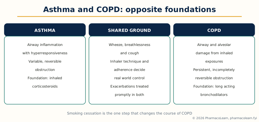 Figure: Opposite foundations. Asthma therapy is built on anti inflammatory control, COPD therapy on bronchodilation and stopping the exposure.
2.3 Drug induced pulmonary diseases
Drug induced pulmonary disease is lung injury caused by a medicine, and it belongs in this unit because the respiratory system is a recognised target of adverse drug reactions and because the pharmacist reviewing therapy is often the professional who connects a new lung picture with a drug timeline. The mechanisms are several and should be classified in an examination answer. Direct cytotoxic injury to alveolar and capillary cells, classically by cytotoxic agents such as bleomycin, whose pulmonary toxicity is its defining dose limiting harm, and by agents including methotrexate and cyclophosphamide, produces pneumonitis and, if injury continues, pulmonary fibrosis. Oxidative injury is the mechanism of bleomycin and of nitrofurantoin chronic lung toxicity and of amiodarone lung injury, amiodarone accumulating in lung tissue over long therapy. Immune and hypersensitivity mechanisms produce eosinophilic pneumonias and hypersensitivity pneumonitis patterns, with nitrofurantoin, sulfonamides and many other agents reported. Drugs also injure the lung functionally rather than structurally: beta blockers and aspirin and other non steroidal anti inflammatory drugs provoke bronchospasm, aspirin sensitive respiratory disease combining asthma, nasal polyps and NSAID sensitivity; opioids and sedatives depress ventilatory drive; ACE inhibitors produce the familiar chronic dry cough by bradykinin accumulation, a benign but adherence destroying effect whose recognition prevents needless investigation; and several agents cause pleural effusions or pulmonary oedema by fluid or permeability mechanisms. Presentation is non specific, new cough, breathlessness, fever, infiltrates on imaging or falling gas exchange appearing during therapy, and that non specificity is the diagnostic problem: drug induced disease is a diagnosis reached by the timeline of exposure, by exclusion of infection, cardiac failure and the underlying disease, and by improvement on withdrawal of the suspect drug, the dechallenge, with lung function tests, imaging, bronchoscopy and occasionally biopsy in support. Management is withdrawal of the offending drug as the essential step, corticosteroid therapy where inflammation is significant and progressive, supportive oxygen and ventilation as needed, documentation and reporting of the reaction, and a permanent record that prevents re exposure; fibrosis established before recognition may not reverse, which is why baseline and interval monitoring is standard for the highest risk agents.
2.4 Diabetes mellitus
Diabetes mellitus is chronic hyperglycaemia resulting from defects in insulin secretion, insulin action, or both. Type 1 diabetes is autoimmune destruction of the pancreatic beta cells, usually presenting in the young with absolute insulin deficiency and a liability to ketoacidosis. Type 2 diabetes, the large majority, combines insulin resistance in liver, muscle and adipose tissue with progressive beta cell failure: the resistant liver overproduces glucose, muscle takes up less, adipose tissue releases free fatty acids that worsen resistance, incretin responses are blunted, and beta cells that first compensate gradually fail. Acutely, marked hyperglycaemia causes osmotic diuresis, dehydration and the emergencies of diabetic ketoacidosis, chiefly in type 1, and the hyperosmolar hyperglycaemic state in type 2. Chronically, hyperglycaemia damages vessels, producing the microvascular complications, retinopathy, nephropathy and neuropathy, and accelerating macrovascular disease, the main cause of death. Type 1 presents rapidly with thirst, polyuria, weight loss and fatigue, sometimes first as ketoacidosis; type 2 is often silent for years and found on testing or through its complications. Diagnosis is by the standard blood glucose and glycated haemoglobin criteria, followed by classification, complication screening and cardiovascular risk assessment.
Pharmacotherapy is built on the physiology, and the classes are best presented as mechanism groups. Insulin replaces the missing hormone, is obligatory and lifelong in type 1 as regimens combining background and mealtime insulin, and is used in type 2 when other therapy no longer controls glycaemia or acutely in illness and pregnancy. Metformin, the usual first drug in type 2, reduces hepatic glucose production, does not cause weight gain or hypoglycaemia alone, and is limited by gastrointestinal intolerance and caution in advanced renal impairment. Sulfonylureas stimulate insulin secretion from functioning beta cells, carrying hypoglycaemia and weight gain as class cautions. Incretin based therapies work through the gut hormone pathway: DPP 4 inhibitors prolong endogenous incretin action and are weight neutral, while GLP 1 receptor agonists enhance glucose dependent insulin secretion, suppress glucagon, slow gastric emptying and reduce appetite, producing weight loss and, for several agents, proven cardiovascular benefit. SGLT2 inhibitors block renal glucose reabsorption, lowering glucose independently of insulin, and have transformed care through heart failure and kidney protection, with genital mycotic infection and volume effects as counselling points. Therapy in type 2 is stepped and individualised: lifestyle and metformin at the foundation, then classes added according to response, weight, hypoglycaemia risk, kidney function and, decisively, the presence of cardiovascular or kidney disease, which directs choice toward the classes with proven organ protection.
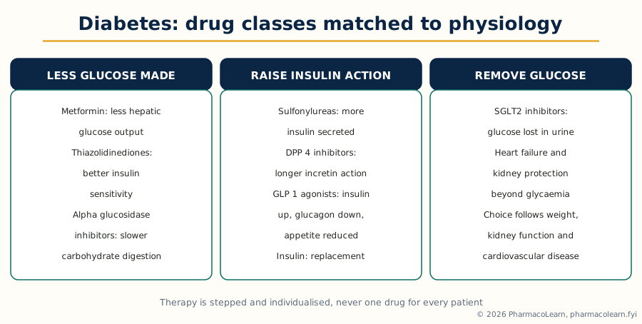 Figure: Diabetes classes matched to physiology. The right combination follows which defect dominates and which organs need protection.
2.5 Thyroid diseases
The thyroid produces thyroxine and triiodothyronine under pituitary thyroid stimulating hormone control by negative feedback, and its pharmacotherapeutic diseases are the overactive and underactive states. Hyperthyroidism is excess thyroid hormone action, most commonly from Graves disease, an autoimmune disorder in which stimulating antibodies activate the TSH receptor, driving overproduction and enlargement, often with Graves eye disease; other causes are toxic multinodular goitre and toxic adenoma, where autonomous nodules escape control, and thyroiditis, where inflamed follicles release stored hormone transiently, a distinction that matters because antithyroid drugs do not treat the release phase. Hypothyroidism is hormone deficiency, most commonly from Hashimoto thyroiditis, autoimmune gland destruction, and otherwise from iodine deficiency, thyroid surgery or radioiodine, and medicines including amiodarone and lithium. Diagnosis in both directions reads TSH and free thyroxine together through the feedback logic, suppressed TSH with raised hormone in primary hyperthyroidism and raised TSH with low hormone in primary hypothyroidism, with antibody tests for the autoimmune causes and uptake scanning where the cause of hyperthyroidism is in doubt.
Pharmacotherapy of hyperthyroidism offers three routes to compare. Thionamide antithyroid drugs, carbimazole and its active metabolite methimazole, and propylthiouracil, block new hormone synthesis; a course may induce lasting remission in Graves disease, and the class carries rash and, rarely, agranulocytosis, whose warning symptoms of sore throat and fever every patient must know, and liver injury, the greater concern with propylthiouracil, reserved mainly for the first trimester of pregnancy and thyroid storm. Beta blockers control the adrenergic symptoms, tremor, palpitations and anxiety, while definitive therapy takes effect. Radioactive iodine destroys thyroid tissue, usually resulting in hypothyroidism that is then replaced; surgery is chosen for large goitres, suspicious nodules and patient preference after preparation with antithyroid drugs and iodide. Hypothyroidism treatment is replacement: levothyroxine restores the hormone pool, is titrated against TSH over weeks because of the long half life, and is lifelong in established primary failure, with counselling on consistent timing in relation to food and on absorption interactions with iron, calcium and other medicines, and dose review in pregnancy and changing therapy. Drug induced thyroid disease, from amiodarone above all, is a recognised monitoring duty linking this section back to Section 2.3.
Unit 3: Gastrointestinal System, First Part
3.1 Peptic ulcer diseases
Peptic ulcer disease is ulceration of the mucosa of the stomach or duodenum extending through the muscularis mucosae, occurring where acid and pepsin overwhelm mucosal defence. The balance is the pathophysiology and the organising image of the topic: on the aggressive side, gastric acid, pepsin, bile reflux and external injurious agents; on the defensive side, the mucus bicarbonate barrier, mucosal blood flow, prostaglandin driven mucus and bicarbonate secretion and rapid cell renewal. The two dominant causes work one on each side. Helicobacter pylori, a spiral bacterium colonising the gastric mucus layer, causes chronic gastritis and duodenal inflammation that ulcerates; it causes most duodenal ulcers and a majority of gastric ulcers. Non steroidal anti inflammatory drugs fall on the defensive side: by inhibiting cyclo oxygenase they deplete the mucosal prostaglandins that maintain mucus, bicarbonate and blood flow, producing ulcers that are often silent until they bleed, the standard drug induced disease of this unit. Diagnosis is by upper gastrointestinal endoscopy, which sees the ulcer, excludes malignancy in gastric ulcers by biopsy and allows H. pylori sampling, supported by the urea breath test and stool antigen test in patients managed without endoscopy; alarm features, weight loss, anaemia, dysphagia, vomiting or older age, mandate endoscopy rather than empirical treatment.
Pharmacotherapy follows the balance: suppress acid so the mucosa can heal, eradicate H. pylori where present so the ulcer does not recur, and remove or protect against NSAID injury. Proton pump inhibitors, which irreversibly block the parietal cell hydrogen potassium ATPase, the final common step of acid secretion, are the most effective suppressants and the healers of choice; histamine H2 receptor antagonists are weaker and now secondary. Eradication therapy combines a proton pump inhibitor with antibacterial agents in the standard multi drug regimens, given as a course whose completion matters more than any single component. Mucosal protectives have defined places: sucralfate forms an adherent barrier over the ulcer base, and misoprostol, a prostaglandin analogue, replaces the defence NSAIDs remove and is the specific prophylactic answer where NSAID therapy must continue at high risk, limited by diarrhoea and by contraindication in pregnancy. For NSAID ulcers the first step is to stop or reduce the NSAID where possible, add a proton pump inhibitor for healing and continuing protection, and consider a cyclo oxygenase 2 selective agent where anti inflammatory therapy is unavoidable, weighing gastrointestinal against cardiovascular risk.
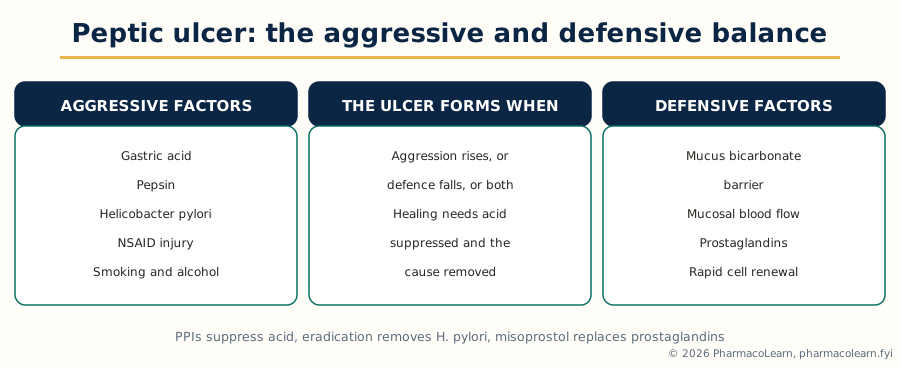 Figure: The peptic balance. An ulcer forms when aggression rises or defence falls, and every therapy in Section 3.1 acts on one side of this balance.
3.2 Reflux esophagitis
Reflux esophagitis is inflammation of the oesophageal mucosa caused by reflux of gastric contents, within the wider gastro oesophageal reflux disease, in which reflux causes troublesome symptoms or complications with or without visible injury. The oesophagus has little defence against acid and pepsin, so reflux that would be harmless lower down injures it. The mechanisms are failures of the antireflux barrier and of clearance: transient relaxations and incompetence of the lower oesophageal sphincter, hiatus hernia, impaired peristalsis that fails to clear refluxate, delayed gastric emptying, and raised intra abdominal pressure from obesity and pregnancy; fatty meals, caffeine, alcohol, smoking and sphincter relaxing medicines such as calcium channel blockers and nitrates contribute. Untreated injury produces erosive esophagitis and, with chronicity, peptic stricture, Barrett oesophagus, in which squamous epithelium is replaced by intestinal type columnar epithelium with malignant potential, and adenocarcinoma arising in that segment. Presentation is heartburn after meals and on lying or bending, acid regurgitation, and dysphagia where stricture narrows the lumen; extra oesophageal presentations include chronic cough and laryngeal symptoms, and chest pain that must first be distinguished from cardiac pain. Diagnosis is usually clinical and confirmed by response to acid suppression; endoscopy grades esophagitis, detects Barrett change and is required for alarm features, and pH monitoring settles doubtful cases.
Pharmacotherapy aims to make the refluxate harmless, since the barrier itself is corrected mechanically, by surgery or endoscopic procedures, rather than by drugs. Proton pump inhibitors are the principal class, healing esophagitis more reliably than H2 receptor antagonists, which retain a place in milder disease and for nocturnal symptoms. Treatment is stepped characteristically: a healing course of full suppression, then step down to the lowest level maintaining control, maintenance therapy where relapse or complications demand it, and on demand therapy for intermittent non erosive symptoms. Lifestyle measures are prescribed with the same seriousness: weight reduction where overweight, smaller and earlier evening meals, head of bed elevation for nocturnal symptoms, and avoidance of individual triggers, smoking and late alcohol.
3.3 Inflammatory bowel diseases
The inflammatory bowel diseases are ulcerative colitis and Crohn disease, two chronic relapsing inflammatory diseases of the gut whose comparison is a fixed examination structure. Both arise from an inappropriate, sustained immune response to the intestinal microbiota in a genetically susceptible host, with environment shaping course: smoking worsens Crohn disease and paradoxically appears protective in ulcerative colitis. Neither is a simple infection and neither is cured by removing a single cause. Ulcerative colitis is continuous inflammation of the colonic mucosa beginning at the rectum, limited to the colon and the mucosal layer, producing a friable, ulcerated surface that bleeds readily. Crohn disease is transmural inflammation that may affect any part of the gut in discontinuous skip lesions, most often terminal ileum and colon, and its depth produces its defining complications: strictures, fistulae, abscesses and perianal disease. Presentation divides accordingly: ulcerative colitis chiefly with bloody diarrhoea, urgency and tenesmus in flares, severe attacks risking toxic megacolon; Crohn disease with pain, less bloody diarrhoea, weight loss and obstructive or fistulating complications.
Pharmacotherapy pursues two goals in sequence, inducing remission in the flare and then maintaining it. Aminosalicylates, delivering 5 aminosalicylic acid to the colonic mucosa, are the foundation in ulcerative colitis for mild to moderate flares and maintenance, by mouth and rectally where disease is distal; their place in Crohn disease is limited. Corticosteroids induce remission in moderate to severe flares of both diseases and are deliberately not maintenance therapy, because dependence and systemic harm forbid it. Immunomodulators, the thiopurines and methotrexate, maintain remission and spare steroids, acting slowly and requiring blood monitoring for marrow and liver toxicity. Biologic therapies, anti TNF agents and the newer biologics against integrins and interleukin pathways, together with oral Janus kinase inhibitors in ulcerative colitis, treat moderate to severe disease failing conventional therapy, under specialist care with infection screening before immunosuppression. Surgery remains part of management: curative of the colonic disease in ulcerative colitis at the price of colectomy, and in Crohn disease reserved and bowel preserving, since recurrence after resection is the rule.
| Feature | Ulcerative colitis | Crohn disease |
|---|---|---|
| Distribution | Colon only, continuous from the rectum | Any part of the gut, discontinuous skip lesions |
| Depth | Mucosal only | Transmural, full thickness |
| Hallmark complications | Bleeding, toxic megacolon, colorectal cancer risk | Strictures, fistulae, abscesses, perianal disease |
| Dominant presentation | Bloody diarrhoea with urgency | Pain, diarrhoea, weight loss, obstructive features |
| Surgery | Colectomy removes the colonic disease | Reserved, bowel preserving, recurrence common |
3.4 Jaundice and hepatitis
Jaundice is the yellow discolouration of skin, sclerae and mucous membranes by accumulated bilirubin, a sign and not a disease, taught by its mechanism in three families. Bilirubin is produced from haem breakdown, carried to the liver bound to albumin, conjugated in hepatocytes and excreted in bile. Pre hepatic jaundice follows overproduction, chiefly haemolysis, with a normal liver. Hepatocellular jaundice follows liver cell injury, in hepatitis and cirrhosis, where uptake, conjugation and excretion fail together. Post hepatic or obstructive jaundice follows blocked bile flow from gallstones, strictures or tumours, with dark urine, pale stools and itch from retained bile salts. The clinical approach reads that pattern with liver enzyme patterns and imaging, ultrasound first for obstruction, to place the patient in a family and then a cause. Hepatitis is inflammation of the liver, and its causes define its pharmacotherapy. The hepatotropic viruses A to E are the classic family: A and E spread faeco orally and cause acute self limiting disease, E dangerous in pregnancy; B and C spread by blood, sexual contact and vertically, and may become chronic and progress to cirrhosis and hepatocellular carcinoma; D occurs only with B. Beyond viruses, hepatitis arises from alcohol, metabolic fatty liver disease, autoimmune hepatitis, inherited copper and iron storage disorders, and drugs, the subject of Section 4.3.
Pharmacotherapy of hepatitis is cause specific, and general measures come first: avoidance of alcohol and of unnecessary hepatotoxic medicines, and no drug therapy for uncomplicated acute viral hepatitis, which resolves with supportive care. Hepatitis A and B are vaccine preventable, and vaccination of contacts and risk groups is pharmacotherapy in preventive form. Chronic hepatitis B is treated with antiviral therapy where viral activity and liver injury criteria are met: nucleoside and nucleotide analogues suppressing replication over long courses, and interferon based therapy in selected patients, aiming to prevent cirrhosis and cancer. Chronic hepatitis C has been transformed by direct acting antivirals, oral combinations inhibiting the viral protease, polymerase and replication complex proteins that cure the great majority of treated patients over short courses, a clear therapeutic advance in this syllabus. Autoimmune hepatitis is treated with corticosteroids and steroid sparing immunosuppression, typically azathioprine. Alcoholic hepatitis is managed by abstinence above all, nutrition, and corticosteroids in selected severe cases under specialist judgement.
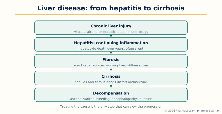 Figure: The road to cirrhosis. Hepatitis is the stage at which treating the cause can still change the destination.
Unit 4: Gastrointestinal System, Second Part, and Hematological Diseases
4.1 Cirrhosis
Cirrhosis is the end stage of chronic liver injury from any cause, in which long standing inflammation and hepatocyte death drive fibrosis that distorts liver architecture into regenerative nodules surrounded by fibrous bands, destroying normal blood flow and liver function together. Its causes are those of chronic hepatitis continued: viral hepatitis B and C, alcohol related disease, metabolic fatty liver disease, autoimmune and cholestatic diseases and inherited storage disorders. Two consequences produce nearly all of its clinical picture. Portal hypertension follows increased resistance to portal flow through the fibrotic liver, compounded by increased splanchnic inflow: it produces splenomegaly, portosystemic collaterals including oesophageal varices that may rupture and bleed catastrophically, and ascites, driven by portal pressure, sodium retention and low albumin. Hepatocellular failure follows lost functioning mass: impaired synthesis of albumin and clotting factors, impaired detoxification producing hepatic encephalopathy, jaundice, and endocrine and nutritional disturbance. Diagnosis combines the cause history, examination for the stigmata of chronic liver disease, blood tests of function, platelets and clotting, imaging and elastography measuring liver stiffness, and endoscopy to find varices.
Pharmacotherapy treats the cause, the portal hypertension and each complication; no drug reverses established fibrosis. Cause directed therapy, antivirals, abstinence support, immunosuppression in autoimmune disease, is the only intervention that can slow progression and is never too late to address. For variceal bleeding prevention, non selective beta blockers reduce portal pressure and are used to prevent first and further bleeding, with endoscopic band ligation as the mechanical partner; acute variceal bleeding is an emergency of resuscitation, vasoactive drug therapy constricting the splanchnic circulation, prophylactic antibiotics and urgent endoscopy. Ascites is managed by sodium restriction and diuretics pairing an aldosterone antagonist, the physiological first agent because secondary hyperaldosteronism drives the retention, with a loop diuretic, monitored by weight, renal function and electrolytes, with paracentesis and albumin cover for tense or diuretic resistant ascites. Hepatic encephalopathy is treated by reducing gut nitrogen production and absorption: lactulose, a non absorbable disaccharide that acidifies the colon, traps ammonium and acts as a laxative, is first therapy, the non absorbable antibiotic rifaximin added for recurrence, and precipitants, infection, bleeding, constipation, dehydration and sedatives, are found and treated.
4.2 Diarrhea and constipation
Diarrhea is an increase in stool frequency, liquidity or volume, acute over days to a few weeks, usually infective, and chronic when it persists, demanding a search for disease. Its mechanisms classify it: secretory, driven by toxins, hormones or bile acids; osmotic, from unabsorbed solute; inflammatory, from mucosal invasion, with blood and pus; and motility disorders speeding transit. Drug induced diarrhoea is common and examinable: antibacterials, including the dangerous overgrowth of Clostridioides difficile after broad spectrum therapy, metformin, magnesium products and many others. The danger is dehydration and electrolyte depletion, most lethal in children and the elderly, so assessment of fluid state governs urgency. Acute diarrhoea is usually diagnosed clinically, stool studies reserved for bloody, severe, prolonged or outbreak cases; chronic diarrhoea is investigated by the route of Section 3.3. Pharmacotherapy is rehydration first and drugs second: oral rehydration solution, whose glucose coupled sodium absorption survives most secretory states, is the life saving therapy, intravenous fluids replacing it where oral replacement fails. Antibacterials are withheld in most acute watery diarrhoea and reserved for dysentery patterns, identified pathogens, severe disease and C. difficile infection, where the offending antibiotic is stopped and targeted therapy given. Chronic diarrhoea is treated by treating its cause, with bile acid sequestrants, pancreatic enzymes and the disease specific therapies of earlier sections as the mechanism dictates.
Constipation is infrequent, hard or difficult evacuation, defined by stool form and symptoms of straining and incomplete emptying rather than frequency alone. Causes divide into functional, slow transit and pelvic floor dysfunction among them, and secondary: low fibre and fluid intake, immobility, medicines, opioids above all, with anticholinergics, iron and calcium channel blockers, metabolic causes including hypothyroidism, neurological disease, and structural disease, new constipation in older patients with weight loss, anaemia or bleeding requiring exclusion of colorectal cancer. Management begins with fibre increased gradually, adequate fluid, exercise and responding to the urge to defecate. Laxative classes then follow a stepped order. Bulk forming agents such as ispaghula swell with water and restore the stool bulk that triggers peristalsis, the gentlest first choice for maintenance. Osmotic laxatives hold water in the lumen: lactulose and the macrogol preparations, the latter now preferred, are standard for continuing treatment and for softening impaction. Stimulant laxatives, senna and bisacodyl, provoke peristalsis and are used for short courses and rescue, and alongside osmotics in opioid induced constipation; peripherally acting opioid antagonists serve resistant opioid induced cases under appropriate care.
4.3 Drug induced liver disease
Drug induced liver disease, or drug induced liver injury, is liver damage caused by a medicine, herbal product or supplement, and it mirrors Section 2.3 as the hepatic counterpart of drug induced lung disease. It is classified by pattern from the liver enzyme picture: hepatocellular injury, with predominant transaminase elevation, cholestatic injury, with predominant alkaline phosphatase and bilirubin elevation and itch, and mixed patterns. Mechanistically it divides into predictable, dose related injury, of which paracetamol overdose is the archetype, its metabolite NAPQI exhausting glutathione and binding hepatocyte proteins to produce centrilobular necrosis, and idiosyncratic injury, unpredictable, dose independent in the usual range and immune or metabolic in origin, for which antitubercular agents such as isoniazid and pyrazinamide, amoxicillin clavulanate, antiepileptics, sulfonamides and many others are repeatedly reported; herbal and dietary supplements are an increasingly recognised source that the drug history must ask for, a link back to the medication history discipline. Presentation ranges from asymptomatic enzyme elevation found on monitoring, through a hepatitis picture indistinguishable from viral hepatitis, to cholestatic jaundice and, rarely, acute liver failure; certain drugs produce characteristic signatures, fatty liver, fibrosis, vascular injury or vanishing bile ducts with chronic use. Diagnosis is by exclusion and timeline: a complete drug and supplement history with start dates, exclusion of viral, autoimmune, metabolic and obstructive causes by the approach of Section 3.4, and improvement after withdrawal, the dechallenge, with causality assessment structured as in pharmacovigilance; rechallenge is diagnostic but generally unethical where injury was significant. Management is withdrawal of the suspect drug as the decisive step, supportive care, and specific antidotes where they exist, N acetylcysteine replenishing glutathione in paracetamol poisoning, where its early use is life saving, and in some other acute liver failure settings; corticosteroids are reserved for hypersensitivity and autoimmune like patterns. Prevention is baseline and interval liver monitoring for the high risk agents, dose adjustment in existing liver disease, avoidance of alcohol and interacting combinations, and patient education on the jaundice and dark urine warning signs, with the reaction documented and reported so that re exposure never occurs.
4.4 Anemia
Anemia is a reduction in haemoglobin and red cell mass below the level needed for oxygen delivery, approached by mechanism in three families. The first is blood loss, acute from haemorrhage and chronic and occult most importantly from the gastrointestinal tract, where finding the source is the treatment priority, and in women from menstrual loss. The second is decreased production: deficiency of iron for haem or of vitamin B12 and folate for DNA synthesis in dividing marrow cells, marrow failure and infiltration, the anaemia of chronic disease, in which inflammation sequesters iron and blunts erythropoietin response, and the erythropoietin deficiency of chronic kidney disease. The third is increased destruction, haemolysis: immune, mechanical, infective and inherited, the haemoglobinopathies and red cell enzyme and membrane defects. The size indices organise the diagnosis: microcytic anaemia points first to iron deficiency, macrocytic to B12 and folate deficiency, their drug causes, alcohol and hypothyroidism, and normocytic to chronic disease, kidney failure, marrow problems and haemolysis; history, blood film, iron studies, B12 and folate levels and reticulocyte response complete the usual work up.
Pharmacotherapy treats the cause and replaces what is missing, in that order. Iron deficiency is treated with oral iron salts over months while the bleeding source is found and treated, absorption improved on an empty stomach and reduced by food, tea and interacting medicines, a standard counselling set; parenteral iron serves intolerance, malabsorption, losses that outrun intake, and kidney disease programmes. B12 deficiency is treated with hydroxocobalamin by injection where absorption is the defect, as in pernicious anaemia, the autoimmune failure of intrinsic factor, and orally where intake is the only problem. Folate deficiency is treated with folic acid, with the cardinal rule that folate is never given alone in undifferentiated megaloblastic anaemia, because it may correct the blood picture while B12 neuropathy progresses, so B12 is secured first or together. Erythropoiesis stimulating agents replace the missing erythropoietin signal in kidney disease and selected settings, always with iron sufficiency ensured. Transfusion is reserved for haemodynamic compromise, severe symptoms and acute blood loss, a restrictive approach being standard.
Anemia: mechanism first, replacement second
  Reduced haemoglobin: find which family
        |
        +-- Blood loss: find and treat the source,
        |   replace iron, transfuse only for compromise
        |
        +-- Underproduction: iron, B12 or folate deficiency,
        |   chronic disease, kidney failure, marrow failure
        |   replace what is missing, secure B12 before folate
        |
        +-- Destruction: haemolysis, immune, drug induced,
            inherited; treat the mechanism, stop oxidant drugs
4.5 Deep vein thrombosis
Deep vein thrombosis is a thrombus in a deep vein, most often of the legs and pelvis, taught as one half of venous thromboembolism, the other half being pulmonary embolism, in which clot breaks away, travels through the right heart and lodges in the pulmonary arteries, causing breathlessness, chest pain, circulatory collapse and death in its major form. Its causes are organised by the Virchow triad, the classification examinations require: stasis of venous flow, from immobility, long travel, paralysis, hospitalisation and surgery; endothelial injury, from trauma, surgery, previous thrombosis and catheters; and hypercoagulability, from inherited thrombophilias such as factor V Leiden, antiphospholipid syndrome, malignancy, pregnancy and the puerperium, oestrogen containing contraceptives and hormone therapy, obesity and acute illness. Presentation is unilateral leg pain, swelling, warmth and tenderness, though many thromboses are silent and found only through their embolism; chronically, damaged valves produce the post thrombotic syndrome of pain, swelling, skin change and ulceration. Diagnosis uses clinical probability assessment with D dimer testing, whose negative result helps exclude thrombosis in low probability patients, and compression ultrasound as the definitive imaging, the pathway designed to avoid both missed thrombosis and unnecessary anticoagulation.
Pharmacotherapy is anticoagulation. Anticoagulants do not dissolve existing clot directly; they prevent extension and embolisation while the body fibrinolytic system removes it, and they prevent recurrence. The classes divide by mechanism and route. Heparins, unfractionated and low molecular weight, potentiate antithrombin and inactivate thrombin and factor Xa, are parenteral and rapid, and are standard for initial treatment, pregnancy, severe renal impairment and hospital care. Warfarin blocks hepatic synthesis of the vitamin K dependent clotting factors, is oral and slow, needs a parenteral overlap at initiation because of its early prothrombotic protein C effect, and demands INR monitoring and interaction vigilance for as long as it is used. Direct oral anticoagulants inhibit factor Xa or thrombin directly, are predictable and monitoring free in routine use, and are now the usual choice for most patients, renal function, interactions and bleeding risk deciding suitability. Duration is individualised by whether the event was provoked by a transient or a persistent factor or was unprovoked, and by bleeding risk; cancer associated thrombosis is managed by its own evidence. Thrombolysis, which actively dissolves clot, is reserved for massive pulmonary embolism with circulatory compromise and a few limb threatening thromboses. Prevention is the other half of the subject: risk assessment of every inpatient, early mobilisation, mechanical methods and pharmacological prophylaxis with low molecular weight heparin where risk justifies it, balanced daily against bleeding.
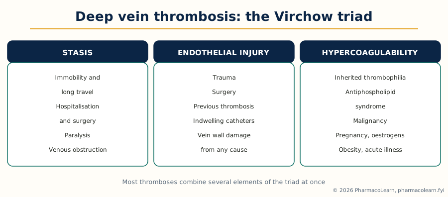 Figure: The Virchow triad. Naming which elements are present in a patient is the first step of both treatment and prevention.
4.6 Drug induced hematological disorders
Medicines injure every line of the blood, and the disorders classify by the element affected. Red cells are injured by immune and oxidant haemolysis, as in Section 4.4, and production is injured by aplastic anaemia, the rare, grave failure of the marrow stem cell compartment classically associated with chloramphenicol and reported with many other agents, presenting with pancytopenia and treated by withdrawal, support and specialist immunosuppression or transplantation. White cells are injured as neutropenia and its extreme, agranulocytosis, the abrupt loss of neutrophils presenting with fever and infection, for which the thionamides of Section 2.5, clozapine with its mandatory blood monitoring programme, cytotoxic agents predictably, sulfonamides and antiepileptics are responsible; management is immediate withdrawal, infection treatment and growth factor support in appropriate cases, and the fever warning given to every patient on an at risk drug is the preventive lesson. Platelets are injured by immune thrombocytopenia, in which drug dependent antibodies destroy platelets, reported with quinine, sulfonamides and anticonvulsants, and by heparin induced thrombocytopenia, the paradoxical prothrombotic reaction in which heparin dependent antibodies activate platelets, so falling platelets during heparin therapy signal new clotting, not bleeding, and demand immediate heparin cessation and a non heparin anticoagulant. Clotting is injured in the opposite direction by the bleeding caused by anticoagulant and antiplatelet therapy itself, the commonest serious haematological drug event in practice. Medicines also disturb formation qualitatively: methotrexate and other folate antagonists produce megaloblastic change, and several agents produce sideroblastic pictures. The pharmacist pattern across all of them is constant: know the at risk drugs, ensure the baseline and monitoring counts the drug demands, connect a new cytopenia or bleeding picture with the drug timeline, stop the suspect drug and escalate urgently, document and report, and counsel the patient on the symptoms, fever, sore throat, bruising, bleeding and unusual pallor, that must be reported at once.
Unit 5: Bone and Joint Disorders, Dermatological Diseases and Ophthalmology
5.1 Rheumatoid arthritis
Rheumatoid arthritis is a chronic systemic autoimmune disease whose central lesion is inflammatory synovitis: the synovial membrane thickens into an invasive tissue called pannus and destroys cartilage and erodes bone at the joint margins, producing deformity and loss of function if inflammation is not controlled. Its aetiology combines genetic susceptibility, notably shared epitope variants of the HLA system, with environmental triggers, smoking the best established, on a background in which autoimmunity to citrullinated proteins develops years before symptoms; rheumatoid factor and anti cyclic citrullinated peptide antibodies are its serological markers, the second the more specific. Synovial inflammation is driven by a cytokine network, tumour necrosis factor and interleukins prominent, recruiting inflammatory cells, activating fibroblasts and osteoclasts, and generating systemic features through circulating inflammation: fatigue, anaemia of chronic disease and accelerated cardiovascular disease, its major cause of premature death. Presentation is classically a symmetric polyarthritis of the small joints of the hands and feet with pain, swelling and prolonged morning stiffness evolving over weeks; over time come the characteristic deformities of ulnar deviation and swan neck and boutonniere fingers. Diagnosis is clinical, supported by inflammatory markers, autoantibodies and imaging showing erosions, and treatment is not delayed awaiting every test result, because early treatment changes the outcome: the window of opportunity principle.
Pharmacotherapy is early, disease modifying and target driven. Disease modifying antirheumatic drugs divide into conventional synthetic, targeted synthetic and biologic groups. Methotrexate is the anchor conventional agent, given weekly with folic acid supplementation, monitored by blood counts and liver tests, avoided in pregnancy, and combined or sequenced with sulfasalazine, leflunomide and hydroxychloroquine according to response and tolerance. Treatment escalates against a target of remission or low disease activity at regular reviews: where conventional therapy fails, biologic agents against tumour necrosis factor, interleukin 6 signalling, B cells or T cell costimulation, or targeted Janus kinase inhibitors, are added under specialist care after screening for latent infection, tuberculosis above all, and with vaccination reviewed. Glucocorticoids are short bridging therapy while DMARDs take effect and for flares, never the long term strategy where avoidance is possible. Non steroidal anti inflammatory drugs relieve symptoms and modify nothing; physiotherapy, occupational therapy and surgery for destroyed joints complete the multidisciplinary answer. Monitoring is the pharmacist visible part: the shared blood monitoring schedule of the DMARDs, infection vigilance under immunosuppression, and counselling on weekly methotrexate, whose accidental daily use is a recognised fatal error this subject expects students to name.
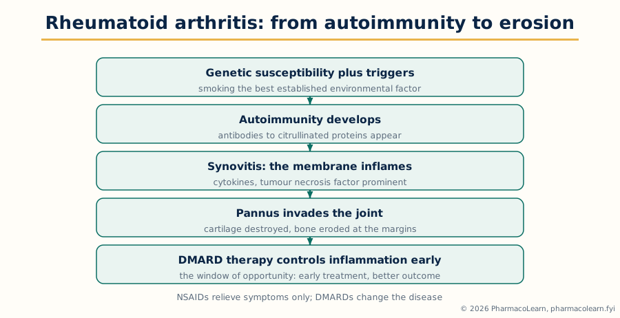 Figure: From autoimmunity to erosion. DMARD therapy works because it interrupts this cascade before the joint is destroyed.
5.2 Osteoarthritis
Osteoarthritis is the commonest joint disease, a disorder of the whole joint in which articular cartilage progressively fails, with changes in underlying bone, synovium, ligaments and muscle. It differs in kind from rheumatoid arthritis: its primary process is cartilage breakdown and failed repair, not autoimmune synovitis. Risk factors are age, obesity, previous joint injury, malalignment, occupational overload and genetic predisposition. Cartilage loses proteoglycan and its collagen network, chondrocyte repair cannot keep pace, the joint space narrows, subchondral bone thickens and forms osteophytes, and pain arises from bone, synovium, ligament and muscle rather than from the aneural cartilage. Presentation is pain on use relieved by rest, brief inactivity stiffness, reduced movement, crepitus and bony enlargement, in the hand the Heberden and Bouchard nodes at the finger joints; inflammatory markers are normal and imaging shows joint space narrowing, osteophytes and subchondral sclerosis. Diagnosis is clinical in the typical older patient, with inflammatory arthritis, infection and malignancy excluded by pattern.
Pharmacotherapy is symptomatic only, no agent modifies structure, and the non drug core takes first place: education, exercise and physiotherapy to strengthen the muscles protecting the joint, weight reduction where overweight, footwear, walking aids and occupational adaptations. Paracetamol has a modest, limited effect. Topical non steroidal anti inflammatory preparations are effective for knee and hand joints with little systemic exposure and are an early choice in older patients. Oral non steroidal anti inflammatory drugs are the most effective symptomatic class, used at the lowest effective exposure for the shortest time, with proton pump inhibitor gastroprotection where gastrointestinal risk requires it, and cardiovascular, renal and gastrointestinal risk assessed in every patient; cyclo oxygenase 2 selective agents trade some gastrointestinal advantage against cardiovascular caution, as in Section 3.1. Intra articular corticosteroid injection gives short term relief of an inflamed flare. Opioids have little place in chronic disease. Joint replacement for end stage knee and hip disease is the definitive treatment, and recognising when conservative therapy has reached its limit is part of pharmacotherapeutics.
5.3 Gout
Gout is arthritis caused by deposition of monosodium urate crystals in and around joints, the clinical expression of chronic hyperuricaemia. Uric acid is the end product of purine metabolism in humans, who lack uricase, and its level is set by production against excretion: most hyperuricaemia is renal under excretion, worsened by kidney disease, diuretics, especially thiazides, low dose aspirin, alcohol, obesity and purine and fructose rich diets; overproduction dominates in rapid cell turnover states and rare enzyme defects. When urate exceeds its solubility, crystals form in cooler peripheral joints and trigger an intense innate immune response through inflammasome activation and interleukin 1 release, producing the acute attack. The acute attack is dramatic: abrupt, exquisitely painful, hot, swollen arthritis, classically of the first metatarsophalangeal joint, podagra, often at night and after a dietary, alcohol, illness or drug provocation, settling over days to weeks even untreated; between attacks the patient is well until the pattern accelerates. Diagnosis is by crystal identification in aspirated joint fluid, the gold standard that also excludes septic arthritis, the dangerous mimic that must always be considered, supported by the clinical pattern and urate measured away from the acute attack, since it may be normal during one.
Pharmacotherapy separates into two questions that must never be merged: treating the acute attack, and lowering urate to prevent attacks and dissolve deposits. The acute attack is treated promptly with anti inflammatory therapy: non steroidal anti inflammatory drugs, colchicine, which disrupts microtubule dependent inflammatory activation and works best early, limited by gastrointestinal intolerance and interactions, or corticosteroids where the first two are unsuitable, with interleukin 1 blockade reserved for refractory cases under specialist care. Urate lowering therapy has its own indications: recurrent attacks, tophi, joint damage, stones and very high urate. Allopurinol, a xanthine oxidase inhibitor blocking uric acid production, is first line, started low and titrated against urate with renal function and its hypersensitivity syndrome in mind; febuxostat is the alternative xanthine oxidase inhibitor, with a cardiovascular caution in its history; uricosuric agents increase renal excretion where under excretion dominates and kidney function and stone risk allow; uricase therapy converting urate to soluble allantoin is a specialist last line for refractory tophaceous disease. Established urate lowering therapy is continued through acute attacks in current teaching, and prophylactic anti inflammatory cover accompanies its initiation, when urate shifts provoke flares.
5.4 Osteoporosis
Osteoporosis is reduced bone mass and microarchitectural deterioration leading to fragility, defined operationally by low bone mineral density on dual energy X ray absorptiometry and clinically by its fractures: vertebral, hip and wrist fractures from falls from standing height or less that healthy bone would withstand. Bone is remodelled continuously, osteoclasts resorbing and osteoblasts forming, coupled under hormonal and cytokine control, the RANKL pathway being the key osteoclast driver that one drug class targets. Peak mass is reached in young adult life; thereafter resorption gradually exceeds formation, and loss of oestrogen at the menopause sharply accelerates resorption, postmenopausal osteoporosis being the commonest form. Other causes must be sought: glucocorticoid therapy, the commonest secondary cause and a classic drug induced disease, hypogonadism, hyperthyroidism, hyperparathyroidism, alcohol, smoking, immobility, low body weight, vitamin D deficiency and malabsorption. The disease is silent until fracture: vertebral crush fractures present with back pain, height loss and kyphosis, and hip fracture dominates its mortality in the elderly. Diagnosis is by bone density measurement, by fracture risk assessment combining density with clinical risk factors, and by blood tests excluding secondary causes; a fragility fracture in an older patient establishes the diagnosis clinically and treatment follows without waiting for density confirmation.
Pharmacotherapy aims to reduce fractures, and its classes act on the remodelling balance from both sides. Foundation measures are adequate calcium and vitamin D, corrected where deficient, weight bearing and muscle strengthening exercise, fall prevention, smoking cessation and alcohol moderation. Bisphosphonates are the first line antiresorptive class: they bind bone mineral, are taken up by resorbing osteoclasts and disable them, reducing spine and hip fractures. The oral agents carry the famous administration discipline, taken fasting with a full glass of water, remaining upright, nothing else by mouth for a period after, because of oesophageal injury risk and near zero absorption with food, a counselling set examinations love; intravenous preparations serve patients who cannot follow or tolerate the oral route. Denosumab, a monoclonal antibody against RANKL, is a potent antiresorptive given by injection at long intervals, with the warning that stopping it without follow on antiresorptive therapy produces rebound bone loss and vertebral fractures. Anabolic agents that build bone, the parathyroid hormone analogues and the sclerostin inhibitor, serve severe and high risk disease under specialist care, followed by an antiresorptive to hold the gain.
5.5 Psoriasis
Psoriasis is a chronic immune mediated skin disease in which keratinocyte proliferation is greatly accelerated and disordered, producing well demarcated, red, scaly plaques. Genetic susceptibility and immune dysregulation are its basis, T cell and cytokine pathways, the interleukin 17 and interleukin 23 axis centrally, driving epidermal hyperplasia so that cells reach the surface in days rather than weeks and pile as silvery scale. Triggers include streptococcal throat infection, classically before guttate psoriasis, skin trauma localising lesions to injury (the Koebner phenomenon), stress, alcohol, smoking and medicines, lithium, beta blockers and antimalarials among the recognised provocations. Patterns include chronic plaque psoriasis of elbows, knees, scalp and lower back, guttate, flexural, pustular and erythrodermic forms, nail disease and associated psoriatic arthritis. Psoriasis is also a systemic inflammatory state with increased cardiovascular and metabolic risk and a heavy psychological burden. Diagnosis is usually clinical, severity graded by extent and by impact on quality of life, which together decide the treatment ladder.
Pharmacotherapy is stepped by severity and site. Emollients are universal foundation therapy. Topical corticosteroids and vitamin D analogues, which normalise keratinocyte proliferation, are the mainstay for limited plaque disease, combined in single preparations for adherence; coal tar and dithranol are older effective agents limited by mess and irritation, and calcineurin inhibitors serve face and flexures where steroids thin skin. Phototherapy treats widespread disease failing topicals. Systemic therapy for moderate to severe disease uses methotrexate, ciclosporin, rapid but limited by kidney and blood pressure effects, and acitretin, a retinoid for pustular forms, teratogenic with a prolonged contraception requirement. Biologic therapies against tumour necrosis factor, interleukin 17 and interleukin 23 pathways, and the oral phosphodiesterase 4 inhibitor for moderate disease, have transformed severe psoriasis under specialist supervision with infection screening. Counselling covers a long term, relapsing course: control, not cure.
5.6 Eczema and scabies
Eczema, or dermatitis, is a pattern of skin inflammation whose commonest important form is atopic eczema, a chronic relapsing itchy dermatosis of childhood onset associated with the atopic family, asthma of Section 2.1 among them. Its pathophysiology combines a defective skin barrier, filaggrin gene defects prominent among its causes, which lets water out and irritants and allergens in, with Th2 type immune inflammation; other eczemas are irritant and allergic contact dermatitis, where patch testing identifies allergic culprits, seborrhoeic, discoid and varicose patterns. Management is barrier repair plus anti inflammatory control: liberal emollients as continuing maintenance and soap substitutes, avoidance of identified triggers, topical corticosteroids stepped by potency to site and severity in defined short courses with fingertip unit guidance, topical calcineurin inhibitors for sensitive sites and steroid sparing maintenance, antibacterials where infection supervenes, and for severe disease phototherapy, systemic immunosuppressants and the biologic and targeted therapies against interleukin 4 and 13 signalling and Janus kinase pathways.
Scabies shares the printed syllabus head with eczema but is a different kind of disease: infestation by the mite Sarcoptes scabiei, transmitted by prolonged skin contact, the female mite burrowing into the stratum corneum and laying eggs, the itching being a hypersensitivity response to the mite and its products. Diagnosis is clinical, supported by mite or egg demonstration on scraping or dermoscopy. Treatment is scabicidal and social at once: permethrin cream, the standard topical agent, applied to the whole body surface as directed and repeated per its schedule, or oral ivermectin in crusted disease, outbreaks and where topical application is impractical, and, decisively, simultaneous treatment of all close contacts with hot washing of bedding and clothing, because untreated contacts reinfest the treated patient. Itch persists for weeks after successful treatment as hypersensitivity settles, must be explained in advance so it is not read as failure, and is treated symptomatically.
5.7 Impetigo
Impetigo is a superficial, highly contagious bacterial skin infection, commonest in children, caused by Staphylococcus aureus, Streptococcus pyogenes, or both. Non bullous impetigo is the classic form, honey coloured crusted erosions around the nose, mouth and limbs arising where minor trauma, insect bites or underlying eczema break the skin; in bullous impetigo, staphylococcal exfoliative toxins split the superficial epidermis into flaccid blisters. Diagnosis is usually clinical, swabs settling doubtful or recurrent cases. Its importance exceeds its depth: it spreads through families, schools and teams, and streptococcal impetigo is associated with post streptococcal glomerulonephritis, a reason to treat rather than merely reassure.
Pharmacotherapy follows extent. Limited lesions in a well patient are treated topically with an antiseptic or a topical antibacterial such as fusidic acid or mupirocin, with crust removal and hygiene. Widespread, bullous, recurrent or outbreak disease, and disease in the immunocompromised, is treated with oral antibacterials active against both organisms per local guidance. Recurrence prompts review for nasal staphylococcal carriage and untreated contacts.
5.8 Drug induced skin disorders
The skin is the most visible target of adverse drug reactions, and drug induced skin disorders range from the common and benign to the rare and life threatening, a range the examination answer must span in order. Exanthematous or maculopapular eruptions are the commonest pattern, a symmetric red rash appearing days into a new drug, most often an antibacterial, and settling on withdrawal, its main task being not to miss the early signs of a severe reaction within it. Urticaria and angioedema follow histamine release by immune and non immune mechanisms, penicillins and NSAIDs prominent, angioedema with airway involvement being an emergency, and ACE inhibitor angioedema, bradykinin mediated and antihistamine resistant, its own named entity. Fixed drug eruption recurs at exactly the same site on each exposure, leaving pigmented patches, a history pattern that makes the diagnosis. Photosensitivity reactions divide into phototoxic, dose related exaggerated sunburn, and photoallergic patterns, with tetracyclines, fluoroquinolones, thiazides and amiodarone among the culprits, managed by withdrawal and photoprotection. The severe cutaneous adverse reactions stand at the head of the topic and must be recognised on sight: Stevens Johnson syndrome and toxic epidermal necrolysis, a spectrum of epidermal necrosis with mucosal involvement and skin detachment, caused above all by allopurinol, sulfonamides, aromatic anticonvulsants and lamotrigine and certain NSAIDs, with high mortality, treated by immediate withdrawal of every suspect drug and specialist burn level supportive care; DRESS, drug reaction with eosinophilia and systemic symptoms, a delayed eruption with fever, facial swelling, eosinophilia and hepatitis and other organ injury, classically from anticonvulsants, allopurinol and sulfonamides, treated by withdrawal and corticosteroids; and acute generalised exanthematous pustulosis, a rapid sterile pustular eruption, usually antibacterial driven, settling on withdrawal. Pharmacogenetic screening prevents some, HLA testing before carbamazepine and abacavir and in defined allopurinol risk groups being the standard examples. The pharmacist discipline across all patterns is the timeline, every new eruption matched against every drug started in the preceding weeks, immediate withdrawal of suspects in severe patterns, documentation and reporting, allergy records that distinguish rash histories precisely, and counselling that a patient who has survived a severe reaction must never receive the culprit again.
5.9 Conjunctivitis
Conjunctivitis is inflammation of the conjunctiva, presenting as a red eye with discharge, irritation and itching, usually with normal vision and little pain, the features that separate it from the dangerous causes of red eye. Infective conjunctivitis is viral, most often adenoviral, watery and highly contagious, or bacterial, with purulent discharge and lids stuck on waking; chlamydial and gonococcal forms demand specific systemic therapy and referral. Allergic conjunctivitis is itchy above all, bilateral, seasonal or perennial with rhinitis. Irritant forms follow chemicals, smoke and medicines themselves, including preservative reactions. Diagnosis is clinical from discharge character, itch, laterality and associated features, with pain, photophobia, reduced vision, corneal involvement, contact lens wear and failure to settle as referral triggers, since keratitis, uveitis and acute glaucoma share the red eye headline and none of them is conjunctivitis.
Pharmacotherapy is cause matched and mostly restrained. Viral conjunctivitis is self limiting, treated supportively with lubricants and hygiene; antibacterials have no action on it. Bacterial conjunctivitis often settles without treatment, topical antibacterial drops or ointment, chloramphenicol and fusidic acid preparations being standard examples, shortening the course where discharge is heavy or return to school or work requires it. Allergic conjunctivitis is treated by allergen avoidance, topical antihistamine and mast cell stabilising drops, often combined in dual acting agents, and oral antihistamines where rhinitis accompanies; topical steroids are specialist directed only, because unsupervised ocular steroids aggravate herpes simplex keratitis and raise intraocular pressure, the caution that links this section to the next. Hygiene counselling, hand washing, no shared towels and no lens wear until settled, completes most episodes.
5.10 Glaucoma
Glaucoma is a group of optic neuropathies in which retinal ganglion cells and their axons are progressively lost, producing characteristic optic disc excavation and visual field loss beginning in the periphery, with raised intraocular pressure the major modifiable risk factor, though damage also occurs at statistically normal pressures. Aqueous humour physiology is the pharmacology of the whole topic: aqueous is produced by the ciliary body, flows through the pupil into the anterior chamber, and drains chiefly through the trabecular meshwork into the canal of Schlemm, with a secondary uveoscleral route; pressure is the balance of production against outflow, and every drug class either reduces production or increases outflow. In primary open angle glaucoma, the commonest form, the angle is open but trabecular resistance rises, pressure climbs slowly and painlessly, and the patient notices nothing until field loss is advanced, so detection is by opportunistic examination of pressure, disc and fields in at risk patients: older age, family history, African ancestry, diabetes and myopia among them. In primary angle closure glaucoma, a shallow anterior chamber lets the iris block the meshwork when the pupil dilates, precipitating an acute attack of severe eye pain, blurred vision with haloes, a red eye, a mid dilated unreactive pupil and nausea, an ophthalmic emergency in which pressure must be lowered at once or sight is lost within days. Diagnosis is by tonometry, gonioscopy, disc assessment and formal visual field testing in combination.
Pharmacotherapy of open angle glaucoma lowers pressure with topical agents in a stepped sequence, laser trabeculoplasty and surgery standing beside drugs in the modern pathway. Prostaglandin analogues are the usual first line class: they increase uveoscleral outflow, are used once daily in the evening, and have the distinctive local effects of iris darkening, lash growth and periorbital fat loss that patients must be warned of so they do not stop a working drug. Beta blocker drops reduce aqueous production and carry their class cautions in miniature, bronchospasm in asthma and bradycardia, since drops drain through the nasolacrimal duct into the systemic circulation, which punctal occlusion after instillation reduces, a practical counselling technique. Alpha agonists reduce production and increase outflow, allergic conjunctivitis limiting them. Topical carbonic anhydrase inhibitors reduce production and are common additions, the oral form reserved for short term and emergency use. Pilocarpine increases trabecular outflow and survives mainly in angle closure management. The acute angle closure attack is an emergency sequence: rapid pressure lowering by combined topical agents and systemic carbonic anhydrase inhibition, analgesia and antiemetics, then the definitive laser peripheral iridotomy that bypasses the blocked pupil pathway, also performed in the fellow eye. Adherence in a symptomless, lifelong, sight preserving therapy is the closing counselling theme of the unit: patients stop the drops that sting, and keep their sight only if the bargain is explained plainly.
Glaucoma: the aqueous balance and where drugs act
  Ciliary body produces aqueous humour
  (beta blockers, alpha agonists and carbonic
   anhydrase inhibitors reduce production here)
        |
        v
  Posterior chamber, through the pupil
  (blocked here in angle closure: iridotomy bypasses it)
        |
        v
  Anterior chamber drains two ways:
  trabecular meshwork (pilocarpine improves this route)
  uveoscleral outflow (prostaglandin analogues
   increase this route, first line therapy)
        |
        v
  Pressure falls, ganglion cell loss slows,
  visual field is preserved
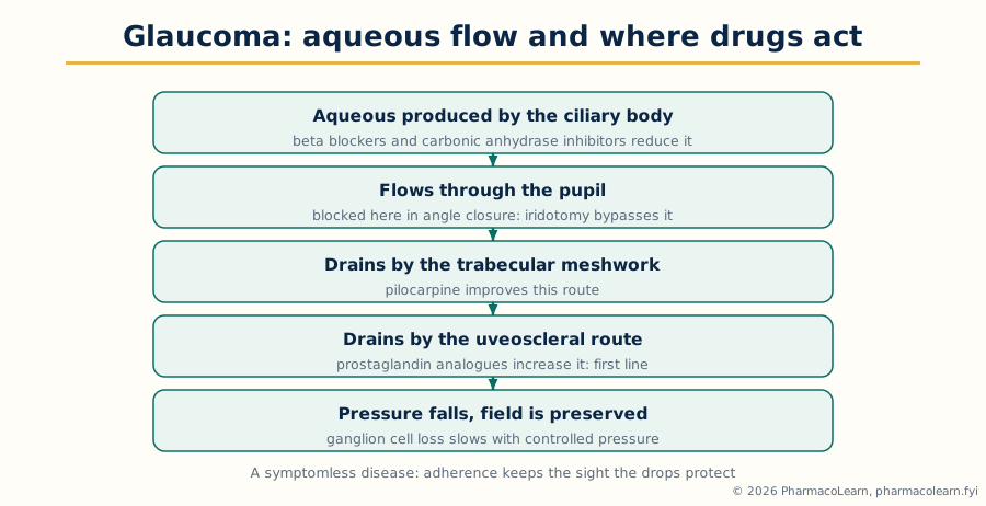 Figure: Aqueous flow and drug action. Every glaucoma class either reduces production or increases one of the two drainage routes.
Frequently Asked Questions
Q: What is the pharmacotherapeutics method this subject teaches for every disease? Begin with etiopathogenesis, then presentation and diagnosis including severity, comorbidity, organ function, other medicines and adherence, then an individualised plan in steps: non drug measures and treatment of causes throughout, first drug classes matched to the mechanism, review against stated goals using clinical and laboratory indices, and adjustment by addition, substitution or escalation, with long term monitoring planned from the start. See Section 1.1.
Q: How is hypertension treated in principle, and which drug classes are first line? Lifestyle measures apply to every patient: weight reduction, salt restriction, a healthy diet, regular exercise, alcohol moderation and stopping smoking. The first line classes are thiazide and thiazide like diuretics, calcium channel blockers, ACE inhibitors and angiotensin receptor blockers, with beta blockers first line chiefly where a compelling cardiac indication coexists. Most patients need complementary classes in combination, and resistant hypertension adds agents such as aldosterone antagonists under specialist care. See Section 1.2.
Q: Which drug classes improve survival in heart failure with reduced ejection fraction, and which only relieve symptoms? Renin angiotensin inhibition, including the angiotensin receptor and neprilysin inhibitor combination, the beta blockers proven in heart failure, aldosterone antagonists and SGLT2 inhibitors improve outcomes, because they block or counter the maladaptive neurohormonal responses. Loop diuretics relieve congestion by removing fluid but are symptom therapy, adjusted to fluid state. See Section 1.3.
Q: What are the two drug aims in acute coronary syndrome, and which classes serve each? To limit necrosis by restoring flow, by reperfusion with intervention or fibrinolysis supported by anticoagulation, and to prevent thrombus extension and recurrence, by dual antiplatelet therapy with aspirin plus a P2Y12 blocker in the acute phase and long term secondary prevention with continued antiplatelet therapy and a statin. Nitrates relieve pain, and beta blockers reduce oxygen demand where not contraindicated. See Section 1.4.
Q: How do asthma and COPD differ in their drug therapy foundations? In asthma the pathology is airway inflammation with hyperresponsiveness, so inhaled corticosteroids are the foundation at every step and long acting bronchodilators are used only in combination with them. In COPD the pathology is persistent airflow limitation from airway and parenchymal damage, so long acting bronchodilators are the foundation, inhaled corticosteroids are added only for selected patients, and smoking cessation is the one intervention that changes the course. See Sections 2.1 and 2.2.
Q: How are the diabetes drug classes matched to the physiology of type 2 diabetes? Metformin reduces hepatic glucose production. Sulfonylureas stimulate insulin secretion and risk hypoglycaemia and weight gain. DPP 4 inhibitors prolong incretin action gently, GLP 1 receptor agonists enhance glucose dependent insulin secretion, reduce appetite and weight and include agents with cardiovascular benefit, and SGLT2 inhibitors lower glucose by renal glucose excretion and protect heart and kidneys. Insulin replaces the hormone and is obligatory in type 1 diabetes. See Section 2.4.
Q: What decides drug therapy in peptic ulcer disease? The balance between aggressive factors, acid, pepsin, Helicobacter pylori and NSAID injury, and defensive factors, the mucus bicarbonate barrier, blood flow and prostaglandins. Proton pump inhibitors suppress acid at the final common step for healing, eradication therapy removes H. pylori so ulcers do not recur, and NSAID ulcers require stopping or reducing the NSAID where possible with proton pump inhibitor healing and protection. See Section 3.1.
Q: How do ulcerative colitis and Crohn disease differ, and what follows for therapy? Ulcerative colitis is continuous mucosal inflammation of the colon only, presenting chiefly with bloody diarrhoea, and colectomy removes the colonic disease. Crohn disease is transmural, discontinuous and may affect any part of the gut, producing strictures and fistulae, and surgery is bowel preserving because recurrence is common. Both are treated by inducing remission, corticosteroids having that role but never the maintenance role, and maintaining remission with aminosalicylates in ulcerative colitis, immunomodulators, and biologic or targeted therapies in moderate to severe disease. See Section 3.3.
Q: What are the principles of anticoagulation in deep vein thrombosis? Anticoagulants prevent extension, embolisation and recurrence while the body removes the clot; they do not dissolve it directly, thrombolysis being reserved for massive pulmonary embolism and a few limb threatening cases. Options are heparins, warfarin with INR monitoring and a parenteral overlap at initiation, and the direct oral anticoagulants, now usual for most patients. Duration is individualised by provoking factors and bleeding risk, and thromboprophylaxis of at risk inpatients is the preventive half of the subject. See Section 4.5.
Q: How do drugs treat glaucoma, in terms of aqueous humour? Aqueous is produced by the ciliary body and drains through the trabecular meshwork and the uveoscleral route, and every class acts on that balance. Prostaglandin analogues increase uveoscleral outflow and are first line. Beta blockers, alpha agonists and carbonic anhydrase inhibitors reduce production, alpha agonists also increase outflow, and pilocarpine increases trabecular outflow. The acute angle closure attack is an emergency treated by rapid pressure lowering followed by laser iridotomy. See Section 5.10.
Complete MPP102T notes, all 5 units in one PDF, free to keep.
Other M.Pharm subjects: MPH101T · MPC101T · MPL101T · MPH102T · MPH103T · MPH104T · MPL102T · MPL103T · MPL104T · MPC102T · MPC103T · MPC104T · MPH201T · MPH202T · MPH203T · MPH204T · MPC201T · MPC202T · MPC203T · MPC204T · MPL201T · MPL202T · MPL203T · MPL204T · MRA101T · MRA102T · MRA103T · MRA104T · MRM301T · MPG101T · MPG102T · MPG103T · MPG201T · MPG202T · MPG203T · MPG104T · MPG204T · MRA201T · MRA202T · MRA203T · MRA204T · MPP101T · MPP102T · MPP103T · MPP104T · MPP201T · MPP202T · M.Pharm