
BP203T — Biochemistry
3 hrs/week · 4 credits · 45 hours · 100 marks (25 internal + 75 end-semester, 3 hr paper). Notes follow the PCI B.Pharm Semester 2 syllabus, unit by unit.
Units in this subject
- Unit 1 — Biomolecules and Biochemical Concepts
- Unit 2 — Bioenergetics and Biological Oxidation
- Unit 3 — Carbohydrate Metabolism
- Unit 4 — Lipid Metabolism
- Unit 5 — Amino Acid Metabolism and Genetic Information
Download BP203T PDF Notes
Free PDFs for this subject, one per unit:
These are original study notes for Biochemistry (BP203T), B.Pharm Semester II. They use simple language and follow the five-unit sequence in the PCI syllabus. The notes explain core definitions, pathways, functions and exam-oriented comparisons without using brand names.
Unit I: Biomolecules and Biochemical Concepts
This unit introduces the molecules found in living systems. Learn each group by its building blocks, major bonds, important functions and one or two representative examples.
Carbohydrates
Carbohydrates are polyhydroxy aldehydes, polyhydroxy ketones, or substances that produce them on hydrolysis. They are classified as monosaccharides, oligosaccharides and polysaccharides.
- Monosaccharides are single sugar units. Examples include glucose, fructose and ribose.
- Disaccharides contain two monosaccharide units. Sucrose contains glucose and fructose. Lactose contains glucose and galactose.
- Polysaccharides contain many sugar units. Glycogen is a storage polysaccharide in animals. Cellulose is a structural polysaccharide in plants.
The glycosidic bond joins sugar units. Reducing sugars have a free anomeric carbon that can participate in a redox reaction. Glucose is a reducing sugar. Sucrose is non-reducing because its anomeric carbons are involved in the glycosidic bond.
Proteins and amino acids
Proteins are polymers of amino acids joined by peptide bonds. An amino acid has an amino group, a carboxyl group, a hydrogen atom and a variable side chain attached to an alpha carbon. Proline is the special case because its side chain forms a ring with its amino nitrogen.
The 20 standard amino acids can be grouped by side-chain properties:
- Nonpolar: glycine, alanine, valine, leucine, isoleucine, methionine, proline, phenylalanine and tryptophan. They commonly contribute to hydrophobic protein cores. Glycine gives flexibility, proline restricts chain rotation, and methionine can begin protein synthesis.
- Polar, uncharged: serine, threonine, cysteine, tyrosine, asparagine and glutamine. They form hydrogen bonds. Cysteine can form disulfide bonds, and serine, threonine and tyrosine can be sites of phosphorylation.
- Acidic: aspartate and glutamate. Their side chains are negatively charged near neutral pH and participate in ionic interactions and acid-base reactions.
- Basic: lysine, arginine and histidine. Their side chains can carry positive charge. Histidine is useful in enzyme active sites because it can accept or donate a proton near physiological pH.
These groupings describe side-chain behaviour, not dietary classification.
Protein structure is described at four levels:
- Primary structure: the amino acid sequence.
- Secondary structure: local folding such as alpha helices and beta sheets.
- Tertiary structure: the overall three-dimensional shape of one polypeptide chain.
- Quaternary structure: association of two or more polypeptide chains.
Hydrogen bonds, ionic interactions, hydrophobic interactions and disulfide bonds help maintain protein structure. Denaturation disrupts higher-order structure and biological activity without normally breaking all peptide bonds.
Lipids
Lipids are water-insoluble or poorly water-soluble biomolecules that dissolve in non-polar solvents. Important groups include fatty acids, triacylglycerols, phospholipids and sterols.
- Saturated fatty acids have no carbon-carbon double bond.
- Unsaturated fatty acids have one or more carbon-carbon double bonds.
- Triacylglycerols contain glycerol esterified with three fatty acids and serve mainly as energy stores.
- Phospholipids contain phosphate and are major components of cell membranes.
Nucleic acids and nucleotides
Nucleotides contain a nitrogenous base, a pentose sugar and one or more phosphate groups. A nucleoside contains a base and sugar but no phosphate. DNA contains deoxyribose and stores hereditary information. RNA contains ribose and participates in gene expression and protein synthesis.
The bases adenine and guanine are purines. Cytosine, thymine and uracil are pyrimidines. DNA usually contains thymine, while RNA usually contains uracil.
In double-stranded DNA, adenine pairs with thymine through two hydrogen bonds, and guanine pairs with cytosine through three hydrogen bonds. The two strands are complementary and antiparallel. In RNA, adenine pairs with uracil in base-paired regions.
Lipid classes in detail
Simple lipids include triacylglycerols and waxes. Triacylglycerols contain glycerol esterified to three fatty acids and are concentrated energy stores. Waxes contain long-chain fatty acids esterified to long-chain alcohols and provide protective coatings.
Compound lipids contain additional groups. Phospholipids contain phosphate and form membrane bilayers. Glycerophospholipids contain glycerol, while sphingophospholipids contain sphingosine. Glycolipids contain carbohydrate and are important in cell recognition, especially in nervous tissue. Lipoproteins contain lipid plus protein and transport hydrophobic molecules in blood.
Derived lipids include fatty acids, sterols and steroid hormones. Cholesterol supports membrane structure and is a precursor for bile acids, steroid hormones and vitamin D. Eicosanoids are signalling molecules formed from polyunsaturated fatty acids.
Enzymes
Enzymes are biological catalysts. They increase reaction rate by lowering activation energy and are not consumed in the overall reaction. The substrate binds at an active site. Enzyme activity depends on factors such as temperature, pH, substrate concentration and inhibitor concentration.
Competitive inhibition can be reduced by increasing substrate concentration in the simple model of enzyme kinetics. In pure non-competitive inhibition, inhibitor binding lowers Vmax without changing Km.
Unit I: Frequently asked questions
Q: What is the difference between a nucleoside and a nucleotide?
A: A nucleoside contains a nitrogenous base and a pentose sugar. A nucleotide contains a nucleoside plus one or more phosphate groups.
Q: What is denaturation of a protein?
A: Denaturation is loss of the normal higher-order structure and activity of a protein caused by conditions such as heat, extreme pH or chemicals. The primary peptide-bond sequence is usually not broken.
Q: Why are enzymes called catalysts?
A: Enzymes increase the rate of biochemical reactions by lowering activation energy and are regenerated at the end of the reaction.
Q: What are the DNA base-pairing rules?
A: Adenine pairs with thymine and guanine pairs with cytosine in double-stranded DNA. In RNA, adenine pairs with uracil in base-paired regions.
Unit I: Key takeaways
- Carbohydrates are classified by the number of sugar units.
- Proteins are amino acid polymers joined by peptide bonds.
- Lipids include storage lipids and membrane lipids.
- DNA and RNA differ in sugar, one base and their main cellular roles.
- Enzymes are biological catalysts with activity affected by conditions such as pH and temperature.
Unit I: Clinical correlation
Clinical correlation: gout. Purine breakdown produces uric acid. Persistent elevation of uric acid can promote urate crystal deposition in joints.
Unit II: Bioenergetics and Biological Oxidation
This unit explains how cells capture, transfer and use energy. Learn the meaning of free energy, oxidation-reduction reactions, high-energy compounds, the electron transport chain and oxidative phosphorylation.
Bioenergetics
Bioenergetics is the study of energy changes in biological systems. A reaction with a negative change in Gibbs free energy is thermodynamically favourable under the stated conditions. ATP couples energy-releasing reactions to energy-requiring reactions.
ATP contains phosphoanhydride bonds. ATP hydrolysis can provide energy for biosynthesis, active transport and mechanical work. ATP is continually formed and used rather than stored as a large long-term energy reserve.
Oxidation-reduction reactions
Oxidation is loss of electrons or hydrogen, or gain of oxygen. Reduction is gain of electrons or hydrogen, or loss of oxygen. Oxidation and reduction occur together in a redox reaction.
NAD+ and FAD are important electron carriers. NADH and FADH2 transfer reducing equivalents to the respiratory chain. The exact ATP yield per molecule of NADH or FADH2 depends on the accounting convention and shuttle assumptions, so use the value specified by the teacher or examination scheme.
Biological oxidation and the electron transport chain
The electron transport chain is located in the inner mitochondrial membrane in eukaryotic cells. Electrons pass through protein complexes and mobile carriers to molecular oxygen, which is reduced to water. Energy released during electron transfer pumps protons across the inner membrane.
The proton gradient stores electrochemical energy. ATP synthase uses proton movement back across the membrane to form ATP from ADP and inorganic phosphate. This process is called oxidative phosphorylation.
Electron transport chain in order
- Complex I, NADH dehydrogenase, accepts electrons from NADH and transfers them to ubiquinone. It pumps protons.
- Complex II, succinate dehydrogenase, accepts electrons from succinate through FADH2 and transfers them to ubiquinone. It does not pump protons.
- Ubiquinone, also called coenzyme Q, is a mobile lipid-soluble carrier that transfers electrons to Complex III.
- Complex III, the cytochrome bc1 complex, transfers electrons from ubiquinol to cytochrome c and pumps protons.
- Cytochrome c is a mobile carrier that transfers electrons from Complex III to Complex IV.
- Complex IV, cytochrome c oxidase, transfers electrons to oxygen and forms water. It pumps protons.
- ATP synthase, sometimes called Complex V, uses the proton-motive force to phosphorylate ADP.
ATP yield accounting
This draft uses the modern convention of approximately 2.5 ATP per mitochondrial NADH and 1.5 ATP per FADH2. Complete oxidation of one glucose is therefore approximately 30 to 32 ATP, depending on the cytosolic NADH shuttle. Older textbooks may use 3 ATP per NADH and 2 ATP per FADH2 and report a higher total. State the convention in a numerical answer.
High-energy compounds
High-energy compounds have hydrolysis reactions with a large negative free-energy change under cellular conditions. ATP, phosphocreatine and some thioesters are examples. Their role is to transfer or provide energy for cellular work.
Unit II: Frequently asked questions
Q: What is oxidative phosphorylation?
A: It is the formation of ATP linked to electron transfer through the respiratory chain. The chain creates a proton gradient, and ATP synthase uses that gradient to make ATP.
Q: What is the role of oxygen in aerobic respiration?
A: Oxygen is the terminal electron acceptor in the mitochondrial respiratory chain. It combines with electrons and protons to form water.
Q: What is the difference between oxidation and reduction?
A: Oxidation is loss of electrons or hydrogen, while reduction is gain of electrons or hydrogen. They occur together in redox reactions.
Q: Which electron transport complexes pump protons?
A: Complexes I, III and IV pump protons. Complex II transfers electrons but does not pump protons.
Unit II: Key takeaways
- ATP couples energy release to cellular work.
- NADH and FADH2 carry reducing equivalents.
- The respiratory chain establishes a proton gradient.
- ATP synthase uses proton flow to form ATP.
- Oxygen is the terminal electron acceptor in aerobic mitochondrial respiration.
- Complexes I, III and IV pump protons, while ubiquinone and cytochrome c are mobile carriers.
Unit II: Clinical correlation
Clinical correlation: ketosis. When carbohydrate use is limited and fatty acid oxidation increases, the liver produces ketone bodies. Accumulation of ketone bodies can lower blood pH in severe insulin deficiency.
Unit III: Carbohydrate Metabolism
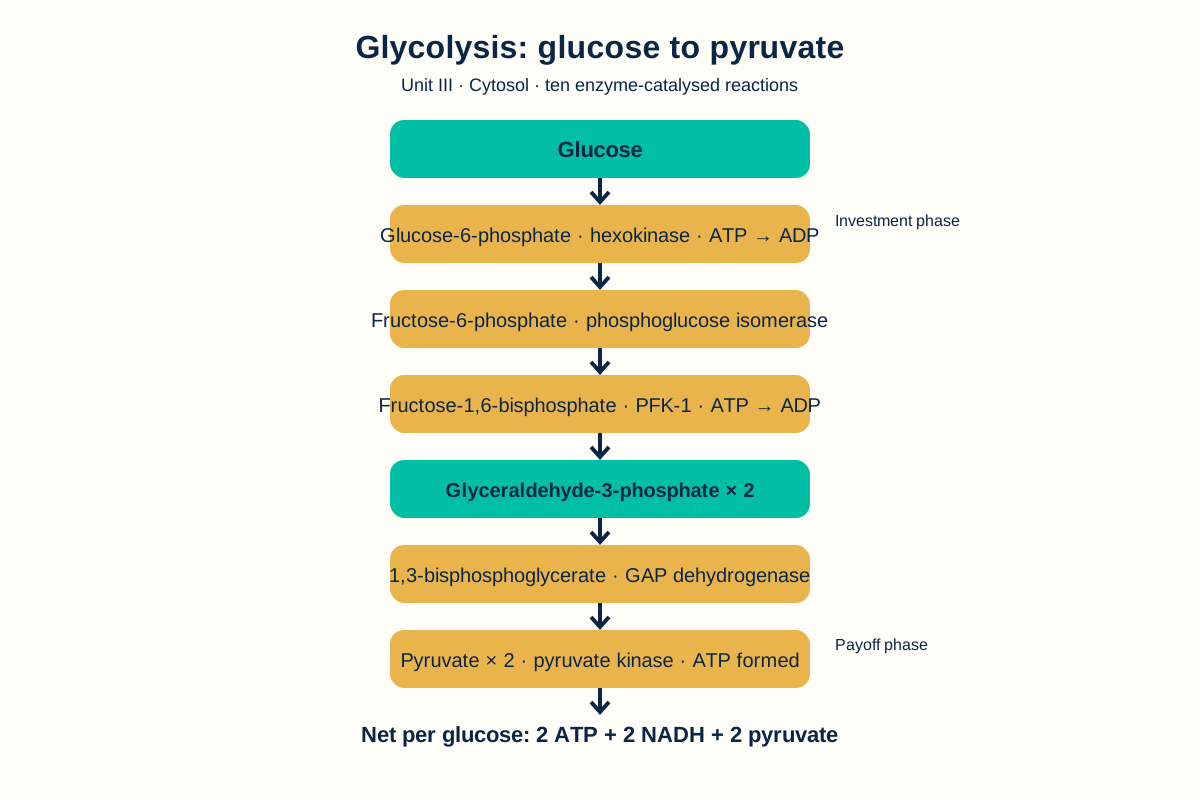
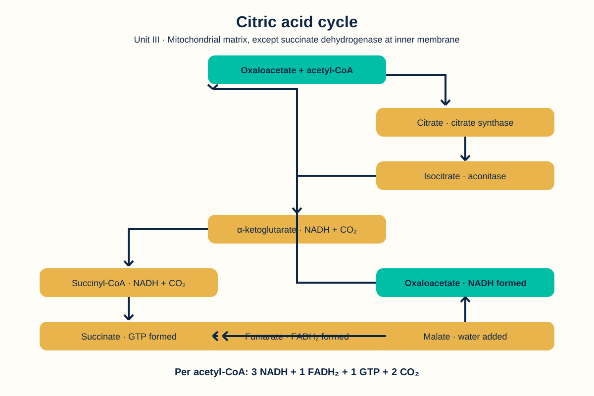
This unit follows the main pathways by which glucose is used, stored and formed. For every pathway, learn the site, purpose, key steps, major regulation points and products.
Glycolysis
Glycolysis is the pathway that converts glucose to pyruvate through a sequence of reactions in the cytosol. It can operate without oxygen, although the fate of pyruvate and the regeneration of NAD+ depend on cellular conditions.
The pathway has an energy-investment phase and an energy-generation phase. It produces ATP by substrate-level phosphorylation and produces NADH when glyceraldehyde-3-phosphate is oxidized.
In aerobic conditions, pyruvate can enter mitochondria and be converted to acetyl-CoA by the pyruvate dehydrogenase complex. In anaerobic conditions, pyruvate can be reduced to lactate, which regenerates NAD+ for continued glycolysis.
Glycolysis: 10 steps and enzymes
- Glucose to glucose-6-phosphate: hexokinase or glucokinase; ATP is used.
- Glucose-6-phosphate to fructose-6-phosphate: phosphoglucose isomerase.
- Fructose-6-phosphate to fructose-1,6-bisphosphate: phosphofructokinase-1; ATP is used and this is a major control step.
- Fructose-1,6-bisphosphate to glyceraldehyde-3-phosphate and dihydroxyacetone phosphate: aldolase.
- Dihydroxyacetone phosphate to glyceraldehyde-3-phosphate: triose phosphate isomerase.
- Glyceraldehyde-3-phosphate to 1,3-bisphosphoglycerate: glyceraldehyde-3-phosphate dehydrogenase; NADH is formed.
- 1,3-Bisphosphoglycerate to 3-phosphoglycerate: phosphoglycerate kinase; ATP is formed.
- 3-Phosphoglycerate to 2-phosphoglycerate: phosphoglycerate mutase.
- 2-Phosphoglycerate to phosphoenolpyruvate: enolase.
- Phosphoenolpyruvate to pyruvate: pyruvate kinase; ATP is formed.
The net yield per glucose is two ATP, two NADH and two pyruvate under the usual accounting conditions.
Citric acid cycle
The citric acid cycle occurs mainly in the mitochondrial matrix in eukaryotic cells. Acetyl-CoA is oxidized to carbon dioxide, and the cycle produces NADH, FADH2 and GTP or ATP, depending on the tissue and nomenclature used.
The reduced coenzymes transfer electrons to the respiratory chain. The cycle also supplies intermediates for biosynthetic pathways.
Citric acid cycle: 8 steps and enzymes
- Oxaloacetate plus acetyl-CoA to citrate: citrate synthase.
- Citrate to isocitrate: aconitase.
- Isocitrate to alpha-ketoglutarate: isocitrate dehydrogenase; NADH and carbon dioxide are formed.
- Alpha-ketoglutarate to succinyl-CoA: alpha-ketoglutarate dehydrogenase complex; NADH and carbon dioxide are formed.
- Succinyl-CoA to succinate: succinyl-CoA synthetase; GTP or ATP is formed.
- Succinate to fumarate: succinate dehydrogenase; FADH2 is formed.
- Fumarate to malate: fumarase.
- Malate to oxaloacetate: malate dehydrogenase; NADH is formed.
Gluconeogenesis
Gluconeogenesis is the formation of glucose from non-carbohydrate precursors such as lactate, glycerol and glucogenic amino acids. It occurs mainly in the liver, with contributions from the kidney during prolonged fasting. It is not simply the reverse of glycolysis because several glycolytic reactions are strongly irreversible and require bypass reactions.
Glycogen metabolism
Glycogenesis is the synthesis of glycogen from glucose. Glycogenolysis is the breakdown of glycogen. Liver glycogen helps maintain blood glucose, while muscle glycogen is used mainly for energy within muscle.
Glycogenesis uses glycogenin to start a primer, glycogen synthase to extend alpha-1,4 chains and a branching enzyme to create alpha-1,6 branches. Insulin promotes activation of glycogen synthase after a carbohydrate-rich meal. Glycogenolysis uses glycogen phosphorylase to release glucose-1-phosphate, a debranching enzyme at branch points and phosphoglucomutase to form glucose-6-phosphate. Liver glucose-6-phosphatase permits free glucose release. Glucagon promotes liver glycogenolysis, while adrenaline can stimulate glycogenolysis in liver and muscle.
Pentose phosphate pathway
The pentose phosphate pathway occurs in the cytosol. It produces NADPH and ribose-5-phosphate. NADPH supports reductive biosynthesis and antioxidant defence. Ribose-5-phosphate is needed for nucleotide synthesis. The pathway does not directly produce ATP. Its oxidative phase is controlled by glucose-6-phosphate dehydrogenase, and its products support fatty acid synthesis, glutathione reduction and nucleotide synthesis.
Unit III: Frequently asked questions
Q: What is the net purpose of glycolysis?
A: Glycolysis converts glucose to pyruvate and captures part of the released energy as ATP and NADH. It occurs in the cytosol.
Q: Why is gluconeogenesis not the exact reverse of glycolysis?
A: Several glycolytic reactions have a large negative free-energy change in cells. Gluconeogenesis bypasses these steps with different enzymes.
Q: What are the main products of the pentose phosphate pathway?
A: The pathway produces NADPH and ribose-5-phosphate. It also produces sugar intermediates that can enter other carbohydrate pathways.
Q: Which enzyme is a major control point in glycolysis?
A: Phosphofructokinase-1 is a major control enzyme and converts fructose-6-phosphate to fructose-1,6-bisphosphate.
Unit III: Key takeaways
- Glycolysis occurs in the cytosol and forms pyruvate, ATP and NADH.
- Pyruvate dehydrogenase links glycolysis with the citric acid cycle.
- The citric acid cycle produces reduced coenzymes and biosynthetic intermediates.
- Glycogenesis stores glucose as glycogen, while glycogenolysis releases usable glucose units.
- The pentose phosphate pathway supplies NADPH and ribose-5-phosphate.
- Glycolysis has 10 enzyme-catalysed steps, and the citric acid cycle has 8.
Unit III: Clinical correlation
Clinical correlation: diabetes mellitus. In diabetes mellitus, inadequate insulin action changes glucose uptake, glycogen metabolism and gluconeogenesis. Persistent hyperglycaemia can be accompanied by increased lipolysis and ketone-body production.
Unit IV: Lipid Metabolism
This unit covers digestion, transport, storage and oxidation of lipids, together with fatty acid synthesis and ketone-body formation. Keep the direction of each pathway clear: synthesis stores energy, while oxidation releases energy.
Digestion and absorption of lipids
Dietary triacylglycerols are emulsified in the intestine and hydrolysed by lipases. Products of digestion form mixed micelles with bile salts and are absorbed by intestinal cells. Long-chain fatty acids are reassembled into triacylglycerols and packaged into lipoprotein particles for transport.
Lipoproteins
Lipoproteins transport lipids through blood. They contain a hydrophobic core and a surface containing phospholipids, cholesterol and apoproteins. Their density and composition differ according to the amount of lipid and protein they contain.
Beta-oxidation of fatty acids
Each beta-oxidation cycle has four reactions: oxidation by acyl-CoA dehydrogenase forms a double bond and FADH2; hydration by enoyl-CoA hydratase adds water; oxidation by beta-hydroxyacyl-CoA dehydrogenase forms NADH; and thiolysis by beta-ketothiolase releases acetyl-CoA. The shortened acyl-CoA repeats the cycle.
Beta-oxidation is the stepwise oxidation of fatty acyl-CoA. In each cycle, the chain is shortened by two carbon atoms and acetyl-CoA is produced. NADH and FADH2 are also formed. Long-chain fatty acids require transport into mitochondria, and the carnitine shuttle is involved in this transport.
Ketone bodies
Ketone-body synthesis occurs in liver mitochondria. Two acetyl-CoA molecules form acetoacetyl-CoA through thiolase. HMG-CoA synthase forms HMG-CoA, HMG-CoA lyase forms acetoacetate, and acetoacetate can be reduced to beta-hydroxybutyrate or spontaneously decarboxylated to acetone. Extrahepatic tissues convert beta-hydroxybutyrate to acetoacetate and then to acetoacetyl-CoA, which thiolase converts to acetyl-CoA. The liver cannot use ketone bodies because it lacks thiophorase.
The liver forms ketone bodies from acetyl-CoA when carbohydrate availability or carbohydrate use is insufficient for the current energy demand. The main ketone bodies are acetoacetate, beta-hydroxybutyrate and acetone. Extrahepatic tissues can use some ketone bodies, but the liver cannot use them as a major fuel because it lacks the required utilization enzyme.
Fatty acid synthesis
Cholesterol synthesis outline
Cholesterol synthesis begins with acetyl-CoA. Acetyl-CoA is converted to HMG-CoA, HMG-CoA is reduced to mevalonate by HMG-CoA reductase, and mevalonate is converted through activated isoprenes to squalene. Squalene is cyclized to lanosterol and then modified to cholesterol. HMG-CoA reductase is a major regulatory step. The pathway uses ATP and NADPH.
Lipoprotein classes
- Chylomicrons transport dietary triacylglycerol from intestine to tissues. Their remnants are taken up by the liver.
- VLDL transports triacylglycerol made by the liver to peripheral tissues. Its metabolism produces remnant particles and then LDL.
- LDL delivers cholesterol to peripheral cells through LDL receptors. Excess LDL is associated with atherogenic risk.
- HDL participates in reverse cholesterol transport from tissues toward the liver and exchanges apoproteins with other lipoproteins.
Fatty acid synthesis occurs mainly in the cytosol and uses acetyl-CoA, ATP and NADPH. Acetyl-CoA is converted to malonyl-CoA, and the fatty acid synthase system adds two-carbon units during chain elongation. Synthesis and beta-oxidation are separated by cellular location and regulated in opposite directions.
Unit IV: Frequently asked questions
Q: What is beta-oxidation?
A: Beta-oxidation is the mitochondrial pathway that removes two-carbon units from fatty acyl-CoA as acetyl-CoA while producing NADH and FADH2.
Q: What are ketone bodies?
A: Ketone bodies are water-soluble products made from acetyl-CoA in the liver. They can provide fuel to extrahepatic tissues during selected metabolic states.
Q: Why are bile salts important in lipid digestion?
A: Bile salts emulsify dietary lipids and help form micelles, increasing the surface available for lipase action and supporting intestinal absorption.
Q: What are the main roles of LDL and HDL?
A: LDL delivers cholesterol to peripheral cells. HDL participates in reverse cholesterol transport toward the liver.
Unit IV: Key takeaways
- Lipids are emulsified, digested and transported in organised particles.
- Beta-oxidation releases acetyl-CoA, NADH and FADH2.
- Ketone bodies are formed mainly in the liver from acetyl-CoA.
- Fatty acid synthesis uses acetyl-CoA and NADPH in the cytosol.
- Lipid synthesis and oxidation are separated and oppositely regulated.
- Chylomicrons carry dietary lipid, VLDL carries liver triacylglycerol, LDL delivers cholesterol and HDL supports reverse transport.
Unit IV: Clinical correlation
Clinical correlation: jaundice. Bilirubin is produced from heme breakdown and is transported to the liver for conjugation and excretion in bile. Accumulation of bilirubin causes jaundice.
Unit V: Amino Acid Metabolism and Genetic Information
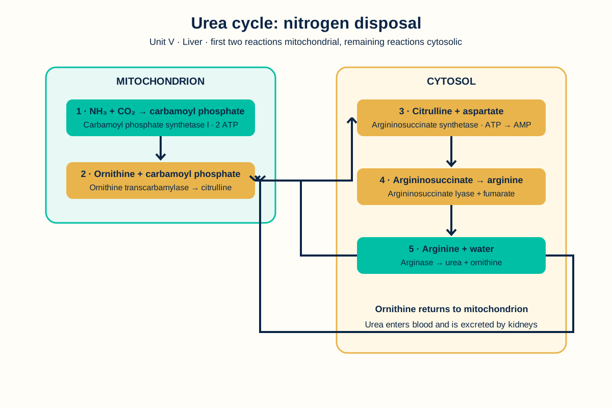
This unit connects amino acid breakdown with nitrogen disposal and explains how genetic information is expressed. Learn the difference between transamination, deamination and the urea cycle, then relate DNA, RNA and protein synthesis.
Transamination and deamination
Examples include alanine aminotransferase transferring an amino group between alanine and alpha-ketoglutarate, and aspartate aminotransferase transferring an amino group between aspartate and alpha-ketoglutarate. Oxidative deamination of glutamate by glutamate dehydrogenase releases ammonia and regenerates alpha-ketoglutarate.
Transamination transfers an amino group from an amino acid to a keto acid. Aminotransferases commonly use pyridoxal phosphate as a coenzyme. Deamination removes an amino group and releases ammonia. These reactions help channel nitrogen toward safe disposal and carbon skeletons toward energy or biosynthetic pathways.
Urea cycle
Urea cycle: 5 steps and locations
- In the mitochondrial matrix, carbamoyl phosphate synthetase I forms carbamoyl phosphate from ammonia, carbon dioxide and ATP.
- In the mitochondrial matrix, ornithine transcarbamylase combines carbamoyl phosphate with ornithine to form citrulline.
- In the cytosol, argininosuccinate synthetase combines citrulline with aspartate and ATP to form argininosuccinate.
- In the cytosol, argininosuccinate lyase splits argininosuccinate into arginine and fumarate.
- In the cytosol, arginase hydrolyses arginine to urea and ornithine. Ornithine returns to the mitochondrion.
The urea cycle converts toxic ammonia nitrogen into urea, which can be excreted in urine. The cycle occurs partly in mitochondria and partly in the cytosol of liver cells. Its carbon and nitrogen sources include carbon dioxide, ammonia and aspartate.
Inborn errors of metabolism
An inborn error of metabolism results from a genetic defect affecting an enzyme, transporter or related protein. The defect can cause accumulation of a substrate, deficiency of a product or formation of an abnormal metabolite. Examples in the standard textbook list used for this draft are phenylketonuria, alkaptonuria, albinism, maple syrup urine disease, homocystinuria and galactosaemia. List source: Harper's Illustrated Biochemistry, sections on inborn errors of metabolism.
DNA replication and transcription
DNA replication copies genetic information before cell division. Each parental strand serves as a template for a new complementary strand. Transcription uses DNA information to produce RNA. Messenger RNA carries coding information, transfer RNA helps deliver amino acids and ribosomal RNA forms part of the ribosome.
Translation
DNA replication uses helicase to separate strands, primase to make RNA primers, DNA polymerase to extend new DNA and DNA ligase to join fragments. Transcription uses RNA polymerase to make RNA from a DNA template. Translation uses ribosomes, messenger RNA, transfer RNA and aminoacyl-tRNA synthetases. In eukaryotic cells, RNA processing includes capping, polyadenylation and splicing before mature messenger RNA is translated.
Translation is the synthesis of a polypeptide using the codon sequence of messenger RNA. Ribosomes read codons in order. Transfer RNA molecules bring amino acids whose anticodons pair with messenger RNA codons. Translation begins at a start codon and ends when a stop codon is reached.
Mutations
A mutation is a change in genetic material. Substitution, insertion and deletion are common categories. The effect can be silent, change an amino acid, introduce a stop codon or alter the reading frame. The biological effect depends on the location and nature of the change.
Unit V: Frequently asked questions
Q: What is transamination?
A: Transamination is transfer of an amino group from an amino acid to a keto acid, forming a new amino acid and keto acid. Pyridoxal phosphate commonly participates in the reaction.
Q: What is the purpose of the urea cycle?
A: The urea cycle converts ammonia nitrogen into urea, a less toxic compound that can be excreted by the kidneys.
Q: What is the difference between transcription and translation?
A: Transcription uses DNA as a template to make RNA. Translation uses messenger RNA as a template to make a polypeptide.
Q: What are the five enzymes of the urea cycle?
A: They are carbamoyl phosphate synthetase I, ornithine transcarbamylase, argininosuccinate synthetase, argininosuccinate lyase and arginase.
Unit V: Key takeaways
- Transamination transfers amino groups, while deamination releases ammonia.
- The liver converts ammonia nitrogen to urea through the urea cycle.
- DNA replication copies genetic information.
- Transcription makes RNA and translation makes a polypeptide.
- Mutations can alter genetic information and protein function.
- The urea cycle has two mitochondrial steps and three cytosolic steps.
Unit V: Clinical correlation
Clinical correlation: jaundice and bilirubin handling. Heme catabolism produces bilirubin, and the liver conjugates bilirubin before biliary excretion. Impaired production, conjugation, hepatic handling or bile flow can produce different jaundice patterns.
Sources
- Pharmacy Council of India. Bachelor of Pharmacy (B.Pharm) Course Regulations and Syllabus, BP203T Biochemistry. https://www.pci.nic.in/pdf/Syllabus_B_Pharm.pdf
- Nelson DL, Cox MM. Lehninger Principles of Biochemistry. NCBI Bookshelf, selected biochemistry chapters. https://www.ncbi.nlm.nih.gov/books/
- Murray RK et al. Harper's Illustrated Biochemistry, sections on amino acids, lipid metabolism, urea cycle and inborn errors of metabolism. McGraw Hill Medical.
- Berg JM, Tymoczko JL, Gatto GJ, Stryer L. Biochemistry, NCBI Bookshelf edition. https://www.ncbi.nlm.nih.gov/books/NBK21154/
- PharmacoLearn. “BP102T: General Pharmacy.” Reference heading pattern and unit order. https://pharmacolearn.fyi/b-pharm/semester-1/bp102t-general-pharmacy/
Complete BP203T notes, all 5 units in one PDF, free to keep.
Other Semester 2 subjects: BP201T · BP202T · BP203T · BP204T · BP205T · BP206T · Semester 2 Hub · MCQ Bank