
BP201T — Human Anatomy and Physiology II
3 hrs/week · 4 credits · 45 hours · 100 marks (25 internal + 75 end-semester, 3 hr paper). Notes follow the PCI B.Pharm Semester 2 syllabus, unit by unit.
Units in this subject
- Unit 1 — Nervous System
- Unit 2 — Digestive System and Energetics
- Unit 3 — Respiratory System and Urinary System
- Unit 4 — Endocrine System
- Unit 5 — Reproductive System and Introduction to Genetics
Download BP201T PDF Notes
Free PDFs for this subject, one per unit:
This draft follows the five units listed above.
These are original study notes for Human Anatomy and Physiology II, B.Pharm Semester II. They use simple language and follow the five-unit sequence in the PCI syllabus.
Unit I: Nervous System
The nervous system receives information, processes it and coordinates responses. Study it through organisation, cells, electrical signals, synapses and the central nervous system.
Organisation of the nervous system
The central nervous system consists of the brain and spinal cord. The peripheral nervous system consists of nerves and ganglia outside the central nervous system. The peripheral system includes sensory pathways that carry information toward the central nervous system and motor pathways that carry commands away from it.
The somatic motor system controls skeletal muscle. The autonomic nervous system regulates smooth muscle, cardiac muscle and glands. The autonomic system is divided into sympathetic and parasympathetic divisions. The enteric nervous system coordinates much of the activity of the gastrointestinal tract.
Neurons and neuroglia
A neuron has a cell body, dendrites and an axon. Dendrites usually receive signals, while an axon conducts signals away from the cell body. Axons may be myelinated or unmyelinated. Myelin increases conduction speed, and gaps in myelin are called nodes of Ranvier.
Neuroglia support neurons. Astrocytes help maintain the chemical environment and contribute to the blood-brain barrier. Oligodendrocytes form myelin in the central nervous system, while Schwann cells form myelin in the peripheral nervous system. Microglia have immune functions in the central nervous system. Ependymal cells line ventricles and are associated with cerebrospinal fluid.
Nerve fibres and electrophysiology
The resting membrane potential results from ion concentration gradients and selective membrane permeability. A stimulus that reaches threshold can generate an action potential. Depolarisation is followed by repolarisation and a brief period of altered excitability called the refractory period.
A nerve impulse propagates along the axon. Myelinated fibres conduct by saltatory conduction, in which the impulse appears to jump between nodes of Ranvier. The action potential follows the all-or-none principle in a single axon, while stimulus intensity can be represented by recruitment of more fibres.
Receptors, synapses and neurotransmitters
Receptors detect changes in the internal or external environment. Mechanoreceptors detect mechanical deformation, thermoreceptors detect temperature, chemoreceptors detect chemical stimuli, photoreceptors detect light and nociceptors respond to potentially damaging stimuli.
A synapse is a junction where a neuron communicates with another neuron, muscle cell or gland cell. At a chemical synapse, an action potential opens calcium channels, vesicles release neurotransmitter and the neurotransmitter binds postsynaptic receptors. Signal termination occurs by enzymatic breakdown, reuptake or diffusion.
Brain, meninges and cerebrospinal fluid
The meninges are dura mater, arachnoid mater and pia mater. The ventricles are connected cavities containing cerebrospinal fluid. Cerebrospinal fluid cushions the central nervous system, supports the chemical environment and helps remove waste. Excess accumulation can increase intracranial pressure.
The cerebrum supports conscious perception, voluntary movement, language and higher cognitive functions. The brain stem contains pathways and centres important for vital functions. The cerebellum coordinates movement, posture and balance.
Spinal cord and reflex activity
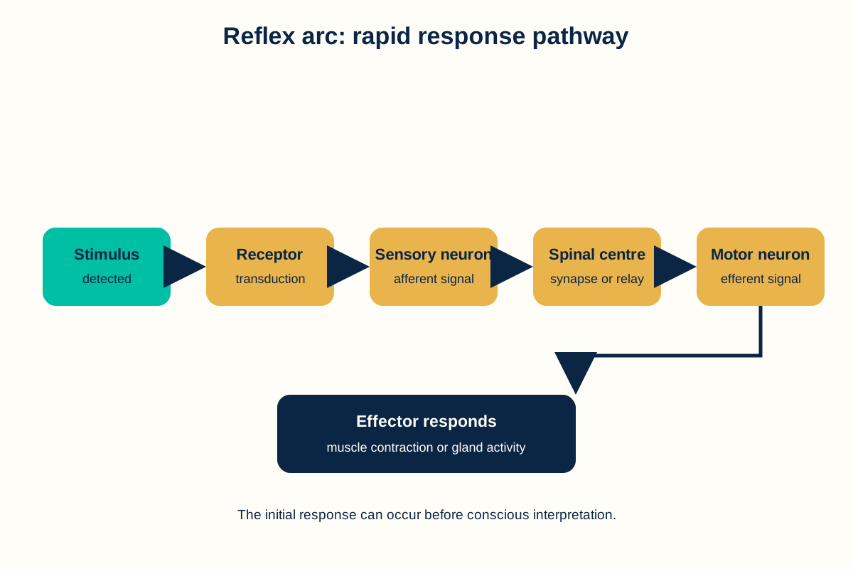
The spinal cord carries ascending sensory and descending motor pathways. A reflex arc includes a receptor, sensory neuron, central synapse or processing region, motor neuron and effector. Reflex activity can occur rapidly because the response does not require conscious processing before the initial action.
Unit I: Frequently asked questions
Q: What is the difference between the central and peripheral nervous systems?
A: The central nervous system consists of the brain and spinal cord. The peripheral nervous system consists of nerves and ganglia outside them.
Q: What is saltatory conduction?
A: Saltatory conduction is rapid impulse transmission along a myelinated axon in which depolarisation is regenerated at the nodes of Ranvier.
Q: What are the three meninges?
A: The meninges are dura mater, arachnoid mater and pia mater.
Unit I: Key takeaways
- The central nervous system is the brain and spinal cord.
- Neurons conduct signals and neuroglia support neural function.
- Action potentials depend on membrane ion gradients and threshold.
- Synapses transfer signals to neurons, muscles or glands.
- Reflex arcs provide rapid patterned responses.
Unit I: Clinical correlation
Clinical correlation: reflex testing. A reflex examination tests the integrity of sensory receptors, peripheral nerves, spinal segments, motor pathways and muscles.
Unit II: Digestive System and Energetics
This unit follows food from the gastrointestinal tract to absorption and explains how cells use ATP, phosphocreatine and basal metabolic rate.
Gastrointestinal tract
The gastrointestinal tract includes the mouth, pharynx, oesophagus, stomach, small intestine, large intestine, rectum and anal canal. Accessory organs include the salivary glands, liver, gallbladder and pancreas.
The stomach stores food, mixes it and begins protein digestion. Parietal cells secrete hydrochloric acid and intrinsic factor. Chief cells secrete pepsinogen, which becomes pepsin in an acidic environment. Gastrin and vagal activity participate in gastric regulation.
The small intestine is the main site of digestion and absorption. Its folds, villi and microvilli increase surface area. The large intestine absorbs water and electrolytes and forms faeces. Peristalsis moves contents forward, while segmentation mixes contents.
Salivary glands, pancreas and liver
Saliva moistens food and begins starch digestion. Pancreatic juice contains digestive enzymes and bicarbonate. Bicarbonate helps neutralise acidic chyme entering the small intestine. The liver produces bile, processes absorbed nutrients and performs metabolic and detoxification functions. Bile salts help emulsify lipids.
Digestion and absorption
Carbohydrates are reduced to monosaccharides, proteins to amino acids and small peptides, and lipids to fatty acids and monoacylglycerols. Monosaccharides and amino acids enter blood capillaries in intestinal villi. Many lipid products enter enterocytes, are reassembled and leave in lipoprotein particles through lymphatic vessels.
Energetics
ATP is the main immediately available energy currency of the cell. Phosphocreatine stores a rapidly transferable phosphate group in muscle and can help regenerate ATP during sudden activity. Basal metabolic rate is the energy expenditure needed to maintain essential functions under defined resting conditions. It is influenced by body size, age, sex, body composition, hormones, temperature and health status.
Unit II: Frequently asked questions
Q: What is the role of hydrochloric acid in the stomach?
A: It creates an acidic environment that supports conversion of pepsinogen to pepsin, helps protein digestion and contributes to defence against swallowed microorganisms.
Q: What is the main site of nutrient absorption?
A: The small intestine is the main site because its folds, villi and microvilli provide a large absorptive surface.
Q: What is basal metabolic rate?
A: Basal metabolic rate is the energy expenditure needed to maintain essential body functions under defined resting conditions.
Unit II: Key takeaways
- The stomach stores, mixes and begins digestion of proteins.
- The small intestine performs most digestion and nutrient absorption.
- The liver produces bile and processes absorbed nutrients.
- ATP supports immediate cellular work, while phosphocreatine helps rapid ATP regeneration.
- Basal metabolic rate changes with biological and environmental factors.
Unit II: Clinical correlation
Clinical correlation: malabsorption. Damage to intestinal mucosa can reduce nutrient absorption and lead to weight loss or deficiency states.
Unit III: Respiratory System and Urinary System
The respiratory system exchanges oxygen and carbon dioxide. The urinary system forms urine, regulates fluid composition and contributes to acid-base balance.
Respiratory anatomy and breathing
The respiratory tract includes the nose, pharynx, larynx, trachea, bronchi, bronchioles and lungs. Gas exchange occurs across the thin respiratory membrane of alveoli. The diaphragm is the main muscle of quiet inspiration. Changes in thoracic volume create pressure changes that move air.
Respiratory centres in the brain stem regulate breathing. Chemoreceptors respond mainly to changes in carbon dioxide, hydrogen ion concentration and oxygen.
Lung volumes and gas transport
Tidal volume is the air moved during a normal quiet breath. Inspiratory reserve volume and expiratory reserve volume are additional volumes that can be moved after a normal breath. Residual volume remains after maximal expiration. Vital capacity is the maximum volume exhaled after maximal inspiration.
Oxygen is transported mainly bound to haemoglobin. Carbon dioxide is transported dissolved in plasma, bound to proteins and mainly as bicarbonate. Gas exchange is driven by partial-pressure gradients.
Artificial respiration and resuscitation
Artificial respiration supports ventilation when spontaneous breathing is inadequate. Resuscitation combines recognition of emergency, airway support, breathing support and circulation support according to current training guidance.
Urinary tract and nephron
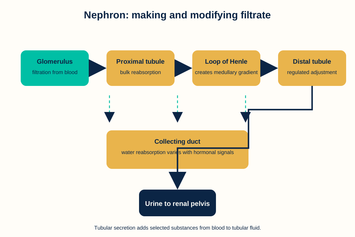
The urinary tract consists of kidneys, ureters, urinary bladder and urethra. The nephron is the functional unit of the kidney. It includes a renal corpuscle, proximal tubule, loop of Henle, distal tubule and collecting duct.
Urine formation involves glomerular filtration, tubular reabsorption and tubular secretion. The kidneys regulate water, electrolytes and acid-base status. The micturition reflex coordinates bladder contraction with relaxation of the outlet. The renin-angiotensin system influences blood pressure, sodium balance and fluid volume.
Unit III: Frequently asked questions
Q: What is the main function of alveoli?
A: Alveoli provide a thin, large surface for diffusion of oxygen into blood and carbon dioxide out of blood.
Q: What are the three processes of urine formation?
A: They are glomerular filtration, tubular reabsorption and tubular secretion.
Q: How do kidneys help acid-base balance?
A: They excrete hydrogen ions, reabsorb filtered bicarbonate and generate new bicarbonate through renal processes.
Unit III: Key takeaways
- Ventilation moves air and alveoli exchange gases with blood.
- Haemoglobin carries most oxygen in blood.
- The nephron filters plasma and modifies the filtrate into urine.
- Kidneys regulate water, electrolytes, blood pressure and acid-base balance.
- The renin-angiotensin system links kidney function with circulatory regulation.
Unit III: Clinical correlation
Clinical correlation: renal failure. Reduced nephron function can impair waste excretion, fluid balance, electrolyte regulation and acid-base control.
Unit IV: Endocrine System
The endocrine system uses hormones released into blood to coordinate growth, metabolism, reproduction, stress responses and homeostasis.
Hormones and mechanisms of action
Hormones may be peptides, proteins, steroids or amino-acid derivatives. Water-soluble hormones usually bind receptors on the cell surface and use intracellular signalling pathways. Lipid-soluble hormones usually cross the cell membrane and bind intracellular receptors that can regulate gene transcription.
Pituitary gland
The pituitary gland has anterior and posterior parts. The anterior pituitary releases growth hormone, prolactin, thyroid-stimulating hormone, adrenocorticotropic hormone, follicle-stimulating hormone and luteinising hormone. The posterior pituitary releases oxytocin and antidiuretic hormone made in the hypothalamus.
Thyroid and parathyroid glands
Thyroid hormones increase metabolic activity and support growth and development. Calcitonin is produced by thyroid parafollicular cells. Parathyroid hormone increases blood calcium through actions on bone, kidney and vitamin D activation.
Adrenal glands, pancreas, pineal gland and thymus
The adrenal cortex produces mineralocorticoids, glucocorticoids and adrenal androgens. The adrenal medulla produces catecholamines. The endocrine pancreas contains islets with cells that release hormones involved in glucose regulation. The pineal gland secretes melatonin, which participates in biological timing. The thymus supports maturation of T lymphocytes, especially during early life.
Unit IV: Frequently asked questions
Q: What is the difference between endocrine and exocrine secretion?
A: Endocrine glands release hormones into blood without ducts. Exocrine glands release products through ducts onto a surface or into a cavity.
Q: What is the role of antidiuretic hormone?
A: Antidiuretic hormone increases water reabsorption in the kidney collecting ducts, helping regulate plasma osmolality and water balance.
Q: Which hormones are released by the anterior pituitary?
A: The principal hormones are growth hormone, prolactin, thyroid-stimulating hormone, adrenocorticotropic hormone, follicle-stimulating hormone and luteinising hormone.
Unit IV: Key takeaways
- Hormones coordinate distant functions and maintain homeostasis.
- Peptide hormones commonly act through membrane receptors.
- Steroid hormones commonly act through intracellular receptors.
- The pituitary links hypothalamic control with several endocrine organs.
- Adrenal, thyroid, parathyroid and pancreatic hormones regulate major body functions.
Unit IV: Clinical correlation
Clinical correlation: diabetes mellitus. Inadequate insulin action raises blood glucose and changes carbohydrate, lipid and protein metabolism.
Unit V: Reproductive System and Introduction to Genetics
This unit covers reproductive anatomy and physiology, sex hormones, gamete formation, pregnancy, parturition and basic genetic information.
Male reproductive system
The male reproductive system includes testes, epididymides, ductus deferens, accessory glands and the penis. Testes produce sperm and testosterone. Spermatogenesis occurs in seminiferous tubules and is supported by Sertoli cells. Leydig cells produce testosterone in response to luteinising hormone.
Female reproductive system
The female reproductive system includes ovaries, uterine tubes, uterus, cervix and vagina. Ovaries produce oocytes and hormones. The ovarian cycle includes follicular, ovulatory and luteal phases. The uterine cycle includes menstrual, proliferative and secretory phases.
Fertilisation, pregnancy and parturition
Fertilisation usually involves fusion of sperm and oocyte genetic material. The early embryo undergoes cleavage and implantation. The placenta supports exchange between maternal and fetal circulations and produces hormones. Parturition involves coordinated uterine contractions and cervical changes.
Genetics
DNA stores hereditary information. A gene is a functional region of genetic material, and chromosomes organise DNA with proteins. Protein synthesis involves transcription of DNA into RNA and translation of messenger RNA into a polypeptide. Mendelian inheritance describes segregation of alleles and independent assortment under stated assumptions. Human inheritance patterns can be more complex than simple Mendelian patterns.
Unit V: Frequently asked questions
Q: Where does spermatogenesis occur?
A: Spermatogenesis occurs in the seminiferous tubules of the testes.
Q: What is the role of the placenta?
A: The placenta supports maternal-fetal exchange and produces hormones that help maintain pregnancy.
Q: What is the difference between a gene and a chromosome?
A: A gene is a functional region of genetic material. A chromosome is a larger organised structure containing DNA and many genes.
Unit V: Key takeaways
- Testes produce sperm and testosterone.
- Ovaries produce oocytes and reproductive hormones.
- Fertilisation begins a developmental sequence supported by the placenta.
- DNA, genes and chromosomes organise hereditary information.
- Protein synthesis uses transcription and translation.
Unit V: Clinical correlation
Clinical correlation: infertility. Infertility can result from factors affecting gamete production, reproductive tract anatomy, endocrine regulation or fertilisation.
Sources
- Pharmacy Council of India. Bachelor of Pharmacy Course Regulations and Syllabus, BP201T Human Anatomy and Physiology II, including Table II and course content. https://www.pci.nic.in/pdf/Syllabus_B_Pharm.pdf
- Hall JE. Guyton and Hall Textbook of Medical Physiology. Elsevier.
- Tortora GJ, Derrickson B. Principles of Anatomy and Physiology. Wiley.
- PharmacoLearn. “BP102T: General Pharmacy.” Reference heading pattern and unit order. https://pharmacolearn.fyi/b-pharm/semester-1/bp102t-general-pharmacy/
Complete BP201T notes, all 5 units in one PDF, free to keep.
Other Semester 2 subjects: BP201T · BP202T · BP203T · BP204T · BP205T · BP206T · Semester 2 Hub · MCQ Bank