
← Biology practicals, Year 1 Practicals
Detailed Study of Frog
1. Aim
To identify major external and internal frog features using an approved model or virtual dissection. The model species, sex, and required structures must be recorded here.
2. Requirements
Instructor-approved frog model, labelled atlas or virtual anatomy resource, notebook, display screen, and pointer or cursor. If preserved specimens are used, their institutional protocol and protective equipment must be recorded here.
3. Theory
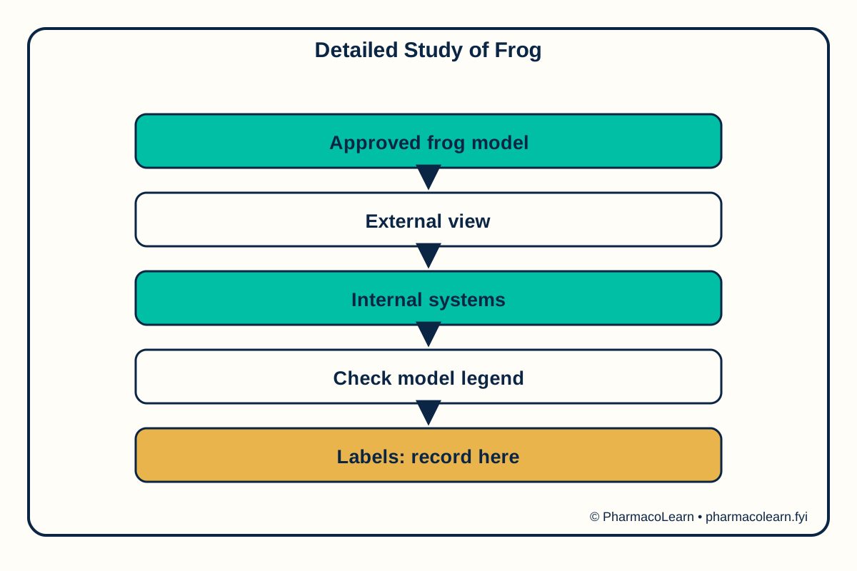
A frog offers a vertebrate model for comparing organ systems. External features can be viewed without opening a specimen. A virtual or preserved internal view can then show digestive, circulatory, and respiratory structures in their anatomical context. Some organs are displaced or hidden when a model layer is removed, and a simplified model may omit small structures. Identification should therefore use position, neighbouring organs, and the model legend together. This article uses a virtual or model-based approach and does not ask students to perform live-animal procedures.
4. Procedure
- Record the species and sex stated by the instructor or model; do not guess either.
- Identify external head, body, limbs, and visible sensory structures on the model.
- Open the virtual internal view and locate the heart, liver, stomach, intestine, and lungs where shown.
- Trace each organ to its organ system using the supplied legend, noting structures hidden or omitted.
- Capture a labelled screenshot or draw a map, then check every label against the atlas.
5. Calculation, graph, or images as appropriate
| Observation | Actual teaching record |
|---|---|
| Model and species | Record here |
| External features | Record here |
| Digestive structures | Record here |
| Circulatory structures | Record here |
| Respiratory structures | Record here |
| Verified labels and omissions | Record here |
No anatomical dimension is invented. Assumed observations for practice only: an assumed virtual model displays a heart above the liver and assumed paired lungs lateral to the heart. These invented observations are a labelling exercise only. Real model features must be recorded here.
6. Result
Record actual observations here when the verified method and record are available. Safety note: Use digital or prepared models. Any preserved-specimen handling needs instructor supervision and institutional chemical and biological controls. No live frog manipulation is described.
7. References
- Pharmacy Council of India, Pharm.D. Regulations, 2008.
- Clarendon College, Frog Laboratory Anatomy Guide.
- Arizona State University, Virtual Reality Frog Dissection Lesson.
Back to Biology practicals, Year 1 Practicals · All Pharm D Practicals · Pharm D
Theory notes: Remedial Mathematics / Biology