
← Biology practicals, Year 1 Practicals
Study of Cell Wall Constituents and Cell Inclusions
1. Aim
To observe prepared plant-cell specimens and distinguish wall features from stored cell inclusions. The assigned specimens and histochemical tests must be recorded here.
2. Requirements
Prepared plant-cell slides or instructor-approved fresh tissue, light microscope, slides, coverslips, mounting medium, labelled reference images, and approved stain or reagent if needed. Exact tissue source, stain, and exposure time are specified in your college lab manual and instructor's method sheet.
3. Theory
The plant cell wall provides a visible boundary and may have primary or secondary thickening. Cells can also contain starch grains, crystals, and other inclusions. A bright particle inside a cell is not identified merely from shape; staining, polarized light, location, and a reference specimen can help. For example, starch is stored in plastids, whereas crystals have distinct optical behaviour. The wall and an inclusion must be drawn as separate structures, avoiding a claim of chemical composition from an unstained field alone.
4. Procedure
- Confirm specimen identity and the precise wall or inclusion features expected by the teaching protocol.
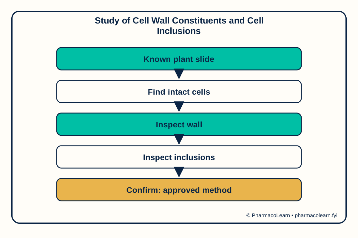
- Examine a prepared slide at low power to locate intact cells before moving to higher magnification.
- Use an approved wet mount or stain only if the instructor has supplied the method; reagent quantity and time are specified in your college lab manual and instructor's method sheet.
- Compare wall outline, thickening, and internal inclusions with the labelled reference.
- Draw representative cells and record whether an inclusion is confirmed, suspected, or not seen.
5. Calculation, graph, or images as appropriate
| Observation | Actual teaching record |
|---|---|
| Plant tissue and slide label | Record here |
| Objective used | Record here |
| Wall feature seen | Record here |
| Inclusion shape and location | Record here |
| Confirmatory test and control | Record here |
| Final interpretation | Record here |
No numerical concentration is needed. Assumed observations for practice only: an assumed field shows intact cell walls and assumed round granules within some cells. The granules are recorded as suspected inclusions until a verified stain or optical test confirms them. Actual cell features must be recorded here.
6. Result
The actual result and any identification or yield conclusion must be recorded here until the teaching method and observed record are confirmed. Safety note: Use blades and stains only under supervision. Do not identify or handle an unknown plant as edible from microscopic appearance.
7. References
- Pharmacy Council of India, Pharm.D. Regulations, 2008.
- University of Wisconsin Botany, Plant Cell Laboratory.
- Prairie View A&M University, Botany Laboratory Manual.
Back to Biology practicals, Year 1 Practicals · All Pharm D Practicals · Pharm D
Theory notes: Remedial Mathematics / Biology