
← Pharmaceutical Microbiology Practicals
Simple, Gram, and Negative Staining
1. Aim
To compare the three PCI-listed staining methods on instructor-approved teaching slides or low-risk cultures. The assigned organism, reagents, and stain timing are specified in your college lab manual and instructor's method sheet.
2. Requirements
Prepared or instructor-approved low-risk smears, clean slides, microscope, approved simple stain, Gram stain set, negative stain, wash station, drying rack, controls, protective equipment, and stain waste. Reagent identities, concentrations, times, and fixation method are specified in your college lab manual and instructor's method sheet.
3. Theory
A simple stain increases contrast but usually colours all cells similarly. Gram staining is differential because cell-wall properties affect retention of the primary stain through the decolorization and counterstain sequence. A negative stain colours the background and leaves cells relatively pale. Smear thickness, culture age, decolorization, and washing can create misleading appearances. A control slide and the approved method are needed before assigning a Gram reaction. No stain result by itself identifies a species.
4. Procedure
- Confirm the risk assessment, approved organism or fixed slide, reagent set, timing, and controls; resolve details with the approved local method.
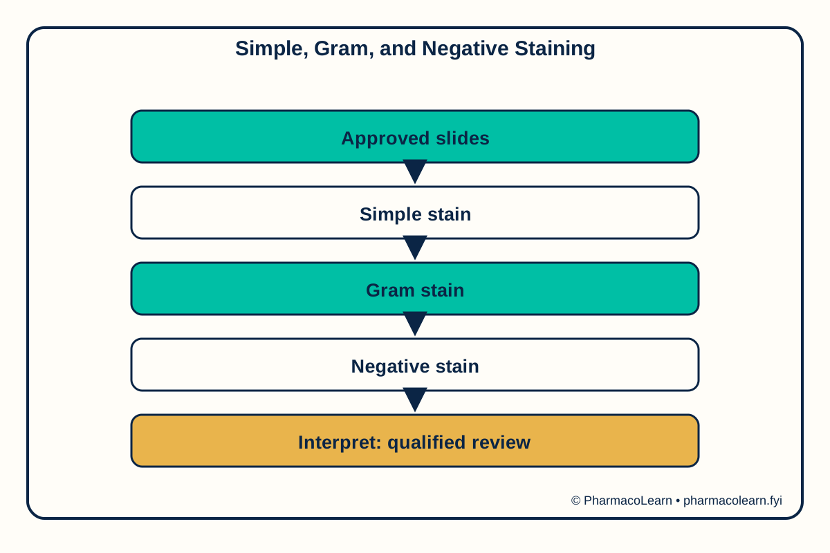
- Label three slides or use a prepared three-slide set for simple, Gram, and negative staining.
- Perform each method only under the instructor’s verified protocol, avoiding aerosols and cross-contamination.
- Examine the dried preparations with the specified microscope method and record colour, shape, and arrangement.
- Compare each with its positive and negative controls; report a staining interpretation, not a species identification.
5. Calculation, graph, or images as appropriate
| Observation | Actual teaching record |
|---|---|
| Sample and control identities | Record here |
| Simple-stain appearance | Record here |
| Gram-stain appearance | Record here |
| Negative-stain appearance | Record here |
| Smear or stain artifacts | Record here |
| Supported interpretation | Record here |
No dye concentration or exposure time is invented. Assumed observations for practice only: an assumed simple stain shows purple rods, an assumed Gram slide shows a consistent control response, and an assumed negative stain shows pale cell outlines. These invented descriptions are not actual results. The real interpretation must follow your college lab manual and instructor's method sheet.
6. Result
Record actual observations here when the verified method and record are available. Safety note: Use instructor-specified low-risk organisms or fixed slides. Handle dyes and glass as directed; decontaminate work surfaces and dispose of cultures through the approved route.
7. References
- Pharmacy Council of India, Pharm.D. Regulations, 2008.
- OpenStax Microbiology, Staining Microscopic Specimens.
- American Society for Microbiology, Biosafety Guidelines for Teaching Laboratories.
Back to Pharmaceutical Microbiology Practicals · All Pharm D Practicals · Pharm D
Theory notes: Pharmaceutical Microbiology