
← Biology practicals, Year 1 Practicals
Preparation of Permanent Slides
1. Aim
To prepare a labelled, durable microscope slide from an instructor-approved specimen using a validated fixation, stain, and mounting workflow. Specific reagents and times are specified in your college lab manual and instructor's method sheet.
2. Requirements
Assigned plant or animal teaching tissue, slides, coverslips, approved fixative and stain, dehydration and clearing reagents if required, permanent mountant, forceps, labels, microscope, hood if required, and waste containers. All identities, concentrations, quantities, and exposure times are specified in your college lab manual and instructor's method sheet.
3. Theory
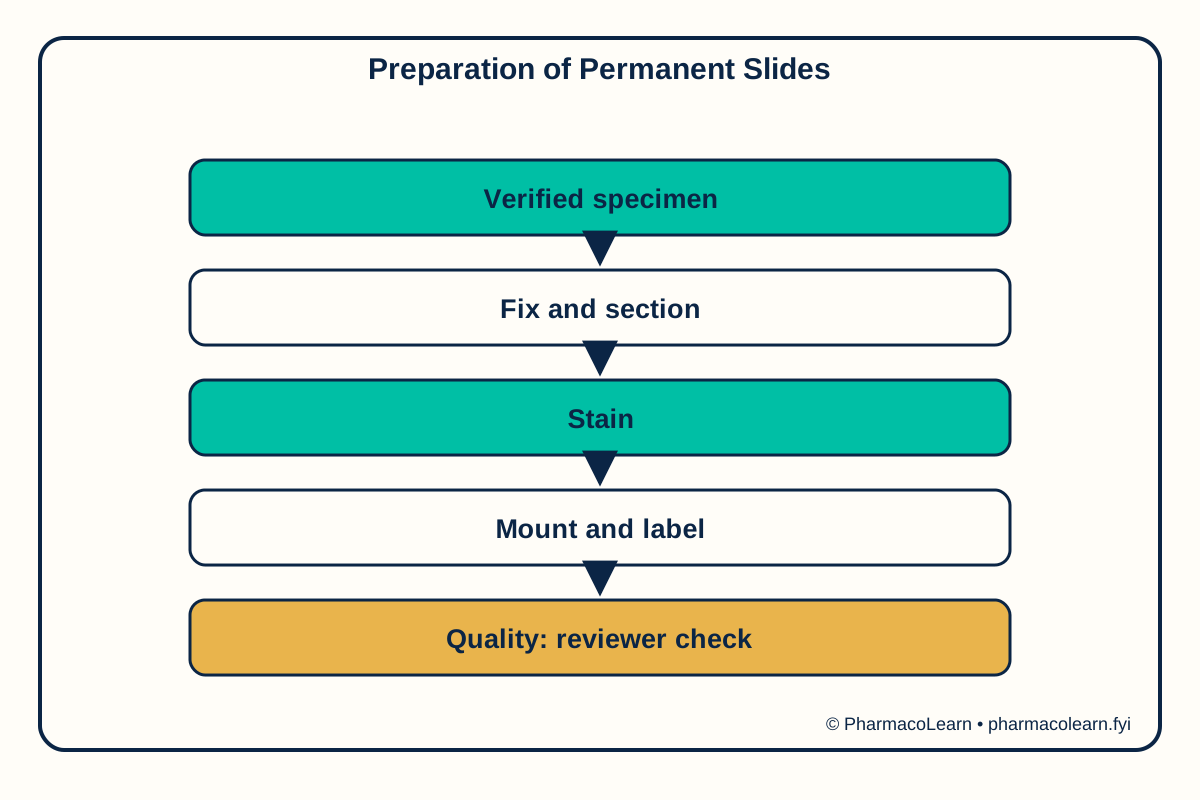
A permanent slide must preserve tissue structure, provide contrast, and retain a coverslip without bubbles or drying artifacts. A typical workflow involves fixation, thin sectioning or placement, staining, any needed dehydration and clearing, mounting, and labelling. Each stage depends on tissue and chemistry. Fixation can introduce artifacts; an excessive stain or thick section can hide diagnostic features. The final slide should be checked under the microscope and linked to its specimen record.
4. Procedure
- Confirm specimen type and the approved fixation, sectioning, stain, mountant, and waste protocol; record missing details as unresolved and consult the approved local method.
- Have trained staff handle hazardous reagents, and prepare the tissue and slide according to the validated method.
- Select a thin, intact section and apply the specified staining sequence.
- Mount beneath a clean coverslip while avoiding bubbles, then allow the mount to cure as prescribed.
- Label specimen, date, stain, and preparer; inspect at low and high power for clarity and artifacts.
5. Calculation, graph, or images as appropriate
| Observation | Actual teaching record |
|---|---|
| Specimen identity | Record here |
| Fixation and section method | Record here |
| Stain and mountant | Record here |
| Coverslip and bubbles | Record here |
| Visible tissue features | Record here |
| Slide label and quality | Record here |
No reagent concentration or exposure time is invented. Assumed observations for practice only: an assumed mounted section is intact and an assumed coverslip has no visible bubbles. These invented conditions illustrate slide-quality criteria. The actual slide quality must be recorded here.
6. Result
The actual result and any identification or yield conclusion must be recorded here until the teaching method and observed record are confirmed. Safety note: Fixatives, solvents, stains, blades, and glass can be hazardous. Use the approved hood and sharps protocol under teaching-lab supervision; never substitute a mountant based on this overview.
7. References
- Pharmacy Council of India, Pharm.D. Regulations, 2008.
- Columbia University, Histology Laboratory Manual.
- University of California Berkeley, Plant and Animal Microtechnique.
Back to Biology practicals, Year 1 Practicals · All Pharm D Practicals · Pharm D
Theory notes: Remedial Mathematics / Biology