
← Human Anatomy and Physiology Practicals
Determination of W.B.C. count of blood
1. Aim
To estimate the total number of white blood cells (W.B.C.s, or leukocytes) per microlitre of blood by manual counting in an improved Neubauer haemocytometer.
2. Requirements
Specimen and chemicals: A correctly labelled anticoagulated blood specimen provided by a trained person; approved W.B.C. diluting fluid; cleaning material for the chamber. The WHO method uses a red-cell-lysing diluent based on acetic acid and crystal violet. The identity, formulation, labelling, and suitability of the diluent available in the teaching laboratory are specified in your college lab manual and instructor's method sheet; check them before use. [1]
Instruments and supplies: Improved Neubauer haemocytometer and its special cover glass, light microscope, calibrated micropipettes and disposable tips, mixing tube, timer, cell counter or tally sheet, and designated biological waste container.
Protective equipment: Laboratory coat, gloves, and eye protection as required by the laboratory's blood-handling risk assessment. [1, 2]
3. Theory
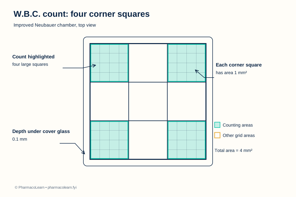
A total W.B.C. count measures the number of leukocytes in a defined volume of blood. Because cells are too crowded in undiluted blood, the specimen is diluted. A suitable W.B.C. diluent clears most red cells while leaving leukocytes visible. The improved Neubauer chamber has a fixed depth when its matching cover glass is correctly seated. Four large corner squares, each 1 mm², provide a total counted area of 4 mm². At a chamber depth of 0.1 mm, their combined volume is 0.4 mm³, or 0.4 µL. The number counted in that volume is corrected for dilution to estimate cells per microlitre of the original blood. [1]
This manual count is a teaching exercise. An actual specimen count is only reportable after the laboratory's specimen, chamber, and quality checks. Compare with your laboratory's current reference interval; do not infer an interval from the illustrative calculation. [1]
4. Procedure
- Under supervision, check the specimen label, the approved diluent and its safety information, and the local procedure. Handle the blood as potentially infectious. Wear the specified protective equipment and never mouth-pipette. [1, 2]
- Clean and dry the chamber and special cover glass. Seat the cover glass over the counting platform. Reject chipped or dirty equipment. [1]
- Gently mix the anticoagulated blood. For the WHO method, add 20 µL blood to 380 µL W.B.C. diluent to make a 1:20 dilution. Mix for 2 to 3 minutes. If the local method differs, its exact volumes, diluent, and mixing time are specified in your college lab manual and instructor's method sheet; check them before proceeding. [1]
- Inspect the diluted sample for obvious clots or debris. Mix again gently, then touch a small drop to the chamber edge and allow capillary action to fill beneath the cover glass. Do not overfill. Reject a chamber with bubbles, overflow, or an incompletely filled grid. [1]
- Allow the cells to settle for 3 to 5 minutes according to the WHO method. Focus on the grid and count leukocytes in the four large corner squares. Keep one boundary rule throughout, for example include cells touching the top or left lines and exclude those touching the bottom or right lines. Do not count debris as cells. [1]
- Record each corner-square count. If the cells appear unevenly distributed or the chamber dries, refill a clean chamber. Compare with your laboratory's current repeat-count and nucleated-red-cell correction rules. [1]
- Dispose of blood-contaminated tips and materials through the designated route, clean the work area as directed, remove gloves, and wash hands. [2]
5. Calculation, graph, or images as appropriate
Observation table. The figures below are invented teaching counts for arithmetic practice, not measurements from a person.
| Large corner square | Example W.B.C.s counted |
|---|---|
| Upper left | 46 |
| Upper right | 51 |
| Lower left | 49 |
| Lower right | 54 |
| Total, N | 200 |
Worked calculation:
- Total area counted = 4 × 1 mm² = 4 mm².
- Counted chamber volume = 4 mm² × 0.1 mm = 0.4 mm³ = 0.4 µL.
- W.B.C.s per microlitre of diluted sample = 200 ÷ 0.4 = 500 cells/µL.
- Correct for the 1:20 dilution = 500 × 20 = 10,000 cells/µL.
- In SI units, 10,000 cells/µL = 10.0 × 10⁹ cells/L.
For this exact grid and dilution, W.B.C. count = N × 50 cells/µL. Recalculate the factor if the dilution or counting area changes. [1]
6. Result
Worked example: The illustrative W.B.C. count is 10,000 cells/µL. Record the actual result here from the student's observed counts, specimen details, and local quality checks. No diagnosis follows from this example.
7. References
- S. M. Lewis and Sudarshan Kumari. Guidelines on Standard Operating Procedures for Haematology, sections 10 and 11. World Health Organization Regional Office for South-East Asia, January 2000. Manual W.B.C. dilution, chamber count, calculation, and sources of error.
- Centers for Disease Control and Prevention. Blood Specimens: Safety. Protective equipment, hand hygiene, and contaminated sharps guidance. Reviewed 3 May 2016.
- Pharmacy Council of India. Pharm.D. Regulations, 2008, Appendix A, first-year Human Anatomy and Physiology practical list. Syllabus authority for this experiment.
Back to Human Anatomy and Physiology Practicals · All Pharm D Practicals · Pharm D
Theory notes: Human Anatomy and Physiology