
← Human Anatomy and Physiology Practicals
Study of appliances used in hematological experiments
1. Aim
To recognize the principal appliances used in the Human Anatomy and Physiology blood practicals, explain each appliance's purpose, and identify the feature that must be checked before use.
2. Requirements
A supervised display or approved images of the laboratory's haemocytometer with its special cover glass, light microscope, calibrated pipette, differential counter, Westergren ESR tube and stand, haemoglobin measurement instrument, and any capillary tube or timing device used for the listed practicals. An observation sheet, pencil, and the local instrument instructions are needed. The exact models and availability of these appliances are specified in your college lab manual and instructor's method sheet in the teaching laboratory. No blood or chemical reagent is needed for this identification exercise. Wear a laboratory coat and any additional protective equipment required locally. [1, 2]
3. Theory
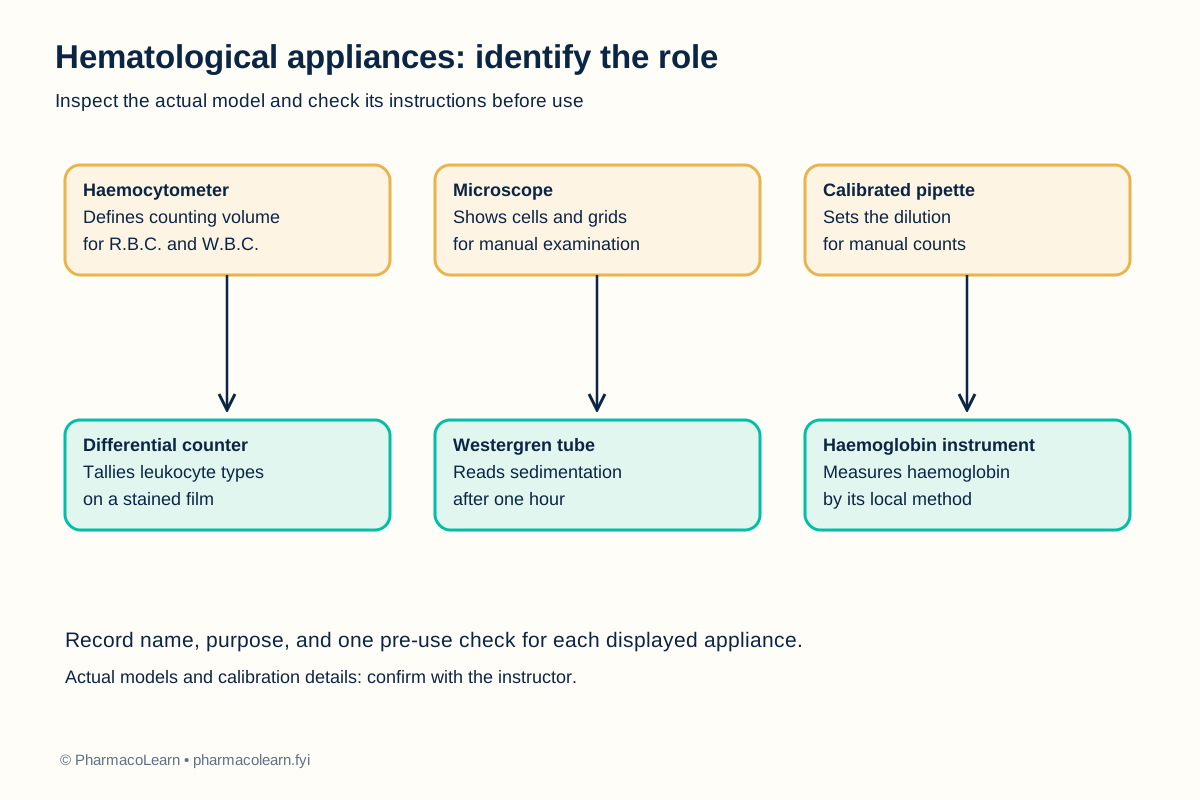
Hematological appliances translate a visible event into a measurement. A haemocytometer has a ruled grid and a defined depth under its matching cover glass. It supports manual cell counts only when its surfaces are clean and the chamber is filled correctly. A microscope makes the grid or a stained blood film visible, while a calibrated pipette controls the dilution that enters a cell-count calculation. A differential counter helps tally leukocyte types in a blood film. [1, 2]
A Westergren tube is marked to measure the distance between the plasma surface and the settled red-cell column after a specified interval. Its stand must keep it vertical and undisturbed. A haemoglobin instrument measures a signal associated with haemoglobin by a method that depends on its design; the local model, method, calibration, and safety instructions are specified in your college lab manual and instructor's method sheet. Capillary tubes and timers are associated with other listed blood practicals, but their precise use depends on the method selected by the instructor. Learning the name alone is insufficient: a student should be able to connect each appliance to its measurement and its main source of error. [1, 2]
4. Procedure
- With the instructor, inspect the available appliances without blood or reagent. Record the name and model from each label. Do not turn on or open an unfamiliar instrument until its local instructions have been reviewed. Record the appliances available in the local teaching set here.
- Examine the haemocytometer and matching special cover glass. Locate the ruled platform and the cover-glass supports. Explain that a damaged or substitute cover glass can change the chamber volume. Handle the glass by its edges. [1, 2]
- Identify the microscope's stage, objective lenses, focusing controls, condenser, and illumination. Demonstrate how a prepared slide is secured and focused at low power under supervision. Clean optical surfaces only with approved lens material. [1]
- Examine a calibrated pipette and its compatible tip. Find the indicated volume setting and explain why accurate dilution matters for a manual count. The local pipette range and calibration status are specified in your college lab manual and instructor's method sheet.
- Locate the differential counter and show how a tally is assigned to a leukocyte category without recording a specimen result. If the laboratory uses a manual tally sheet instead, record that choice. [1]
- Inspect the Westergren tube and stand. Locate the graduation marks and identify how the stand keeps the tube vertical. Do not insert a tube containing blood during this identification exercise. [2]
- Identify the haemoglobin instrument, any capillary tubes, and the timer used locally. Record their intended practical and one required pre-use check from the instructor or equipment instructions. Model-specific operating steps are specified in your college lab manual and instructor's method sheet and must not be inferred from another appliance. [1, 2]
- Complete the table below, return all items to their storage positions, and report cracked glass or damaged equipment rather than attempting to use it.
5. Calculation, graph, or images as appropriate
No calculation is required. The table records appliance identification and a functional check. Fill the observed model and condition columns from the appliances actually displayed. [1, 2]
| Appliance | Main role in this practical sequence | Pre-use feature to inspect | Observed model and condition |
|---|---|---|---|
| Haemocytometer and special cover glass | Defines area and depth for manual cell counts | Clean grid, intact matched cover glass | Record during practical |
| Light microscope | Enlarges chamber grid or blood-film detail | Clean optics and stable illumination | Record during practical |
| Calibrated pipette | Measures sample and diluent volumes | Correct range, tip fit, calibration status | Record during practical |
| Differential counter | Tallies leukocyte categories | Working keys or a clear tally sheet | Record during practical |
| Westergren tube and stand | Holds a vertical blood column for ESR | Clean tube, legible marks, vertical rack | Record during practical |
| Haemoglobin instrument | Measures haemoglobin by its stated method | Local method and calibration status | Record during practical |
| Capillary tube and timer | Support selected blood tests | Intact tube and working timer | Record during practical |
6. Result
The appliances are identified by their purpose and pre-use checks. Record the actual models, their condition, and the instructor's accepted identifications here. No patient or specimen measurement is made in this study.
7. References
- World Health Organization. Manual of Basic Techniques for a Health Laboratory, second edition, sections 2.5 and 9. Laboratory instruments and their use in haematology. 2003.
- S. M. Lewis and Sudarshan Kumari. Guidelines on Standard Operating Procedures for Haematology, sections 6, 10, 11, and 14. World Health Organization Regional Office for South-East Asia, January 2000. Counting chamber, blood films, and Westergren ESR apparatus.
- Pharmacy Council of India. Pharm.D. Regulations, 2008, Appendix A, first-year Human Anatomy and Physiology practical list. Syllabus authority for this study item.
Back to Human Anatomy and Physiology Practicals · All Pharm D Practicals · Pharm D
Theory notes: Human Anatomy and Physiology