
← Human Anatomy and Physiology Practicals
Determination of R.B.C. count of blood
1. Aim
To estimate the number of red blood cells (R.B.C.s, or erythrocytes) per microlitre of a blood sample by manual counting in an improved Neubauer haemocytometer.
2. Requirements
Apparatus: Improved Neubauer haemocytometer; matching special cover glass; light microscope; calibrated micropipettes and tips; clean dilution tube; mixing device; timer; tissue or lint-free paper; suitable biohazard waste container.
Reagents and specimen: A properly labelled, anticoagulated blood specimen supplied under supervision; a laboratory-approved isotonic R.B.C. diluent. Follow your college lab manual and your instructor's method sheet for the diluent identity, formulation, preparation, expiry, and the blood and diluent volumes used with the available calibrated pipettes. Do not substitute a reagent on the basis of this article. [1, 2]
3. Theory
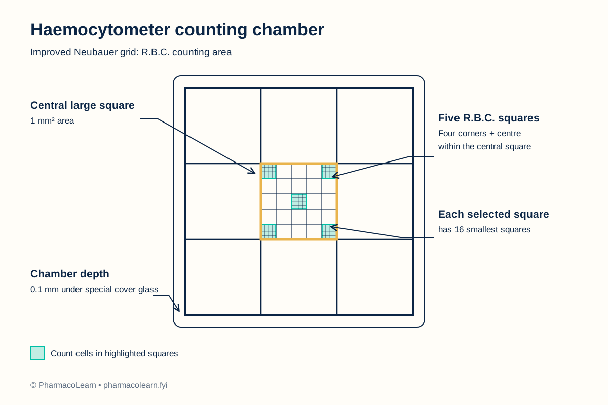
A measured blood sample is diluted so that individual red cells can be distinguished in the counting chamber. The chamber grid defines a known area, and its cover glass establishes a known depth. Cells counted in the four corner groups and the centre group of the large central square occupy a calculable volume. Multiplying the observed count by the dilution factor and dividing by that volume gives the estimated R.B.C. concentration in the original sample. This manual exercise illustrates the counting method; a real specimen result must be checked under the teaching laboratory's quality procedures. [1, 2]
4. Procedure
- Under instructor supervision, confirm the specimen identity, the approved diluent, the chamber type, and the laboratory's specimen-handling instructions. Wear the required protective equipment. [3]
- Clean and dry the haemocytometer and its matching cover glass. Seat the cover glass over the counting platform as instructed for the chamber. [1, 2]
- Gently mix the anticoagulated blood specimen. Prepare a 1:200 dilution with the approved isotonic diluent using calibrated pipettes. Record the actual blood and diluent volumes in the student record below, following the local protocol. Mix the diluted sample thoroughly without vigorous shaking. [1, 2]
- Touch a small drop of the mixed dilution to the chamber edge and let capillary action fill the space beneath the cover glass. Avoid bubbles, overflow into the gutters, and an incompletely filled grid. If filling is poor, clean and recharge the chamber. [1, 2]
- Allow the cells to settle for the period specified in your college lab manual and instructor's method sheet. Focus first at low power to locate the large central square, then use an appropriate higher-power objective to distinguish individual cells. [1, 2]
- In the large central square, count R.B.C.s in five of its 25 medium squares: the four corner squares and the centre square. Count each cell once. Apply one consistent boundary rule, such as including cells touching the top or left boundary and excluding cells touching the bottom or right boundary. Record the five counts. [1, 2]
- If the distribution is visibly uneven, the cells are clumped, or the chamber has dried or overflowed, discard that count and prepare a fresh chamber. Compare with your laboratory's current repeat-count rule. [1, 2]
Precautions
- Use the special cover glass supplied for the haemocytometer; an ordinary cover slip may change the chamber depth. [1, 2]
- Mix the specimen before dilution and mix the dilution before charging the chamber, because settling can distort the count. [1, 2]
- Charge by capillary action without pressing liquid into the chamber. Reject a grid with bubbles, overflow, drying, or obvious uneven distribution. [1, 2]
- Use the same boundary rule in all five squares so edge cells are not counted twice. [1, 2]
- Check the pipette calibration and actual dilution against your college lab manual and instructor's method sheet. Compare with your laboratory's current repeat-count rule. [1, 2]
Safety note
This is a supervised teaching-laboratory method, not an invitation to collect or test blood independently. A trained person should obtain and provide any human specimen under the institution's approved protocol. Treat every blood sample as potentially infectious. Wear gloves, a lab coat, and eye protection as directed; never mouth-pipette; use a designated sharps container if collection equipment is involved; disinfect the work area; and wash hands after removing gloves. The instructor must confirm the diluent's safety information and the waste route before the exercise. [3, 5]
5. Observation and calculation
| Counting area within the large central square | R.B.C.s counted |
|---|---|
| Upper-left medium square | 92 |
| Upper-right medium square | 95 |
| Centre medium square | 94 |
| Lower-left medium square | 93 |
| Lower-right medium square | 96 |
| Total, N | 470 |
These figures are assumed values for a worked teaching example, not observations from a patient specimen. For an actual exercise, replace them with the counts observed at the microscope.
| Student record field | Entry |
|---|---|
| Blood volume and diluent volume | Record here |
| Five observed square counts | Record here |
| Calculated specimen result | Record here |
| Supervisor check | Record here |
Calculation
The large central square has an area of 1 mm² and contains 25 equal medium squares. The five selected squares therefore cover 5/25 = 0.2 mm². With a chamber depth of 0.1 mm, the volume counted is 0.2 × 0.1 = 0.02 mm³, which equals 0.02 µL. [1, 2]
- Add the five example counts: N = 92 + 95 + 94 + 93 + 96 = 470 cells.
- Find the count per microlitre of diluted blood: 470 ÷ 0.02 = 23,500 cells/µL.
- Correct for the 1:200 dilution: 23,500 × 200 = 4,700,000 cells/µL.
- Express in scientific notation: 4.70 × 10⁶ cells/µL, equivalent to 4.70 × 10¹² cells/L.
Thus, for this chamber and dilution, R.B.C. count = N × 10,000 cells/µL. The factor is derived from the chamber volume and dilution, not a number to use automatically with another chamber or dilution. [1, 2]
6. Result
Worked example using assumed values: The calculated R.B.C. count is 4.70 × 10⁶ cells/µL of the original sample. This is an illustrative result only. Record the real specimen result in the student record below after the laboratory's quality checks. Compare with your laboratory's current reference interval and interpretation guidance for the relevant population; neither is inferred from the example. [1, 4]
7. References
- University of Pennsylvania, Robertson Laboratory. Cell Counts with a Hemacytometer, section “B. Red Blood Cell Counts”. Procedure and five-square calculation. Accessed 1 November 2026.
- bioanalytic GmbH. Neubauer Counting Chamber: Calculation Examples. Manufacturer guide for improved Neubauer grid, 1:200 dilution, and 10,000 factor. Accessed 1 November 2026.
- World Health Organization. Laboratory Biosafety Manual, fourth edition. Risk-based laboratory biosafety framework. 2020.
- OpenStax. Clinical Nursing Skills, section 24.3, Complete Blood Count. Reference ranges vary by context; no interval is adopted in this pilot. Accessed 1 November 2026.
- Centers for Disease Control and Prevention. Blood Specimens: Safety. Precautions for blood handling and sharps. Reviewed 3 May 2016.
Back to Human Anatomy and Physiology Practicals · All Pharm D Practicals · Pharm D
Theory notes: Human Anatomy and Physiology