
← Human Anatomy and Physiology Practicals
Study of the urinary system using charts, models, and specimens
1. Aim
To identify the kidneys, ureters, urinary bladder, and urethra on charts, models, or approved specimens and trace the path of urine from formation to exit. [1, 2]
2. Requirements
A labelled urinary-tract chart; an unlabelled torso or urinary-system model; a kidney section or nephron chart if available; an observation sheet; and gloves if the instructor's specimen rules require them. The available model, whether internal kidney structures are displayed, specimen-handling rules, and assessed landmarks are specified in your college lab manual and instructor's method sheet. No specimen collection or physical examination is required. [1, 2]
3. Theory
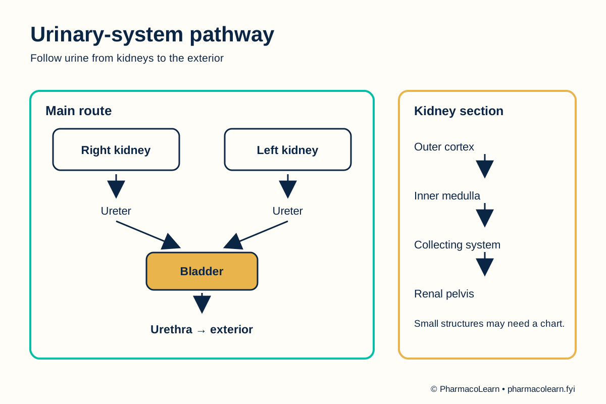
The kidneys filter blood and make urine from wastes and excess fluid. Urine drains from each kidney into a ureter, enters the urinary bladder for storage, and leaves the body through the urethra. In a kidney section, the outer cortex and inner medulla surround a collecting region that drains into the renal pelvis. The nephron is the functional filtering unit; a separate chart may be needed to show its small parts. The urinary pathway should be distinguished from the blood vessels entering and leaving the kidney. [2, 3, 4]
4. Procedure
- Ask the instructor which chart, model, or specimen may be handled. Confirm the model orientation and check for loose or fragile parts. Local handling instructions are specified in your college lab manual and instructor's method sheet. [1]
- On the overview chart, locate the right and left kidneys relative to the spine. Find the ureter leaving each kidney and follow both ureters toward the bladder. [2]
- Locate the bladder in the pelvis and the urethra below it. Trace the complete route: kidney, ureter, bladder, urethra. State which structures make, transport, store, and release urine. [2]
- If a kidney-section model is available, identify the cortex, medulla, and renal pelvis. Trace the collecting route toward the ureter. If these details are not visible, record not displayed rather than guessing from the external kidney shape. Record whether a kidney-section model is available here. [3]
- If the chart includes a nephron, locate the glomerular region and tubule as microscopic structures within the kidney. Explain that a whole-organ model cannot show them at true scale. [3]
- Cover the labels and repeat the route on an unlabelled chart. Record one positional clue and one role for each structure. Ask the instructor to resolve any uncertain identification. [2]
- Replace removable pieces correctly, follow the local cleaning rule, and wash hands after handling shared material. [1]
5. Calculation, graph, or images as appropriate
There is no calculation. Complete the table using the assigned teaching material. [2, 3]
| Structure | Positional clue | Role | Shown on local material? |
|---|---|---|---|
| Kidney | Paired organs beside the spine | Forms urine | Record here |
| Ureter | Tube from each kidney to bladder | Transports urine | Record here |
| Bladder | Pelvic reservoir | Stores urine | Record here |
| Urethra | Outlet below bladder | Releases urine | Record here |
| Cortex, medulla, renal pelvis | Features of a kidney section | Filtration region and drainage pathway | Record here |
Worked tracing example: Start at a kidney on the chart, follow its ureter → bladder → urethra, and identify the bladder as the storage point. Record the labels visible on the assigned model here. [2]
6. Result
Urinary structures identified and urine pathway traced on the assigned material: Record here. Record any structure omitted by the model separately from an uncertain identification. [2]
7. References
- Pharmacy Council of India. Pharm.D. Regulations, 2008, Appendix A. Lists urinary-system study with charts, models, and specimens.
- National Institute of Diabetes and Digestive and Kidney Diseases. The Urinary Tract and How It Works. Urinary organs and urine pathway.
- National Institute of Diabetes and Digestive and Kidney Diseases. Your Kidneys and How They Work. Kidney filtration and nephron context.
- Garza FA, Leslie SW. Anatomy, Abdomen and Pelvis: Kidneys. StatPearls. Cortex, medulla, and collecting system.
Back to Human Anatomy and Physiology Practicals · All Pharm D Practicals · Pharm D
Theory notes: Human Anatomy and Physiology