
← Human Anatomy and Physiology Practicals
Study of the nervous system using charts, models, and specimens
1. Aim
To distinguish central and peripheral nervous-system structures on charts, models, or approved specimens and trace a simple sensory-to-motor information path. [1, 2]
2. Requirements
A labelled nervous-system chart; an unlabelled brain and spinal-cord model; a peripheral-nerve chart; an observation sheet; and gloves if the instructor's specimen rules require them. The available models, removable brain regions, specimen-handling rules, and assessed landmarks are specified in your college lab manual and instructor's method sheet. No neurological examination of a person is required. [1]
3. Theory
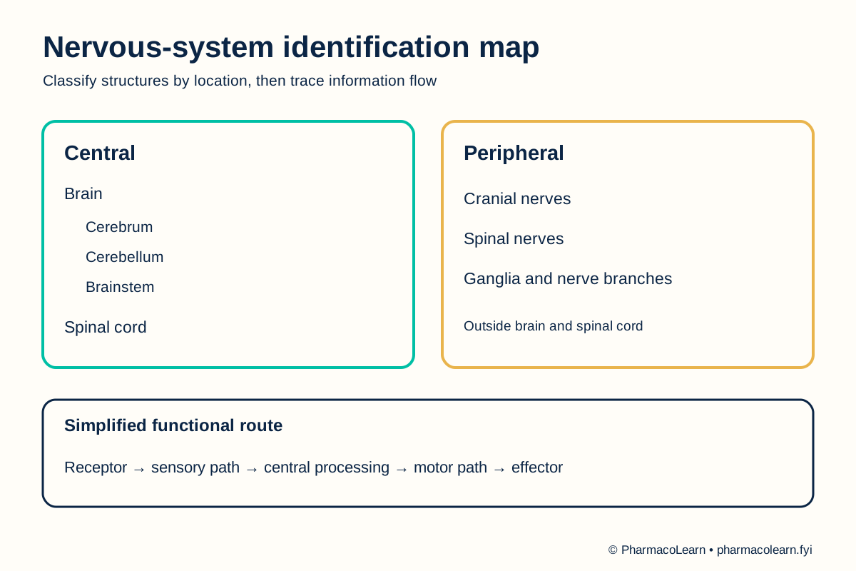
The central nervous system consists of the brain and spinal cord. The peripheral nervous system includes neural structures outside them, notably cranial nerves, spinal nerves, and ganglia. The cerebrum, cerebellum, and brainstem are broad regions of the brain; the spinal cord connects with peripheral nerves through roots and pathways. Sensory information reaches the central nervous system from receptors, while motor output travels from it toward effectors. This is a functional teaching route, not a claim that every response uses the same anatomical path. [2, 3]
4. Procedure
- Confirm with the instructor which chart, model, or specimen may be handled. Check orientation and any fragile removable parts. Local handling instructions are specified in your college lab manual and instructor's method sheet. [1]
- On the overview chart, distinguish the brain and spinal cord as central structures from cranial and spinal nerves outside them. Mark the boundary between central and peripheral regions on the observation sheet. [2, 3]
- On the brain model, identify cerebrum, cerebellum, and brainstem by position. Locate the brainstem's continuity with the spinal cord. Record a structure not shown by the model as not displayed. [2, 3]
- On the spinal-cord chart, identify the cord inside the vertebral column and find at least one spinal nerve leaving it. Use the peripheral-nerve chart to trace a nerve into a body region. [2]
- On a suitable teaching diagram, trace a simple functional route: receptor, sensory pathway, central processing, motor pathway, effector. State that the chart simplifies the many possible circuits. The exact diagram available is specified in your college lab manual and instructor's method sheet. [3]
- Cover the labels and repeat the central-versus-peripheral classification. For each structure, record one visible positional clue and one broad role. Ask the instructor to check uncertain labels. [2, 3]
- Replace model parts correctly, follow the local cleaning rule, and wash hands after shared material. [1]
5. Calculation, graph, or images as appropriate
There is no calculation. Complete the table from the assigned chart or model. [2, 3]
| Structure | Division | Identification clue | Shown on local material? |
|---|---|---|---|
| Cerebrum | Central | Large upper brain region | Record here |
| Cerebellum | Central | Posterior region beneath cerebrum | Record here |
| Brainstem | Central | Connects brain with spinal cord | Record here |
| Spinal cord | Central | Runs within vertebral canal | Record here |
| Cranial and spinal nerves | Peripheral | Extend outside brain and cord | Record here |
Worked classification example: A nerve drawn leaving the spinal cord and extending into the arm is a peripheral spinal nerve, while the cord itself is central. Record the nerve shown on the assigned chart here. [2, 3]
6. Result
Central and peripheral structures identified on the assigned material: Record here. Record any model omissions and unresolved identifications. This practical is an anatomy study, not a patient assessment. [2]
7. References
- Pharmacy Council of India. Pharm.D. Regulations, 2008, Appendix A. Lists nervous-system study with charts, models, and specimens.
- National Library of Medicine. Nervous System, MeSH. Central and peripheral structures.
- Purves D, et al. Neuroscience, Neural Systems. NCBI Bookshelf. Broad central and peripheral organization and information flow.
Back to Human Anatomy and Physiology Practicals · All Pharm D Practicals · Pharm D
Theory notes: Human Anatomy and Physiology