
← Human Anatomy and Physiology Practicals
Study of the digestive system using charts, models, and specimens
1. Aim
To identify the principal gastrointestinal tract organs and accessory digestive organs on charts, models, or approved specimens, and trace the path of food through the tract. [1, 2]
2. Requirements
A labelled digestive-system chart; an unlabelled torso or gastrointestinal model; a chart showing the liver, gallbladder, pancreas, and their duct connections; an observation sheet; and gloves if required for approved specimens. The available models, removable parts, specimen-handling rules, and assessed structures are specified in your college lab manual and instructor's method sheet. No sampling, ingestion, or dissection is required. [1]
3. Theory
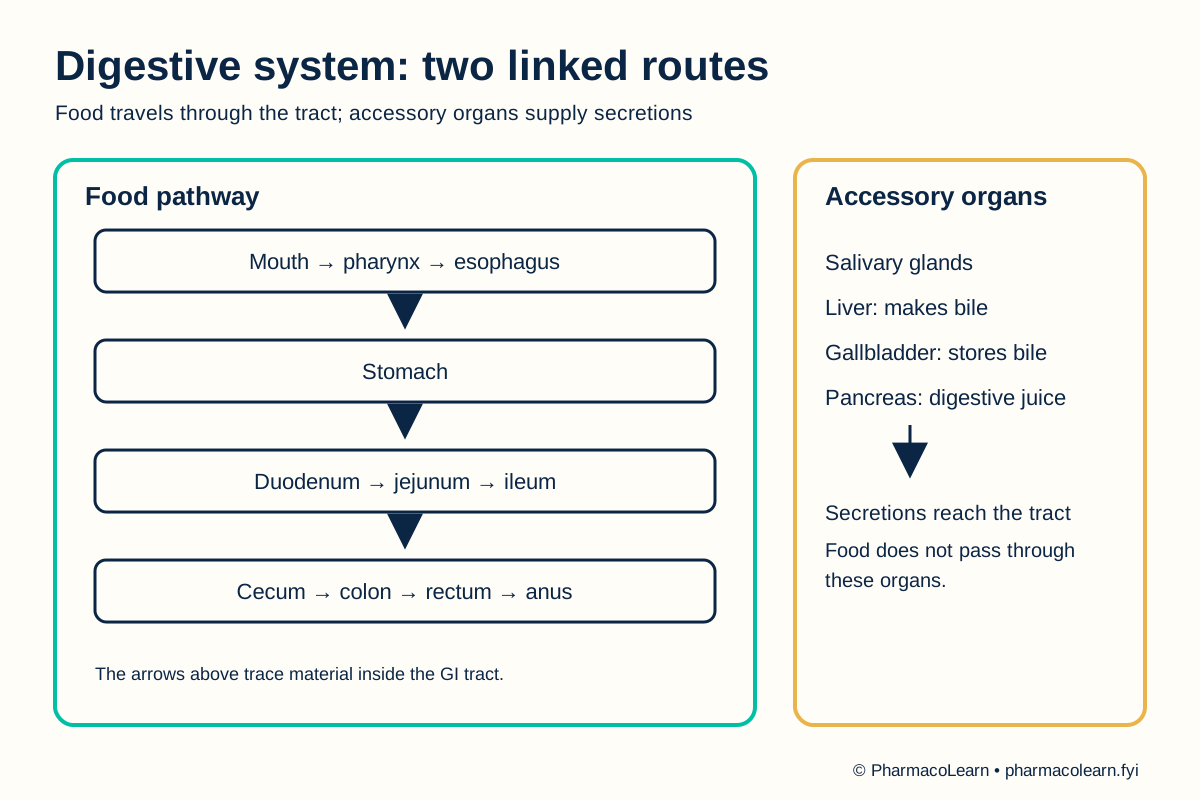
The gastrointestinal tract forms a continuous passage from mouth to anus. Food moves through the mouth, esophagus, stomach, small intestine, large intestine, and rectum before elimination. Digestion combines mechanical movement with digestive secretions; most nutrient absorption occurs in the small intestine, and the large intestine reclaims water. The salivary glands, liver, gallbladder, and pancreas contribute secretions or storage functions without being part of the route that food itself travels. The liver makes bile, the gallbladder stores it, and the pancreas delivers digestive enzymes to the small intestine. [2]
4. Procedure
- Ask the instructor which chart, model, or specimen may be handled. Orient the torso model before removing any part. Check for fragile attachments and follow the local handling rule: follow your college lab manual and your instructor's method sheet for quantities, editions, and instruments. [1]
- Start at the mouth on the overview chart. Trace the food path through the pharynx and esophagus into the stomach. Find these structures on the model and note where the esophagus enters the stomach. [2]
- Trace the route from the stomach into the small intestine. On a chart that shows its subdivisions, identify duodenum, jejunum, and ileum. Record any subdivision absent from the local model as not displayed. [2]
- Continue into the large intestine. Identify the cecum, colon, rectum, and anus on the chart; use the model to locate whichever of these are visible. [2]
- Locate salivary glands, liver, gallbladder, and pancreas on the accessory-organ chart. Use arrows to distinguish the food pathway from the routes by which bile and pancreatic secretions reach the small intestine. Do not describe food as passing through these accessory organs. [2]
- Cover the labels and retrace the entire food pathway. At each step, record one visible clue and a brief function. Ask the instructor to check any uncertain identification. [2]
- Replace model parts correctly, follow the local cleaning rule, and wash hands after shared equipment. [1]
5. Calculation, graph, or images as appropriate
There is no calculation. Complete the table from the assigned chart or model. [2]
| Region | Structures to identify | Main role | Shown on local material? |
|---|---|---|---|
| Entry and transport | Mouth, pharynx, esophagus | Ingestion and movement | Record here |
| Stomach | Stomach | Mixing and digestion | Record here |
| Small intestine | Duodenum, jejunum, ileum | Digestion and absorption | Record here |
| Large intestine and exit | Cecum, colon, rectum, anus | Water recovery and elimination | Record here |
| Accessory organs | Salivary glands, liver, gallbladder, pancreas | Supply or store digestive secretions | Record here |
Worked tracing example: On a labelled chart, trace stomach → duodenum → jejunum → ileum → cecum. The duodenum is the first part of the small intestine, and the cecum begins the large intestine. Record the labels visible on the assigned model here. [2]
6. Result
Digestive tract and accessory structures correctly identified on the assigned material: Record here. Record absent model features separately from uncertain identifications. [2]
7. References
- Pharmacy Council of India. Pharm.D. Regulations, 2008, Appendix A. Lists digestive-system study with charts, models, and specimens.
- National Institute of Diabetes and Digestive and Kidney Diseases. Your Digestive System and How It Works. Digestive tract, accessory organs, movement, digestion, and absorption.
Back to Human Anatomy and Physiology Practicals · All Pharm D Practicals · Pharm D
Theory notes: Human Anatomy and Physiology