
← Human Anatomy and Physiology Practicals
Study of tissues of the human body: connective and nervous tissue
1. Aim
To identify connective and nervous tissues in prepared microscope slides and relate their visible organization to support and communication functions.
2. Requirements
Prepared, labelled slides or approved digital slide images of connective and nervous tissues; light microscope for glass slides; observation sheet; pencil; lens paper; and the instructor's slide key. The exact examples, stains, and available magnifications are specified in your college lab manual and instructor's method sheet for the local slide collection. No chemical reagent is needed when prepared slides are used. Wear a laboratory coat and other protective equipment specified by the teaching laboratory. [1, 2, 3]
3. Theory
Connective tissue contains cells dispersed within an extracellular matrix. The amount and arrangement of fibres and ground substance vary widely. Loose connective tissue has relatively open spaces and scattered fibres; dense regular connective tissue has closely packed, parallel collagen bundles. Adipose tissue is dominated by large fat-storing cells. Cartilage contains cells in spaces called lacunae within a firm matrix, while bone has a mineralized matrix with osteocytes in lacunae. Blood is also classified as connective tissue, although its fluid matrix makes its appearance very different from a section of tendon or cartilage. [1, 3]
Nervous tissue contains neurons, which receive and transmit information, and supporting glial cells. A neuron commonly has a large cell body with a nucleus and processes extending from it. In a section of a peripheral nerve, most visible structures are bundles of nerve fibres and their surrounding connective tissue rather than whole neuron cell bodies. Slide orientation and stain affect what can be seen, so a single process or a round profile should not be identified by guesswork. [2, 3]
4. Procedure
- Confirm each prepared slide's label with the instructor and record the supplied specimen name separately from the features you observe. The slide identity, stain, and section orientation are specified in your college lab manual and instructor's method sheet for the actual teaching set. Handle glass slides by their edges.
- Inspect a connective tissue slide at low power to see the overall arrangement, then at a suitable higher power to compare cells, fibres, and matrix. The objective sequence for the local microscope is specified in your college lab manual and instructor's method sheet.
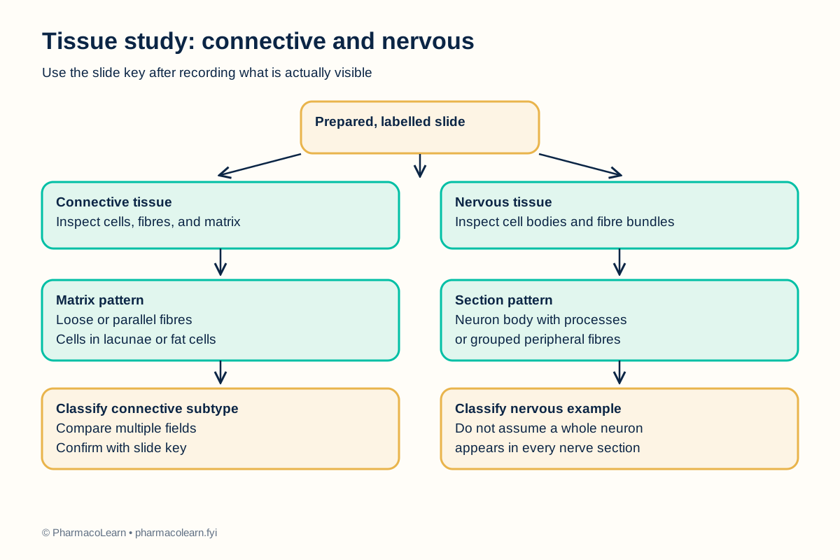
- For each connective tissue example, record whether the matrix appears abundant, whether fibres are loose or parallel, and whether cells occupy lacunae or dominate the field. Compare multiple regions before selecting a subtype. [1, 3]
- On a nervous tissue slide, locate a suitable region at low power. At higher power, look for a neuron cell body and processes if the preparation contains them. On a peripheral nerve section, look for grouped fibres and surrounding connective tissue. Do not assume both patterns will be present on the same slide. [2, 3]
- Use the instructor's slide key to distinguish genuine structures from folds, tears, stain deposits, or empty spaces caused by preparation. If a feature is not visible, record it as not seen rather than inventing it.
- Complete the observation table and the labelled identification flowchart. Record the findings and final identification for each slide here for instructor review.
- Return slides to storage, clean the microscope as directed, and report damaged glass. Do not pick up broken fragments with bare fingers.
5. Calculation, graph, or images as appropriate
No numerical calculation is required. Use the table as a record of actual visual evidence. The clues describe common appearances and do not guarantee that every slide will show each feature. [1, 2, 3]
| Tissue example | Clues to inspect | Student's observed features | Identification |
|---|---|---|---|
| Loose connective tissue | Scattered fibres and cells in relatively open matrix | Record during practical | Record after review |
| Dense regular connective tissue | Closely packed, largely parallel fibre bundles | Record during practical | Record after review |
| Adipose tissue | Large fat-storing cell profiles with little visible matrix | Record during practical | Record after review |
| Cartilage or bone | Cells in lacunae within a firm matrix | Record during practical | Record after review |
| Nervous tissue with neuron cell bodies | Large soma, nucleus, and processes where visible | Record during practical | Record after review |
| Peripheral nerve section | Bundles of nerve fibres with connective tissue sheaths | Record during practical | Record after review |
6. Result
For each supplied slide, state the identified tissue or subtype and cite at least two visible clues. Record the actual slide findings and identifications here; this article reports no observation from a specimen.
7. References
- OpenStax. Anatomy and Physiology 2e, section 4.3, Connective Tissue Supports and Protects. Connective tissue matrix, cells, and classes.
- OpenStax. Anatomy and Physiology 2e, section 4.5, Nervous Tissue Mediates Perception and Response. Neurons and glial cells.
- Robert L. Sorenson and T. Clark Brelje, Histology Guide. Tissue types overview and peripheral nerve slide. Slide-based tissue and nerve context.
- Pharmacy Council of India. Pharm.D. Regulations, 2008, Appendix A, Human Anatomy and Physiology practical list. Syllabus authority for this study item.
Back to Human Anatomy and Physiology Practicals · All Pharm D Practicals · Pharm D
Theory notes: Human Anatomy and Physiology