
← Human Anatomy and Physiology Practicals
Study of tissues of the human body: epithelial and muscular tissue
1. Aim
To identify epithelial and muscular tissues in prepared microscope slides and explain how their visible features relate to their functions.
2. Requirements
Prepared, labelled slides or approved digital slide images of epithelial and muscular tissues; light microscope when glass slides are used; observation sheet; pencil; lens paper; and the instructor's slide key. The slides, stain, and available magnifications are specified in your college lab manual and instructor's method sheet for the teaching laboratory's set. No chemical reagent is needed when prepared slides are used. Wear a laboratory coat and any additional protective equipment required locally. [1, 2, 3]
3. Theory
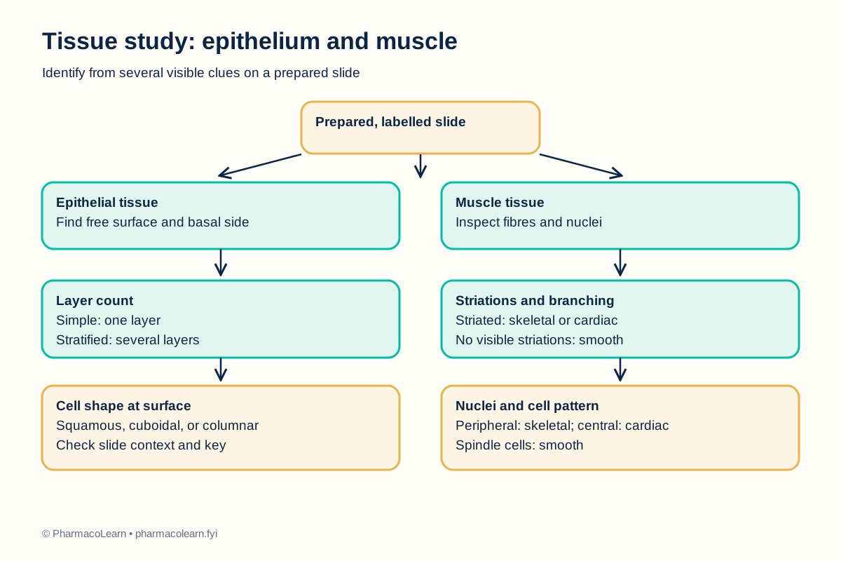
Epithelium forms closely packed sheets that cover surfaces, line spaces, and contribute to glands. It has a free, or apical, surface and an attached basal surface. Classification begins with the number of layers: a single layer is simple, while several layers are stratified. The surface cell shape then helps distinguish squamous, cuboidal, and columnar epithelium. Pseudostratified epithelium looks multilayered because nuclei lie at different heights, although its cells contact the basal layer. A slide must be interpreted with its tissue context because an oblique section can change the apparent shape. [1, 3]
Muscle is specialized for contraction. Skeletal muscle usually shows long, parallel, striated fibres with multiple nuclei near their edges. Cardiac muscle has striated, branching cells with centrally placed nuclei and intercalated discs where they join. Smooth muscle lacks visible cross-striations and has elongated, spindle-shaped cells with central nuclei. A small field may not reveal every feature, so identification should use several fields and the slide key. [2, 3]
4. Procedure
- Under instructor guidance, confirm each slide label and record its supplied tissue name without treating it as an observation. The slide identities and stains in the local set are specified in your college lab manual and instructor's method sheet. Handle glass slides by their edges and keep the microscope bench clear.
- Start with the lowest-power objective. Focus using the coarse adjustment only at low power, then move to a higher suitable magnification using fine adjustment. The objective sequence for the available microscope is specified in your college lab manual and instructor's method sheet.
- On each epithelial slide, find the free surface and underlying tissue. Record whether the cells form one layer or several, the shape of the surface cells, the position of nuclei, and any visible special feature such as cilia. Compare the pattern with the slide key. [1, 3]
- On each muscle slide, inspect more than one field. Record whether fibres run in parallel or branch, whether striations are visible, and the approximate position and number of nuclei in a cell profile. Look for intercalated discs only where the preparation makes them visible. [2, 3]
- Identify the tissue type from the combined clues. If a field is folded, damaged, too thick, or unclear, move to a better region or ask the instructor for a second slide. Do not infer an absent feature from a poor field.
- Enter observations in the table below and make a labelled sketch or use the supplied flowchart. Record the observed findings and final identifications here, then check them against the actual slides.
- Return slides to their labelled storage slots, clean the microscope as instructed, and report any broken glass. Do not handle fragments with bare fingers.
5. Calculation, graph, or images as appropriate
No numerical calculation is required. Use the table to connect a feature you actually see to a tissue identification. The comparison clues are teaching guides, not substitute observations. [1, 2]
| Tissue to examine | Distinguishing clues to look for | Student's observed features | Identification |
|---|---|---|---|
| Simple epithelium | One cell layer, with squamous, cuboidal, or columnar shape | Record during practical | Record after review |
| Stratified epithelium | Several layers, classified by surface cell shape | Record during practical | Record after review |
| Skeletal muscle | Parallel striated fibres, nuclei toward fibre edges | Record during practical | Record after review |
| Cardiac muscle | Striated branching cells, central nuclei, possible intercalated discs | Record during practical | Record after review |
| Smooth muscle | No visible cross-striations, spindle-shaped cells, central nuclei | Record during practical | Record after review |
6. Result
After the slides are examined, record the epithelial type and the muscle type for each labelled preparation, with at least two visible features supporting each identification. Record the actual slide findings and final identifications here; none are claimed from a specimen in this article.
7. References
- OpenStax. Anatomy and Physiology 2e, section 4.2, Epithelial Tissue. Epithelial layers, cell shapes, and surface organization.
- OpenStax. Anatomy and Physiology 2e, section 4.4, Muscle Tissue and Motion. Skeletal, cardiac, and smooth muscle features.
- Robert L. Sorenson and T. Clark Brelje, Histology Guide. Tissue types overview. Slide-based overview of the four basic tissues.
- Pharmacy Council of India. Pharm.D. Regulations, 2008, Appendix A, Human Anatomy and Physiology practical list. Syllabus authority for this study item.
Back to Human Anatomy and Physiology Practicals · All Pharm D Practicals · Pharm D
Theory notes: Human Anatomy and Physiology