
← Human Anatomy and Physiology Practicals
Study of the respiratory system using charts, models, and specimens
1. Aim
To identify the main air passages, lungs, alveolar region, and diaphragm on charts, models, or approved specimens, then trace the route of inhaled air. [1, 2]
2. Requirements
A labelled respiratory-system chart; an unlabelled airway and lung model; an alveolar-region chart or model; an observation sheet; and gloves if the instructor provides specimens that require them. The available models, removable parts, specimen-handling rules, and assessed landmarks are specified in your college lab manual and instructor's method sheet. No breathing test or contact with a person is required. [1]
3. Theory
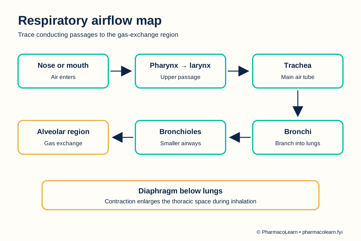
Air passes from the nose or mouth through the pharynx, larynx, trachea, bronchi, and smaller bronchioles toward the alveoli. The earlier passages conduct and condition air. The thin alveolar region is where oxygen and carbon dioxide are exchanged with nearby capillary blood. The diaphragm is a major muscle of ventilation; its contraction enlarges the thoracic space during inhalation. A model may simplify or omit small branches, so a chart of the entire path should be used alongside it. [2, 3]
4. Procedure
- Confirm with the instructor which charts, models, or specimens may be handled. Check how the model is oriented and whether any part is loose or fragile. Local handling instructions are specified in your college lab manual and instructor's method sheet. [1]
- On the overview chart, locate the nasal cavity, oral cavity, pharynx, larynx, and trachea. Follow the air route in that order and note where the pathway divides toward the two lungs. [2]
- On the lung model, identify the right and left main bronchi and at least one progressively smaller airway branch. Use the alveolar chart to continue from bronchioles to the gas-exchange region. Record any branch not shown on the model as not displayed. [2, 3]
- Find the lungs within the thoracic cage and locate the diaphragm below them. On a suitable chart, compare the diaphragm's position during inspiration and expiration. Do not treat a static model as a measurement of breathing movement. [2, 3]
- On the alveolar chart, point to the air space and the surrounding capillary region. Explain that gas exchange occurs across their thin boundary, while the trachea and larger bronchi mainly conduct air. [3]
- Cover the labels and retrace the pathway from nasal cavity to alveolar region. For each structure, note one positional clue or relationship visible on the assigned material. Ask the instructor to resolve uncertain labels. [2]
- Return the model to its original arrangement and follow the local cleaning and hand-hygiene rule. [1]
5. Calculation, graph, or images as appropriate
There is no calculation. Use the table for observations from the assigned chart or model. [2, 3]
| Region | Structure to identify | Role to explain | Shown on local material? |
|---|---|---|---|
| Entry and upper passage | Nasal cavity, pharynx, larynx | Receives and directs air | Record here |
| Main conducting passage | Trachea, main bronchi | Carries air into lungs | Record here |
| Small airway | Bronchioles | Distributes air toward alveoli | Record here |
| Gas-exchange region | Alveoli and surrounding capillaries | Exchanges respiratory gases | Record here |
| Ventilation muscle | Diaphragm | Changes thoracic volume | Record here |
Worked tracing example: On an airway chart, follow trachea → main bronchus → smaller bronchi → bronchiole → alveolar region. Identify each transition by its position and branching pattern. Record the structures visible on the assigned model here. [2, 3]
6. Result
Respiratory structures identified and air pathway traced on the assigned material: Record here. Record any structure omitted from the model separately from a label that remains uncertain. [2]
7. References
- Pharmacy Council of India. Pharm.D. Regulations, 2008, Appendix A. Lists respiratory-system study with charts, models, and specimens.
- StatPearls. Anatomy, Airway. Upper and lower airway structures.
- Kia'i N, Bajaj T. Histology, Respiratory Epithelium. StatPearls. Conducting and gas-exchange regions.
Back to Human Anatomy and Physiology Practicals · All Pharm D Practicals · Pharm D
Theory notes: Human Anatomy and Physiology