
← Human Anatomy and Physiology Practicals
Study of the cardiovascular system using charts, models, and specimens
1. Aim
To identify the heart chambers, major valves and vessels, and the pulmonary and systemic circuits on teaching charts, models, or approved specimens. [1, 2, 3]
2. Requirements
A labelled cardiovascular chart; an unlabelled heart model with removable sections if available; a circulation chart showing lungs and body tissues; an observation sheet; and gloves where local specimen rules require them. The exact model, whether valves are visible, specimen-handling rules, and assessed structures are specified in your college lab manual and instructor's method sheet. No examination of a person or dissection is needed. [1]
3. Theory
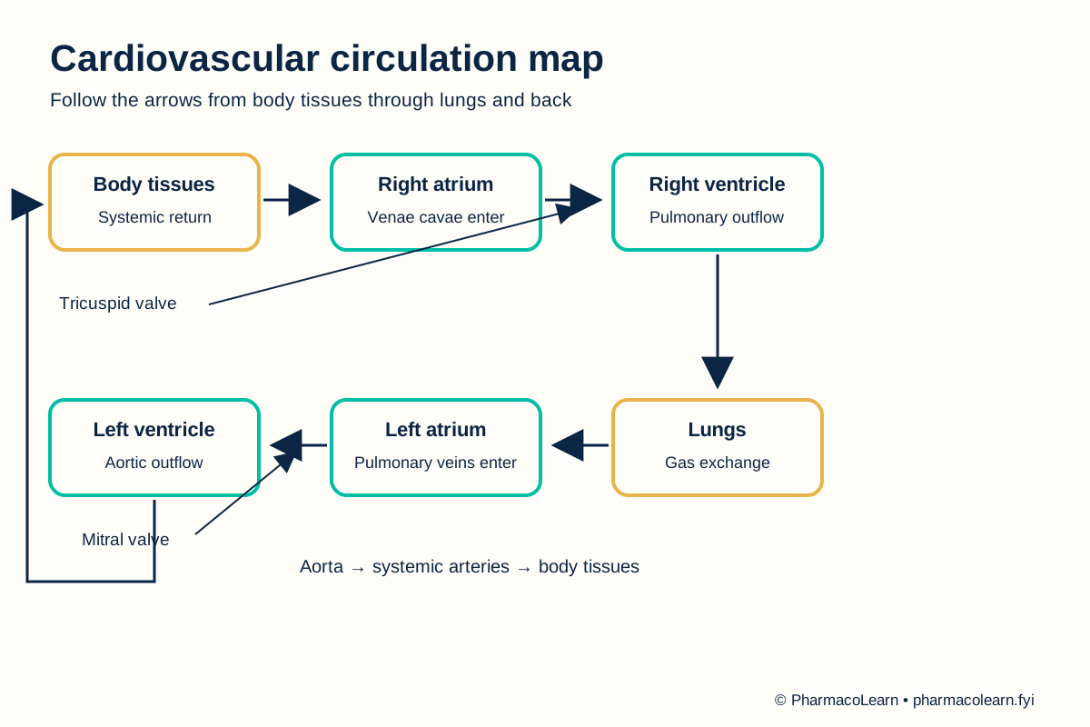
The cardiovascular system consists of the heart and blood vessels. The right side of the heart receives systemic venous blood and sends it through the pulmonary arteries to the lungs. Pulmonary veins return oxygenated blood to the left side, which sends it through the aorta into systemic arteries. The four chambers are right and left atria and ventricles. Tricuspid and mitral valves lie between atria and ventricles; pulmonary and aortic valves guard the ventricular outflow tracts. Arteries carry blood away from the heart and veins return it, regardless of oxygen content. Capillaries provide the main exchange surface in tissues. [2, 3, 4]
4. Procedure
- Confirm with the instructor which chart, model, or specimen may be handled. Note the orientation, removable parts, and cleaning rule. Local handling instructions are specified in your college lab manual and instructor's method sheet. [1]
- On the overview chart, locate the heart between the lung regions and identify the pulmonary and systemic circuits. Follow the arrows rather than relying on colour alone. [2, 3]
- On the heart model, locate right and left atria, right and left ventricles, and the septum. Check the model's anatomical right and left before naming chambers. [2]
- Trace systemic venous return through the superior and inferior vena cavae into the right atrium, then through the tricuspid valve to the right ventricle. Continue through the pulmonary valve and pulmonary trunk toward the lungs. If a structure is hidden, use the chart and record not displayed for the model. [2]
- Trace return from the lungs through pulmonary veins to the left atrium. Continue through the mitral valve to the left ventricle, then through the aortic valve and aorta toward body tissues. [2, 3]
- Point out one artery, one vein, and a capillary bed on the circulation chart. Explain each by direction of flow and role. Cover the labels and retrace the complete path aloud or on the observation sheet. [3, 4]
- Record each identified structure, its visible clue, and any model limitation. Return the model parts in the instructor's required arrangement and wash hands after shared equipment. [1]
5. Calculation, graph, or images as appropriate
There is no calculation. Use the table to record what the assigned teaching material actually shows. [2, 3]
| Structure or circuit | Identification clue | Visible on local material? |
|---|---|---|
| Right atrium and right ventricle | Receive systemic venous blood; send blood toward lungs | Record here |
| Pulmonary trunk and arteries | Leave right ventricle toward lungs | Record here |
| Pulmonary veins | Return from lungs to left atrium | Record here |
| Left atrium and left ventricle | Receive pulmonary return; send blood into aorta | Record here |
| Aorta and systemic arteries | Leave left ventricle toward body tissues | Record here |
| Four heart valves | Located at atrioventricular and ventricular outflow openings | Record here |
Worked tracing example: On a labelled teaching chart, follow right ventricle → pulmonary trunk → lungs → pulmonary veins → left atrium. This is the pulmonary circuit. Record the labels visible on the assigned chart here. [2, 3]
6. Result
Heart chambers, great vessels, valves, and the two circulation paths identified: Record here. Note structures that were not visible on the assigned model. This is an anatomical identification exercise, not a clinical assessment. [2, 3]
7. References
- Pharmacy Council of India. Pharm.D. Regulations, 2008, Appendix A. Lists cardiovascular-system study with charts, models, and specimens.
- Bamalan OA, Jozsa F, Soos MP. Anatomy, Thorax, Heart Great Vessels. StatPearls. Great-vessel connections and flow.
- Matienzo D, Bordoni B. Anatomy, Blood Flow. StatPearls. Pulmonary and systemic circuits.
- Sundjaja JH, Bordoni B. Anatomy, Thorax, Heart Pulmonic Valve. StatPearls. Heart chambers and valve roles.
Back to Human Anatomy and Physiology Practicals · All Pharm D Practicals · Pharm D
Theory notes: Human Anatomy and Physiology