
← Human Anatomy and Physiology Practicals
Study of the reproductive system using charts, models, and specimens
1. Aim
To identify the principal structures shown on typical female and male reproductive-system charts or models and trace the broad routes of an oocyte and sperm. [1, 2]
2. Requirements
Labelled and unlabelled female and male pelvic charts or models; an observation sheet; and gloves if the instructor provides specimens that require them. The available models, specimen-handling rules, and assessed structures are specified in your college lab manual and instructor's method sheet. No examination of a person, sampling, or dissection is required. [1]
3. Theory
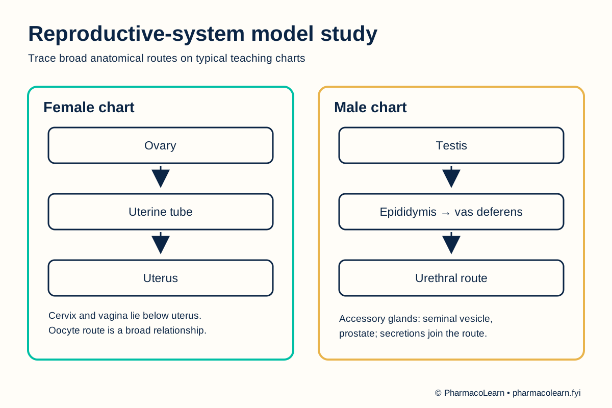
Typical female reproductive anatomy includes ovaries, uterine tubes, uterus, cervix, vagina, and external genital structures. The ovaries produce oocytes; an oocyte released at ovulation may enter a uterine tube, and the tube opens toward the uterus. Typical male reproductive anatomy includes testes, epididymides, vasa deferentia, accessory glands, urethra, and penis. The testes produce sperm, which pass through the epididymis and vas deferens before reaching the urethral route. Charts show common anatomical patterns; real bodies vary, and an anatomy model cannot establish any person's reproductive function. [2, 3, 4]
4. Procedure
- Ask the instructor which charts, models, or specimens may be handled. Orient each pelvic model and check whether sections are removable. Local handling instructions are specified in your college lab manual and instructor's method sheet. [1]
- On the female chart, identify ovaries, uterine tubes, uterus, cervix, and vagina. Locate the uterus relative to the bladder and rectum if those neighboring organs are shown. [3]
- Trace the broad oocyte route from ovary toward uterine tube and then toward uterus. Explain that the chart shows anatomical relationships, not a guaranteed event in any individual cycle. [3]
- On the male chart, identify testes, scrotum, epididymides, vasa deferentia, seminal vesicles, prostate, urethra, and penis. Record any missing structure as not displayed rather than guessing its location. [2, 4]
- Trace the broad sperm route from testis to epididymis, vas deferens, and urethra. Identify accessory glands as contributing secretions rather than as the site where sperm are produced. [2, 4]
- Cover the labels and repeat both routes. For each named structure, record one positional clue and one basic role. Ask the instructor to check uncertain identifications. [2, 3]
- Replace model sections correctly, follow the local cleaning rule, and wash hands after shared equipment. [1]
5. Calculation, graph, or images as appropriate
There is no calculation. Complete the observation table from the assigned charts or models. [2, 3, 4]
| Chart or model | Structure group | Role or relationship to explain | Shown locally? |
|---|---|---|---|
| Female | Ovary and uterine tube | Oocyte production and route toward uterus | Record here |
| Female | Uterus and cervix | Uterine cavity and lower opening | Record here |
| Female | Vagina | Passage below cervix | Record here |
| Male | Testis and epididymis | Sperm production and maturation | Record here |
| Male | Vas deferens and urethra | Route of sperm transport | Record here |
| Male | Seminal vesicle and prostate | Accessory secretions | Record here |
Worked identification example: On a labelled female pelvic chart, the narrow structure extending from an ovary toward the uterus is the uterine tube. Confirm its connection and position before naming it. Record whether the corresponding structure is shown on the assigned model here. [3]
6. Result
Structures and broad reproductive routes identified on the assigned material: Record here. Note any model omissions or unresolved labels. This exercise does not assess fertility, pregnancy, or health status. [2, 3]
7. References
- Pharmacy Council of India. Pharm.D. Regulations, 2008, Appendix A. Lists reproductive-system study with charts, models, and specimens.
- Gurung P, Yetiskul E, Jialal I. Physiology, Male Reproductive System. StatPearls. Male structures and sperm route.
- Ameer MA, Peterson DC. Anatomy, Abdomen and Pelvis: Uterus. StatPearls. Uterine and pelvic anatomy.
- Reproductive Concepts, section 8.2. Nursing Health Promotion, NCBI Bookshelf. Female and male reproductive structures and basic roles.
Back to Human Anatomy and Physiology Practicals · All Pharm D Practicals · Pharm D
Theory notes: Human Anatomy and Physiology