
← Human Anatomy and Physiology Practicals
Determination of differential count of blood
1. Aim
To determine the percentage distribution of the five main leukocyte types in a stained peripheral blood film by manual differential counting.
2. Requirements
Specimen and chemicals: A correctly labelled anticoagulated blood specimen supplied under supervision, or an approved prepared teaching slide; a laboratory-approved Romanowsky stain and its matching buffer. This article follows the Leishman stain option described by WHO. The exact stain product, buffer preparation, safety instructions, and any local alternative are specified in your college lab manual and instructor's method sheet; check them before use. [1]
Instruments and supplies: Clean, dry microscope slides; smooth-edged spreader slide; staining rack or tray; light microscope; cover glass or mounting arrangement suitable for the objective; differential cell counter or tally sheet; timer; and designated waste containers.
Protective equipment: Laboratory coat, gloves, and eye protection according to the local risk assessment. Stains containing methanol require supervised handling away from ignition sources. [1, 2]
3. Theory
A differential count describes the proportion of leukocytes that are neutrophils, lymphocytes, monocytes, eosinophils, and basophils. A thin blood film spreads cells so that their nuclei, cytoplasm, and granules can be examined after Romanowsky staining. The stain helps distinguish the cell types, but the smear must be thin enough for cells to retain their shape and the staining must be even. The result is a percentage for each type, not a total W.B.C. concentration. If a separately validated total W.B.C. count is available, an absolute number for each type can be calculated from its percentage. [1]
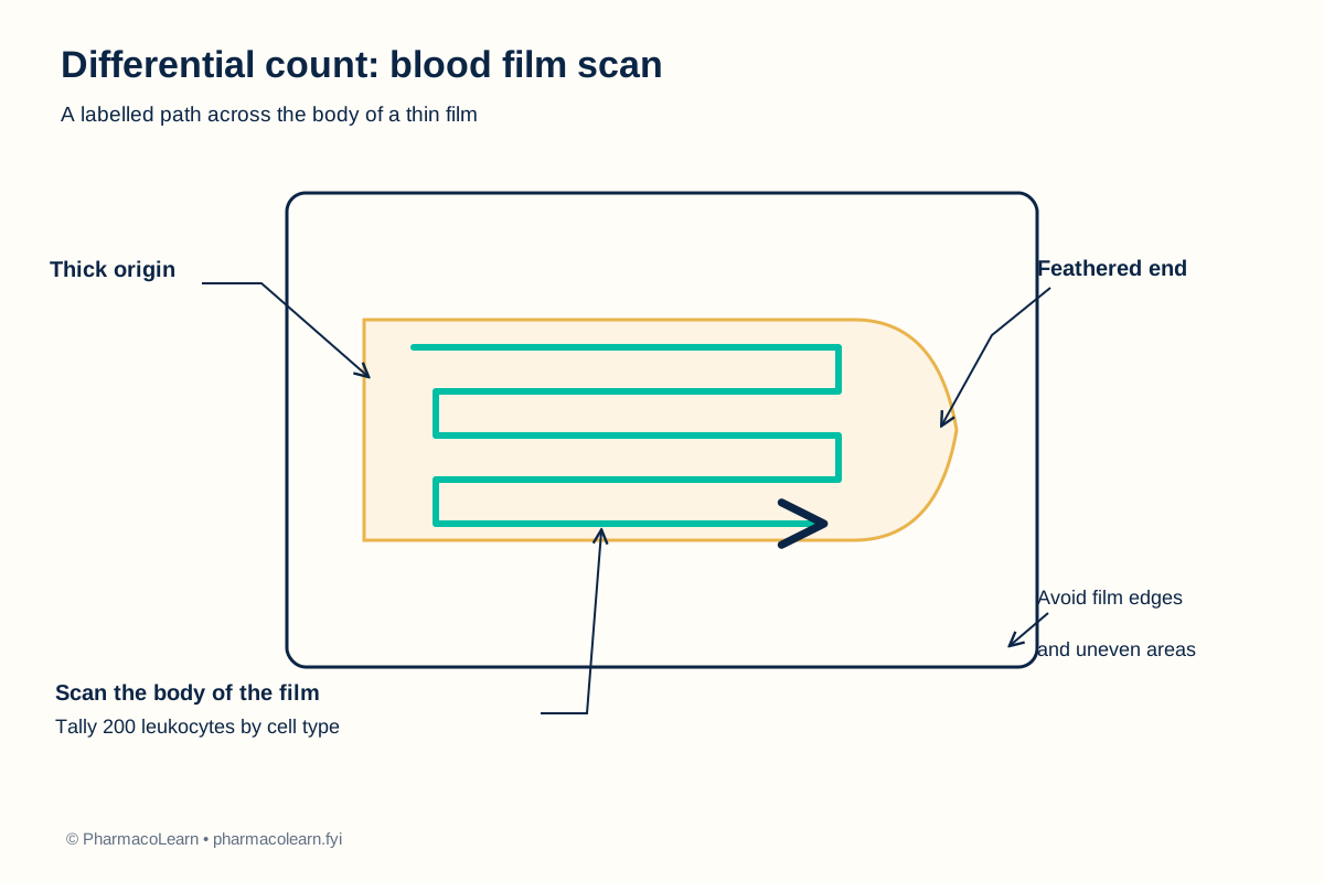
The WHO haematology SOP describes a 200-cell differential and a broad transverse scan across the body of the film, avoiding its edges. This sample count gives each category's percentage as its tally divided by 200 and multiplied by 100. Compare with your laboratory's current review rule and reference interval. [1]
4. Procedure
- Under instructor supervision, confirm the specimen or teaching-slide identity and the stain instructions. Treat human blood and used slides as potentially infectious. Put on the required protective equipment. [1, 2]
- If preparing a film, gently mix the anticoagulated blood. Place a small drop near one end of a clean slide. Draw a smooth spreader back to touch the drop, let the blood run along its edge, then push the spreader forward in one steady motion to form a thin film with a feathered end. Air-dry promptly and label the slide. A prepared teaching slide can be used when the laboratory does not authorize student film preparation. [1]
- For the WHO Leishman method, cover the dry film with neat stain for 3 minutes. Carefully add an equal amount of pH 6.8 buffered water, mix gently on the slide, and leave for about 12 minutes. Rinse gently, flood with buffer for 1 minute, then dry. The precise stain volume and any product-specific adaptation are specified in your college lab manual and instructor's method sheet. A trained instructor must manage the stain's chemical hazards and waste. [1]
- Survey the entire film at low power. Choose a well-stained region where red cells are evenly distributed and mainly touch without overlapping. Reject a film with thick ridges, stain deposits, a poor feathered edge, or markedly uneven leukocyte distribution. [1]
- Examine the selected region with an appropriate higher-power objective. Use the WHO broad transverse scan across the body of the film, staying clear of the edges. Tally 200 leukocytes, classifying each as neutrophil, lymphocyte, monocyte, eosinophil, or basophil. Use oil immersion when finer detail is needed and according to the microscope setup. [1]
- Record any cell that cannot be classified confidently and ask the instructor to review it. Unusual or immature cells must not be silently forced into a normal category. Compare with your laboratory's current rule for review and repeat counts of these cells. [1]
- Check that the five category tallies sum to 200, calculate the percentages, clean the microscope and work area as instructed, dispose of contaminated materials correctly, remove gloves, and wash hands. [1, 2]
5. Calculation, graph, or images as appropriate
Observation table. These are invented teaching tallies, not observations from a person.
| Leukocyte type | Example cells in 200-cell count | Percentage |
|---|---|---|
| Neutrophils | 114 | 57% |
| Lymphocytes | 60 | 30% |
| Monocytes | 16 | 8% |
| Eosinophils | 8 | 4% |
| Basophils | 2 | 1% |
| Total | 200 | 100% |
Worked calculation: For neutrophils, 114 ÷ 200 × 100 = 57%. Repeat the same calculation for each row and check that the percentages total 100%. If a separately validated total W.B.C. count were 10,000 cells/µL for this same illustrative specimen, the illustrative absolute neutrophil count would be 10,000 × 57 ÷ 100 = 5,700 cells/µL. That total count is example data, not a measured or normal value. [1]
6. Result
Worked example: The illustrative differential is 57% neutrophils, 30% lymphocytes, 8% monocytes, 4% eosinophils, and 1% basophils. Record the actual result here from the student's own film and tally. Interpretation requires the laboratory's reference information and clinical context; neither is supplied by this teaching example.
7. References
- S. M. Lewis and Sudarshan Kumari. Guidelines on Standard Operating Procedures for Haematology, section 11. World Health Organization Regional Office for South-East Asia, January 2000. Blood-film preparation, Leishman staining, scan pattern, and 200-cell differential.
- Centers for Disease Control and Prevention. Blood Specimens: Safety. Blood-handling precautions. Reviewed 3 May 2016.
- Pharmacy Council of India. Pharm.D. Regulations, 2008, Appendix A, first-year Human Anatomy and Physiology practical list. Syllabus authority for this experiment.
Back to Human Anatomy and Physiology Practicals · All Pharm D Practicals · Pharm D
Theory notes: Human Anatomy and Physiology