
← Human Anatomy and Physiology Practicals
Study of the axial skeleton using charts, models, and specimens
1. Aim
To identify the main regions of the axial skeleton on teaching charts, models, or approved specimens and relate each region to its protective or supporting role. [1, 2]
2. Requirements
A labelled overview chart for orientation; an unlabelled axial-skeleton chart or model for identification; skull and vertebral-column models; a rib-cage model; an observation sheet; and protective gloves if the instructor provides actual specimens. The available models, any specimen-handling rule, and the required list of fine landmarks are specified in your college lab manual and instructor's method sheet for the local teaching collection. No dissection is needed. [1, 2]
3. Theory
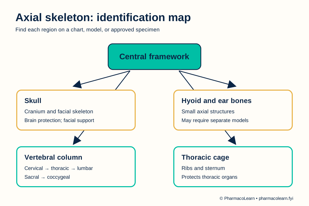
The axial skeleton forms the central framework of the head and trunk. It includes the skull, hyoid bone, auditory ossicles, vertebral column, and thoracic cage. The skull protects the brain and supports facial structures. Vertebrae surround the spinal canal and support the trunk. Ribs and sternum help form a cage around thoracic organs while permitting breathing movements. The appendicular skeleton contains the limb and girdle bones and is studied separately in the next practical. [2, 3]
4. Procedure
- Ask the instructor which chart, model, or specimen may be handled. Check its orientation and any fragile or sharp features. Apply gloves only where the local rule requires them. The collection and handling rule are specified in your college lab manual and instructor's method sheet. [1]
- On the overview model, locate the midline axis from skull to vertebral column and thoracic cage. Distinguish the axial region from the shoulder and pelvic girdles and limbs. [2]
- At the head, identify the cranium and facial skeleton. Locate the hyoid on a suitable model if visible. Record any structure absent from the collection as not displayed, not as absent from the human body. [2, 3]
- Trace the vertebral column from superior to inferior. Identify cervical, thoracic, lumbar, sacral, and coccygeal regions by their positions. On a separate vertebra model, locate the body, vertebral arch, and vertebral foramen if the model shows them. [2, 3]
- On the thoracic-cage model, identify ribs and sternum and show their relation to thoracic vertebrae. Use the chart to name the costal cartilages if the model omits them. [2, 3]
- Cover or turn away the labels and identify each region again from a second view. Record the observed landmark, the model used, and one function for each. Ask the instructor to resolve any uncertain identification before submitting the record. [2]
- Return every model or specimen to its assigned position. Clean hands after handling shared material. [1]
5. Calculation, graph, or images as appropriate
There is no calculation. Use this observation table with the actual teaching material. [2, 3]
| Axial region | Landmark to identify | Structure shown on local model? | Main role to explain |
|---|---|---|---|
| Skull | Cranium and facial skeleton | Record here | Protects brain; supports face |
| Hyoid and auditory ossicles | Hyoid; ear bones if displayed | Record here | Supports tongue; transmits sound |
| Vertebral column | Cervical, thoracic, lumbar, sacral, coccygeal regions | Record here | Supports trunk; protects spinal cord |
| Thoracic cage | Ribs and sternum | Record here | Protects thoracic organs; aids breathing |
Worked identification example: A model segment below the cervical vertebrae, bearing rib articulations, is identified as the thoracic vertebral region. State the visible clue before naming the region. Record the landmarks visible on the assigned model here. [3]
6. Result
Record the axial regions correctly identified and any landmarks that the local collection did not show: Record here. The completed observation sheet should distinguish an unseen structure from an incorrectly identified one. [2]
7. References
- Pharmacy Council of India. Pharm.D. Regulations, 2008, Appendix A. Lists the axial skeleton as part I of the chart, model, and specimen study.
- Wisconsin Technical College System. Medical Terminology 2e, Chapter 13: Skeletal System Terminology. Axial regions and major structures.
- DeSai C, Reddy V, Agarwal A. Anatomy, Back, Vertebral Column. StatPearls. Updated 2023. Vertebral regions and axial framework.
Back to Human Anatomy and Physiology Practicals · All Pharm D Practicals · Pharm D
Theory notes: Human Anatomy and Physiology